

Identifying inhibitors of intracellular protein fibrillization
a technology of intracellular protein fibrillation and inhibitors, which is applied in the direction of biochemistry apparatus and processes, instruments, peptide/protein ingredients, etc., can solve the problem of impracticality of most high-throughput screening uses
- Summary
- Abstract
- Description
- Claims
- Application Information
AI Technical Summary
Benefits of technology
Problems solved by technology
Method used
Image

Examples
example 1
Assays for Tau Fibrillization
[0084]Materials. Recombinant His-tagged htau40 was prepared as described previously. AD-derived PHFs prepared as in were generously supplied by Dr. Lester I. Binder (Northwestern University Medical School, Chicago, Ill.). Arachidonic acid (AA) was obtained from Cayman Chemicals (Ann Arbor, Mich.), and stored at −80° C. under argon until used. Alkyl sulfate detergents (12, 18, and 20 carbons) were obtained from Mallinckrodt (Paris, Ky.), Lancaster Synthesis (Pelham, N.H.), and Research Plus (Bayonne, N.J.), respectively, as sodium salts. Glutaraldehyde, uranyl acetate, and 300 mesh carbon-coated copper grids were from Electron Microscopy Sciences (Ft. Washington, Pa.). N-Phenyl-1-naphthylamine, ThS, Protamine (grade IV from salmon) and histone (type II-A from calf thymus) were from Sigma (St. Louis, Mo.). Stocks of protamine and histone were made in water at 800 μM and used the same day. Carboxylate conjugated polystyrene microspheres of defined nominal d...
example 2
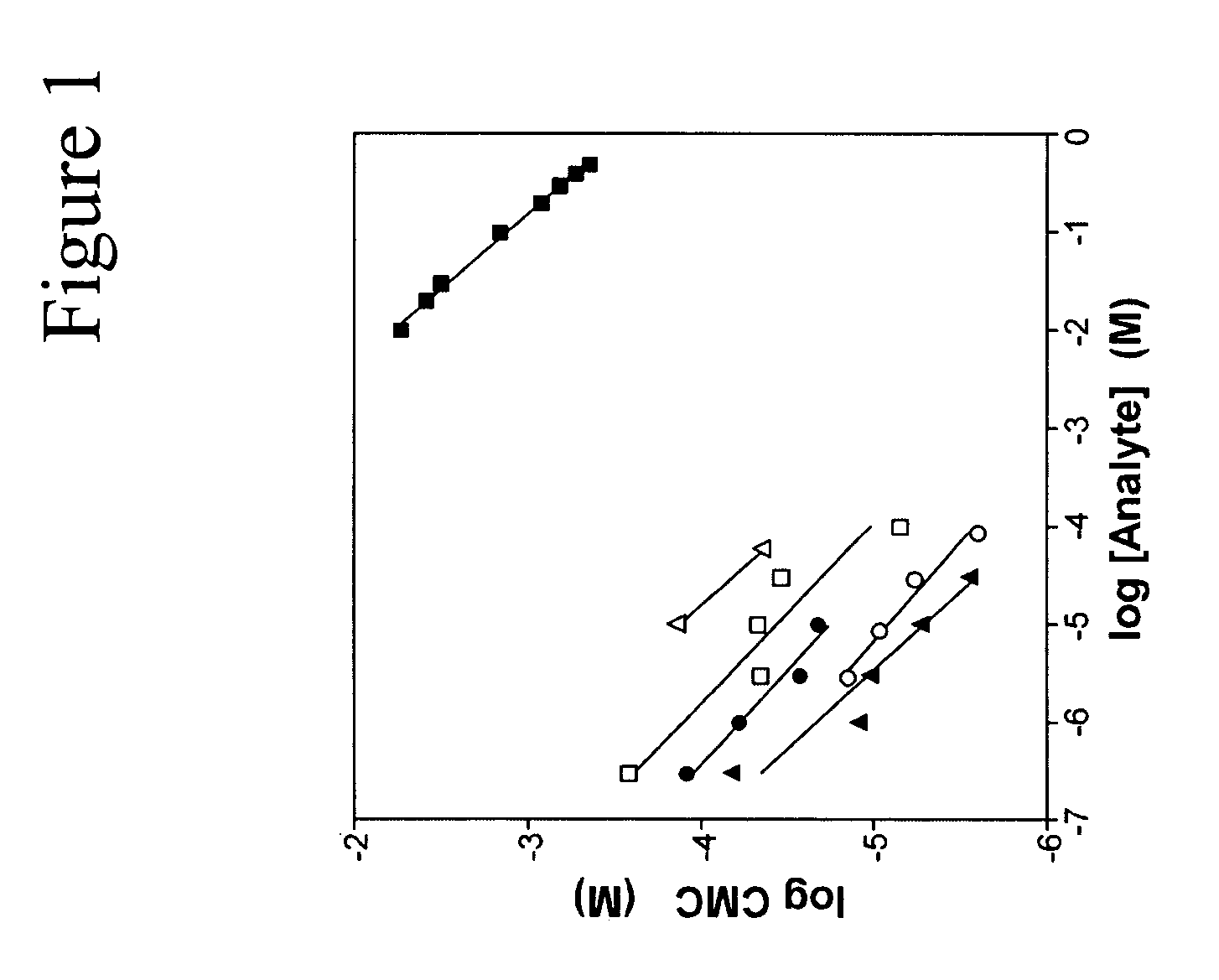
CMC Measurements.
[0091]CMC values for SDS were estimated at 37° C. using N-phenyl-1-naphthylamine as described previously, except that various concentrations of protamine, histone, α-synuclein, or htau40 were included with the samples. Detergent stock solutions were prepared in 1:1 water isopropanol.
[0092]Analytical Methods. Dependence of detergent CMC on protein concentration was fit to the empirical Corrin-Harkins equation:
log CMC=−kCH(log n)+bCH (1)
where n is the molar concentration of protein or salt and kCH and bCH are constants. The slope of these curves, kCH, approximates the proportion of detergent micelles that are ionized.
[0093]Sigmoidal reaction progress curves were fit to the sigmoidal logistic equation:
[0094]y=y0+A(1+ⅇ-kapp(t-t50))(2)
where y is total filament length (50 nm cutoff) measured at time t, yo is total filament length at time zero, t50 is time to 50% maximum fibrillization, A is the maximum total filament length at equilibrium, and kapp is the apparent first...
example 3
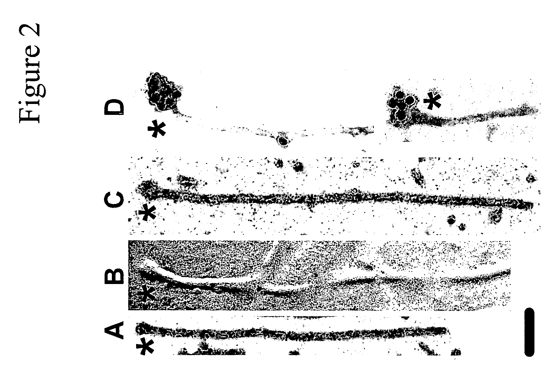
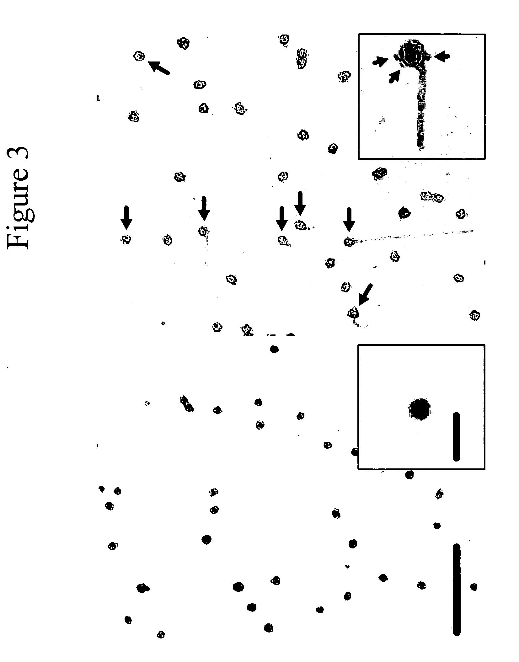
Micelles Serve as Nucleation Centers for Tau Fibrillization: Identification of Anionic Bodies on Protein Filaments.
[0102]When tau is incubated with preassembled anionic phospholipid vesicles, fibrillization proceeds from the vesicle surface, suggesting a key role for such surfaces in filament nucleation. However, it has been shown that when oppositely charged protein / surfactant pairs are coassembled, hierarchical structures more complicated than simple micelles or vesicles can form. To clarify the structural relationship between surfactant and tau in coassembly reactions, tau was fibrillized in the presence of AA and alkyl sulfate detergents (C18H37NaSO4 and C20H41NaSO4) and viewed by electron microscopy. These inducers were chosen because their long alkyl chains yield the largest micelles of any surfactants known to fibrillize tau in vitro. Low concentrations (18H37NaSO4 (FIG. 2B) and C20H41NaSO4 (FIG. 2C). Typically, swellings were seen at only one end of a filament (FIGS. 2A, C, ...
PUM
| Property | Measurement | Unit |
|---|---|---|
| diameter | aaaaa | aaaaa |
| diameter | aaaaa | aaaaa |
| diameter | aaaaa | aaaaa |
Abstract
Description
Claims
Application Information