Method and system for using ultrasound in cardiac diagnosis and therapy

a technology for cardiac diagnosis and therapy, applied in the field of techniques, can solve the problems of less efficient blood pumping, inconvenient operation, and inability to quickly electrically constrict the ventricle, and achieve the effects of convenient viewing of the left ventricle, reduced operating costs, and reduced operating costs

Inactive Publication Date: 2007-05-01
ST JUDE MEDICAL ATRIAL FIBRILLATION DIV
View PDF6 Cites 61 Cited by
  • Summary
  • Abstract
  • Description
  • Claims
  • Application Information

AI Technical Summary

Benefits of technology

[0009]A new therapy to treat heart failure is bi-ventricular pacing, or “resynchronization” therapy, where both ventricles of the heart are paced with an implantable pulse generator, commonly known as an artificial pacemaker. Normal pacing for a slow heart is performed via an implanted electrode in the right ventricle. The conduction myofibers (Purkinje fibers) conduct the electrical pulse and the ventricles contract synchronously in an inward direction, resulting in blood being pumped efficiently from the heart. In heart failure, the left ventricle becomes enlarged and conduction through the tissue of the left ventricular wall often becomes slow, so that the upper part of the left ventricle conducts as much as 200 to 250 milliseconds behind the apex area of the ventricles. This leads to poor and discoordinated contraction, and in many cases, an outward movement of the heart muscle, so that blood sloshes around rather than being squeezed out of the ventricle. Thus, an ideal location to place a pacing electrode in the left ventricle is in the area of slowest conduction, which can be a rather large area of the left ventricle, and may not always be the area that has the largest conduction. The problem facing physicians today is to locate the optimal spot for the permanent fixation of the pacing electrode. The thrust of this invention is to provide a method and device to optimize the location of the electrode.
[0023]4. Often times the heart failure patient has a number of co-morbidities showing symptoms at the same time, such as atrial fibrillation, ventricular tachycardias, and renal failures, among others. Atrial fibrillation and ventricular tachycardia can be brought under control via electrical shock cardioversion, either internally with catheters, or externally, although with much higher energy, with patches or paddles. A cardioversion device which could utilize the same electrodes that are otherwise introduced into the heart for pacemaker implantation, would be advantageous if also integrated with the overall electrophysiology system. In this manner, inadvertent shocks could be avoided as the trigger mechanism would come from the ventricular signal from the internal electrode. Thus, in one embodiment, the ultrasound imaging system of the present invention also comprises an integral defibrillation system whereby, if needed, internal cardiac defibrillation can be implemented quickly and easily. The integrated defibrillation electrode or system may be incorporated into the ultrasound imaging catheter, attached to the ultrasound imaging catheter, or as a separate electrode system or catheter which is inserted along with the ultrasound imaging catheter.

Problems solved by technology

The heart tissue can absorb fluid, the left ventricle does not allow quick electrical conduction, becomes enlarged, does not contract well, and becomes less efficient at pumping blood.
Heart failure leads to an extremely diminished lifestyle, and, left untreated, can be a major cause of mortality.
This leads to poor and discoordinated contraction, and in many cases, an outward movement of the heart muscle, so that blood sloshes around rather than being squeezed out of the ventricle.
The problem facing physicians today is to locate the optimal spot for the permanent fixation of the pacing electrode.
Capture may not be the best parameter to use.
Furthermore, advancing the electrode to the proper spot may not be easy.

Method used


Image

  • Method and system for using ultrasound in cardiac diagnosis and therapy
  • Method and system for using ultrasound in cardiac diagnosis and therapy
  • Method and system for using ultrasound in cardiac diagnosis and therapy

Examples


Embodiment Construction

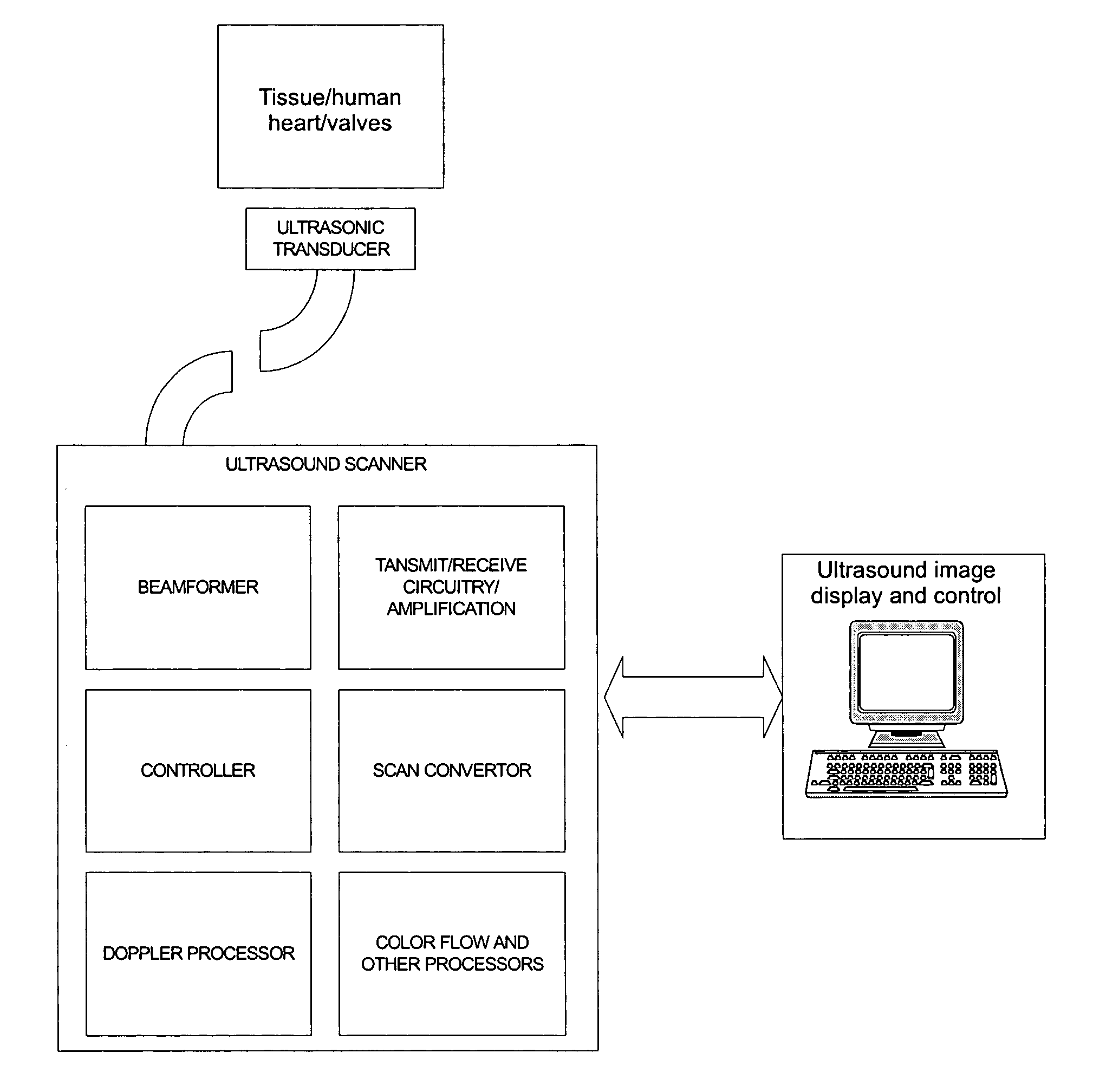
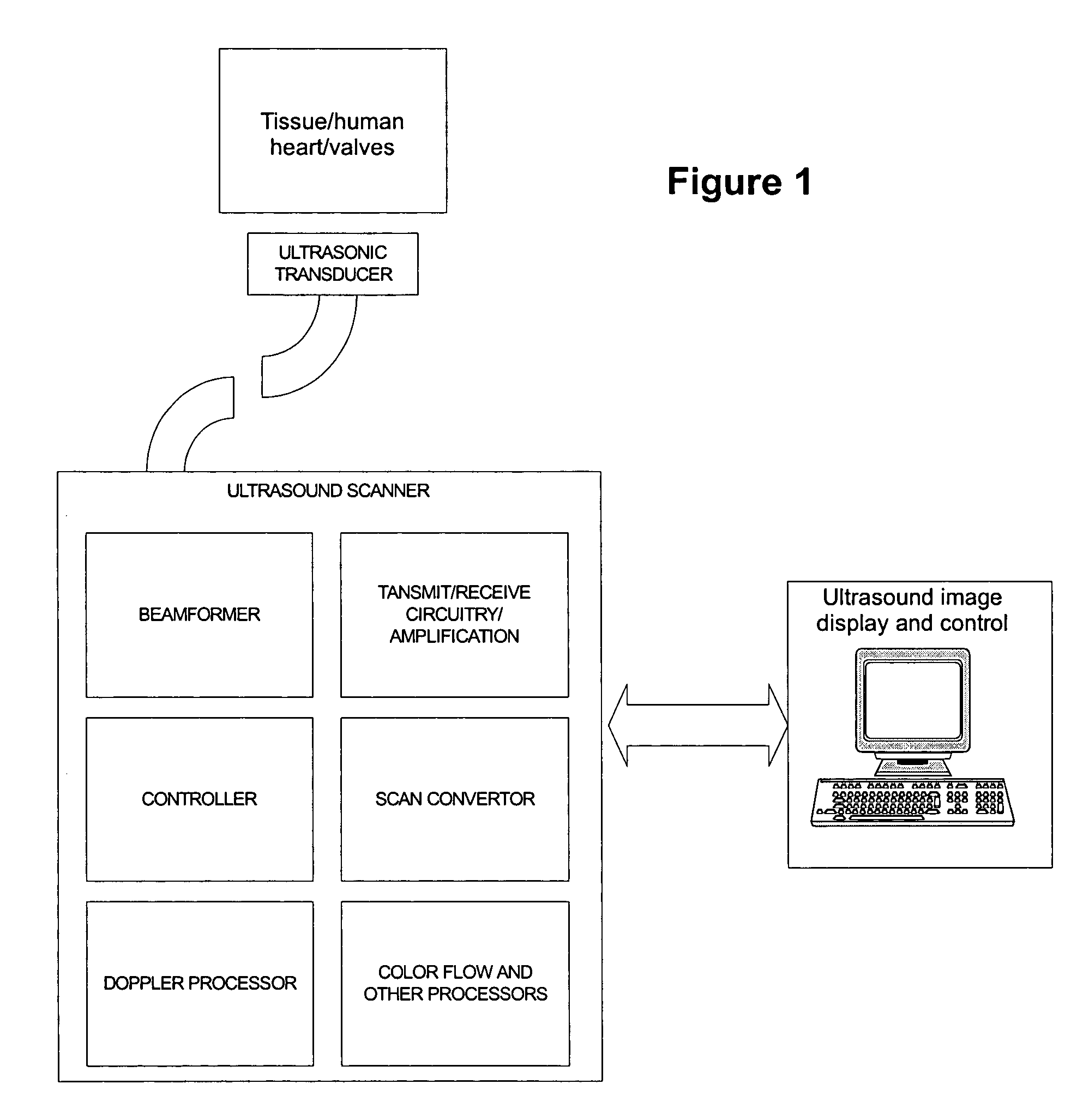
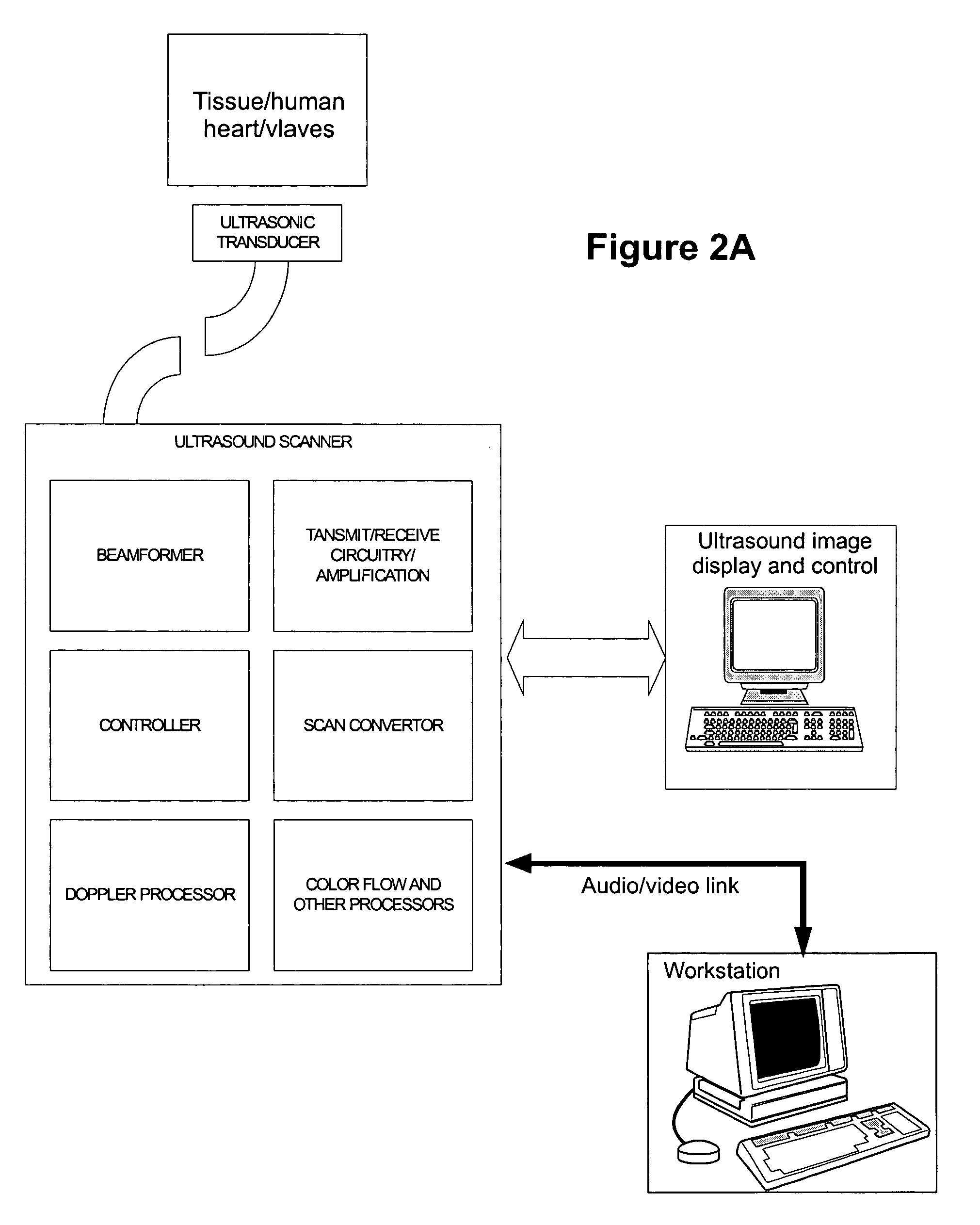
[0036]The present invention provides an ultrasound imaging system suitable for measuring cardiac output of a patient's heart, said system comprising:

[0037](1) an ultrasound imaging catheter comprising at least one transducer utilizing piezoelectric properties to generate acoustic signals from electrical signals in order to obtain ultrasound signals, wherein the at least one transducer is suitable for insertion into the patient's heart and to obtain ultrasound signals associated with an area of the patent's heart in which cardiac output is to be measured;

[0038](2) digital and / or analog electronics capable of generating and processing ultrasound signals from the at least one transducer to generate B-mode, M-mode, or Doppler representations of the cardiac output of the patient's heart; and

[0039](3) an associated computer that can generate and process the ultrasound signals in order to measure the cardiac output in the patient's heart.

[0040]This invention also provides a method of placi...


PUM


Abstract

A method and system for estimating the volume of blood ejected from the left ventricle of the heart to a succeeding chamber / conduit, or vice-versa, while imaging the intersection of such structures is described. The process utilizes either the M-mode to estimate volume differences in a view of the ventricle over time or Doppler processing techniques to obtain flow profiles across intersections (e.g., valves), or blood vessels, which are then utilized to calculate output. This process can be combined with ECG guidance / triggering to measure / track changes in output from beat to beat, or during the course of an evaluation or therapeutic procedure. This process can be specifically used for the placement of permanent pacemaker electrodes.

Description

RELATED APPLICATION[0001]This application is based on, and claims benefit of, U.S. Provisional Application Ser. No. 60 / 397,653, filed on Jul. 22, 2002, which is herby incorporated by reference.FIELD OF THE INVENTION[0002]The present invention relates to a technique for measuring cardiac output (i.e., total volume of blood ejected by the left ventricle in one cardiac cycle). It is achieved either by measuring volumetric flow at the entrance of the aorta, or by measuring the change in volume of the left ventricle through a cardiac cycle, using ultrasonic imaging and pulse wave Doppler techniques.BACKGROUND OF THE INVENTION[0003]Volumetric output of blood from the heart and / or circulatory system are of interest in various diagnostic and therapeutic procedures. Such measurements are of significant interest during electrophysiological evaluation / therapy to first evaluate the extent of dysfunction due to arrhythmia and subsequently to judge the effect / effectiveness of any ablations / therap...

Claims


Application Information

Patent Timeline
Patent Type & Authority Patents(United States)
IPC IPC(8): A61B8/06
CPCA61B8/065A61B8/0883A61B8/543A61B8/12
Inventor DALA-KRISHNA, PRAVEENBYRD, CHARLES BRYANJENKINS, DAVIDBYRD, CHARLES LEE
Owner ST JUDE MEDICAL ATRIAL FIBRILLATION DIV
Why Eureka
Try Eureka