Ultrasound color characteristic mapping

a color characteristic and ultrasonic technology, applied in the field of ultrasonic machines, can solve the problems of difficult or impossible direct assessment of new parameters, unacceptable inter-observer variability, and hampered evaluation of cardiac wall function, and achieve the effect of easy visualization of tissue motion parameter information

Active Publication Date: 2007-07-17
GE MEDICAL SYST GLOBAL TECH CO LLC
View PDF15 Cites 52 Cited by
  • Summary
  • Abstract
  • Description
  • Claims
  • Application Information

AI Technical Summary

Benefits of technology

[0015]Certain embodiments of the present invention afford an approach to more easily visualize tissue motion parameter information, such as tissue velocity and strain rate, in a two-dimensional ultrasound image such that more of the tissue motion parameter information is broken out and observed.

Problems solved by technology

Evaluation of cardiac wall function has been hampered by a lack of well-established parameters that may be used to increase the accuracy and objectivity in the assessment of, for example, coronary artery diseases.
It has also been shown that inter-observer variability between echo-centers is unacceptably high due to the subjective nature of the wall motion assessment.
Many of the new parameters have been difficult or impossible to assess directly by visual inspection of the ultrasound images generated in real-time.
The quantification has typically required a post-processing step with tedious, manual analysis to extract the necessary parameters.
However, the non-adaptive color schemes in the prior art are not well suited for visual determination of other parameters, such as quantitative velocities or strain rates.
Quantitative assessment of parameters such as velocities or strain rates from 2-D images has been difficult, even in lucky situations, with a good spread of measured imaging parameters.
None of the foregoing patents, however, describe or suggest any color mapping technique for generating an ultrasound display of moving structure that uses the full dynamic range of the color map.

Method used


Image

  • Ultrasound color characteristic mapping
  • Ultrasound color characteristic mapping
  • Ultrasound color characteristic mapping

Examples


Embodiment Construction

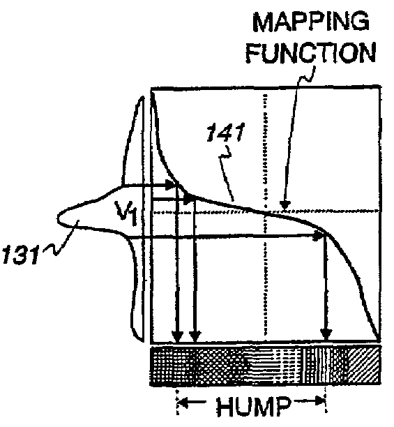
[0026]An embodiment of the present invention enables adaptive color mapping of moving tissue structure based on the distribution of movement parameter data. As used in this specification and claims, structure means non-liquid and non-gas matter, such as cardiac wall tissue. An embodiment of the present invention offers improved, real-time visualization and assessment of wall tissue function. The moving structure is characterized by a movement parameter, which means a parameter derived from movement of the structure, such as velocity or strain rate.

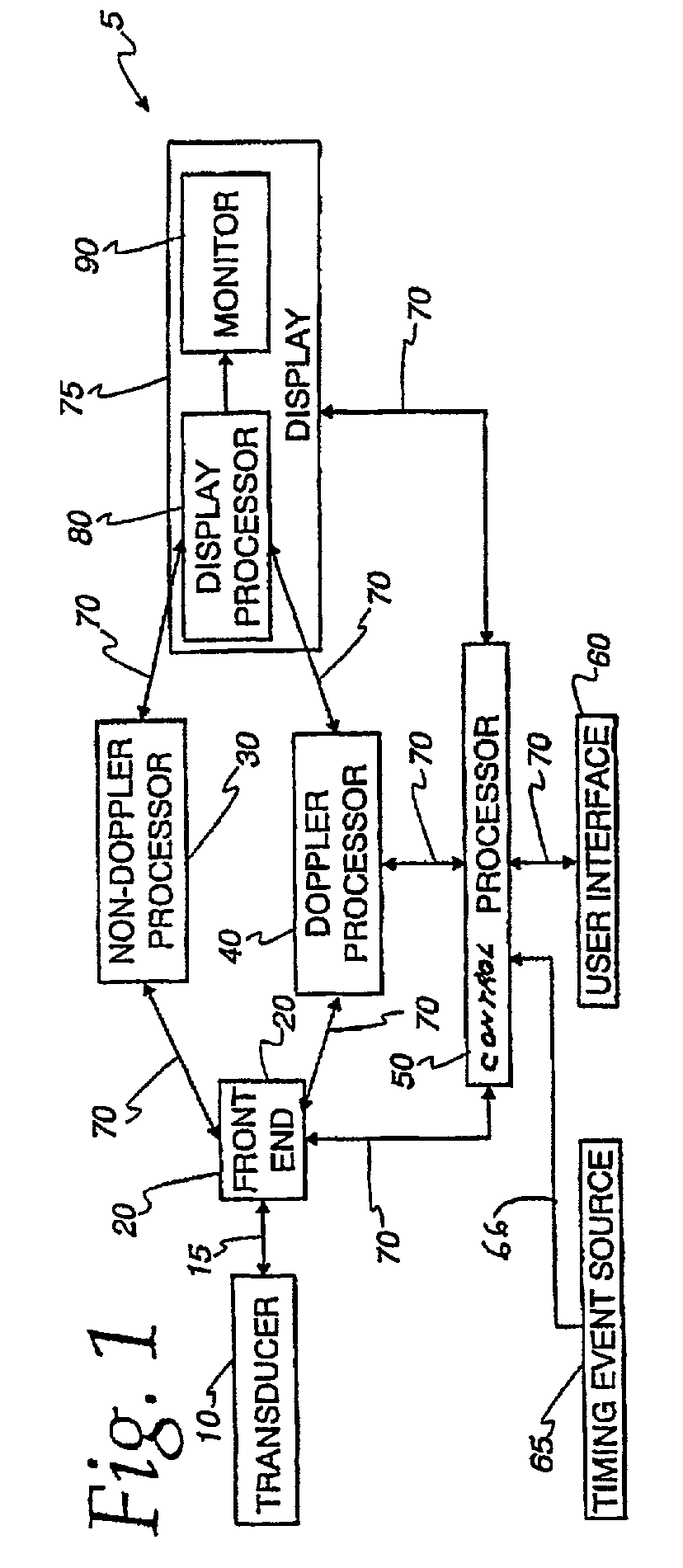
[0027]FIG. 1 is a schematic block diagram of an embodiment of the present invention comprising an ultrasound machine 5. A transducer 10 is used to transmit ultrasound waves into a subject by converting electrical analog signals to ultrasonic energy and to receive ultrasound waves backscattered from the subject by converting ultrasonic energy to analog electrical signals. A front-end 20 comprising a receiver, transmitter, and beamformer, is...


PUM


Abstract

An ultrasound machine is disclosed that displays a color representation of moving structure, such as a cardiac wall tissue, within a region of interest on a monitor. The color representation is generated by displaying at least one color characteristic corresponding to a movement parameter of the structure, such as velocity or strain rate. The movement parameter is mapped to the color characteristic by apparatus comprising a front-end that generates received signals in response to ultrasound waves. A Doppler processor generates a set of parameter signals representing values of the movement parameter within the structure. A control processor adaptively generates a mapping function based on the distribution of the parameter signals to map the parameter signals to a set of color characteristic signals. A display processor applies the mapped values of the color characteristic legend to the values of the movement parameter representing the moving structure, to display a color representation on the monitor in response to the mapping function.

Description

CROSS REFERENCE TO RELATED APPLICATIONS[0001]The applicants claimed priority based on provisional application No. 60 / 297,572 filed Jun. 12, 2001 in the names of Bjorn Olstad, Steinar Bjaerum, and Kjell Kristoffersen.BACKGROUND OF INVENTION[0002]Certain embodiments of the present invention relate to an ultrasound machine for generating and displaying an image of moving structure. More particularly, certain embodiments relate to adaptively color mapping an image of moving structure such as heart tissue.[0003]Echocardiography is a branch of the ultrasound field that is currently a mixture of subjective image assessment and extraction of key quantitative parameters. Evaluation of cardiac wall function has been hampered by a lack of well-established parameters that may be used to increase the accuracy and objectivity in the assessment of, for example, coronary artery diseases. Stress echo is such an example. It has been shown that the subjective part of wall motion scoring in stress echo...

Claims


Application Information

Patent Timeline
Patent Type & Authority Patents(United States)
IPC IPC(8): G06K9/00A61B5/352A61B8/08G01S7/52G01S15/89
CPCG01S15/8988A61B8/0883A61B8/463A61B8/485A61B8/488G01S7/52034G01S7/52042G01S7/52066G01S7/52071G01S7/52087G01S7/52088G01S15/8979G01S15/8981A61B8/08A61B5/0456A61B5/352
Inventor BJAERUM, STEINAROLSTAD, BJORNKRISTOFFERSON, KJELL
Owner GE MEDICAL SYST GLOBAL TECH CO LLC
Why Eureka
Try Eureka