Periocular drug delivery for diabetic retinopathy

a diabetic retinopathy and periocular drug technology, applied in the direction of extracellular fluid disorder, prosthesis, metabolic disorder, etc., can solve the problems of macular edema, blurred vision, vessels can bleed, etc., to maintain retinal cell function and survival, and achieve safe and effective

Inactive Publication Date: 2007-07-24
PENN STATE RES FOUND
View PDF4 Cites 39 Cited by
  • Summary
  • Abstract
  • Description
  • Claims
  • Application Information

AI Technical Summary

Benefits of technology

[0009]Accordingly, there is a great demand for safe and effective methods for delivering agents effective in treating diabetic retinopathy. In particular, there is a need in the art for treatment methods that maintain retinal cell function and survival in the face of persistent hyperglycemia.

Problems solved by technology

Diabetic retinopathy is an illness that occurs when diabetes damages tiny blood vessels in the retina, affecting vision, and is a leading cause of blindness.
When the fluid accumulates in the center of the retina (i.e., the macula) it leads to macular edema.
The fluid makes the macula swell, which blurs vision.
These new blood vessels can bleed, cloud vision, and destroy the retina unless treated.
However, in the preclinical phase sensitive tests reveal reduced contrast sensitivity, electrical responses with an electroretinogram, or color vision.
However, none of these treatment approaches have proven successful in addressing the primary metabolic disorder or in preventing retinopathy.
Conventional diabetic retinopathy treatments are limited to controlling the diabetic state with systemic insulin administration or oral hypoglycemic agents.
The problem with these systemic approaches is that they do not restore normal physiologic metabolic control or provide overall effective levels of the drug to the eye.
These methods are palliative in nature, and none of these methods is sufficiently effective to prevent or cure the disease.
Although diabetic retinopathy is extensively studied in the art, the direct effects of insulin or insulinomimetics on diabetic retinopathy are limited.
Moreover, insulin has been shown to be a survival factor for retinal neurons in culture, and excess hexosamines impair insulin's survival-promoting effects.
However, the ability to administer systemically sufficient insulin or other insulinomimetic agents to be effective for prevention of retinopathy is limited by the risk of hypoglycemia.

Method used


Image

  • Periocular drug delivery for diabetic retinopathy
  • Periocular drug delivery for diabetic retinopathy
  • Periocular drug delivery for diabetic retinopathy

Examples


example 1

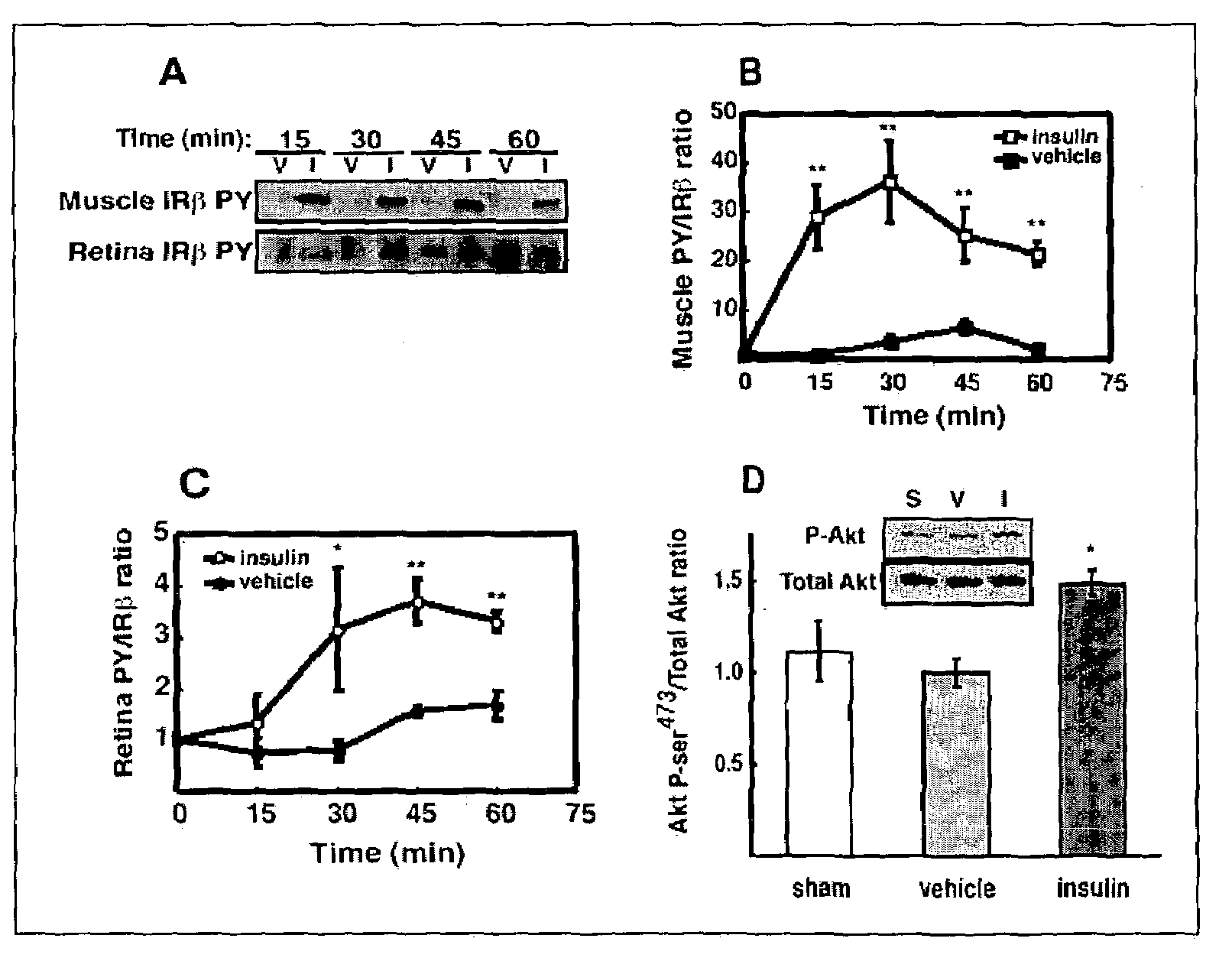
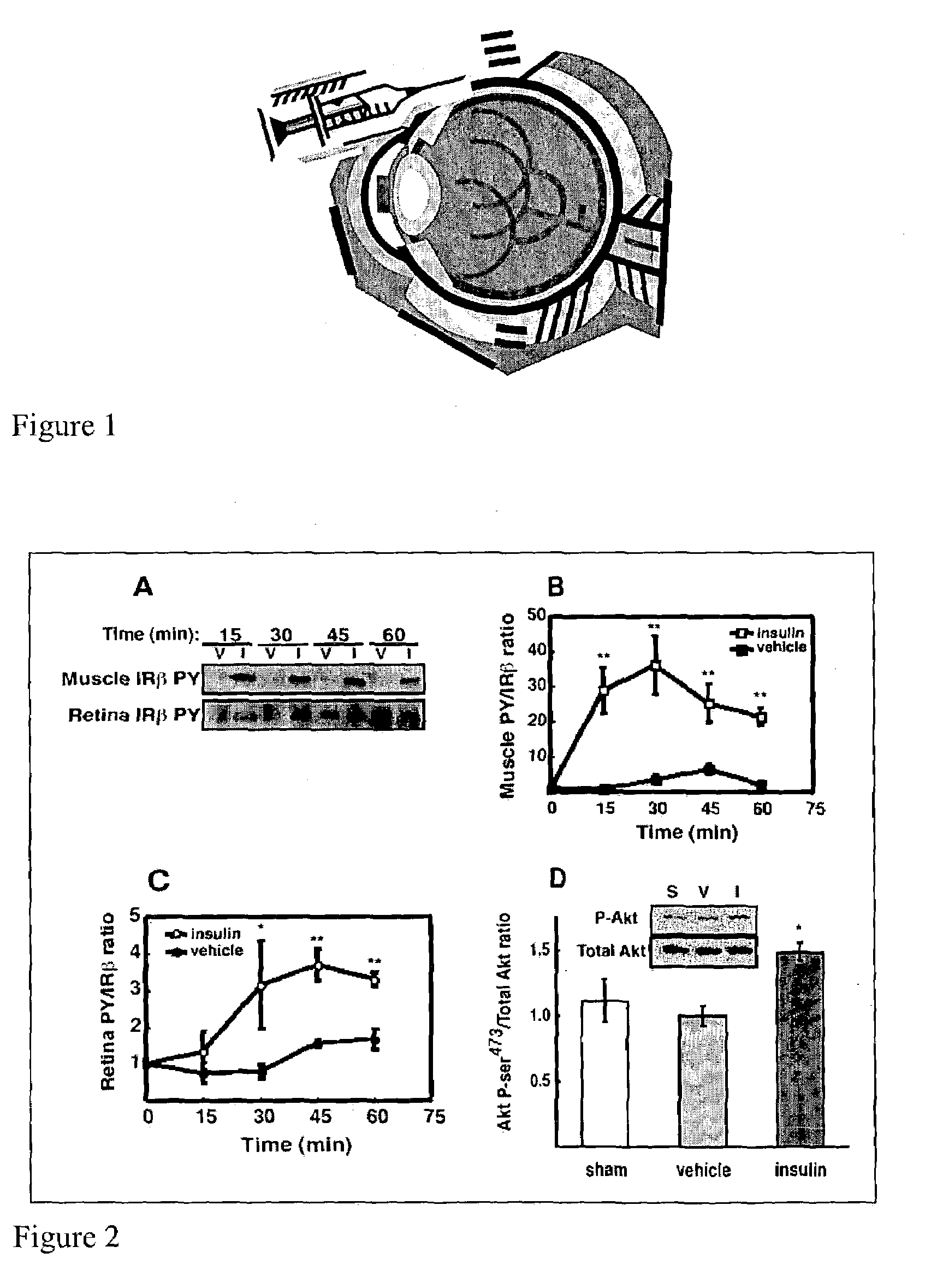
[0038]Intraportal insulin injection was performed to determine if a single bolus insulin injection, and therefore an acute elevation in circulating insulin, could activate retinal insulin receptors (IR) in vivo as it does in other tissues. Intraportal insulin injection was conducted as follows. Male Sprague-Dawley rats (Charles River, Mass.) weighing 200–350 g were fasted 18 hours prior to being anesthetized with a 10:1 ketamine:xylazine cocktail (53.5 mg / kg ketamine and 5.33 mg / kg xylazine) administered by intramuscular injection. The fasted rats were administered a 500 μg bolus of insulin, nothing (sham), or vehicle (0.9% saline) via the portal vein. At 15, 30, 45 and 60 minutes post injection (shown in FIG. 2A), hindquarter skeletal muscle and retina were snap-frozen under liquid nitrogen, and then stored at −80° C. pending analysis by immunoprecipitation and immunoblotting. There was no difference in insulin receptor beta-subunit (IRβ) phosphorylation between vehicle (V) and ins...

example 2

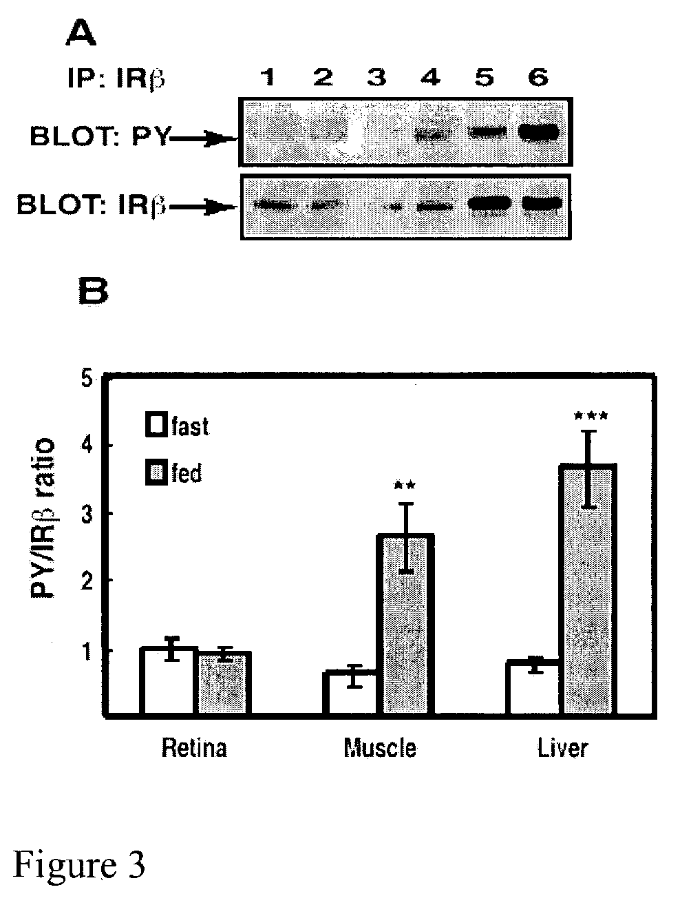
[0045]Tyrosine phosphorylation in retinal IRβ and changes therein was compared with other insulin responsive tissues under freely fed and moderately fasted conditions as follows.

[0046]Six male Sprague-Dawley rats (Charles River) were fasted (18 hours) and were compared to 6 rats that were freely fed overnight. All rats weighed 200–350 g and were anesthetized with a 10:1 ketamine:xylazine cocktail as described in Example 1. Upon loss of motor reflex in the rats' retina, tissue samples from liver and hindquarter skeletal muscle were obtained, homogenized and immunoprecipitated as described in Example 1 and then subjected to Western blot analysis, also as described in Example 1. The blot in FIG. 3A shows that IRβ phosphorylation was unaltered in retina, but increased in muscle and liver compared to fasted rats. Phosphotyrosine-probed immunoblots were then reprobed for total IRβ content with the immunoprecipitating antibody as described in Example 1 to normalize the Western blot data.

[0...

example 3

[0049]Because tyrosine phosphorylation of the IR does not directly measure enzymatic activity, an IR kinase assay was developed.

[0050]The IRβ from fasted and fed rats was immunoprecipitated from retina and liver and analyzed by PY immunoblotting for autophosphorylation with (+) and without (−) the addition of ATP to the kinase reaction. IRβ immunoprecipitates obtained as described in Example 2 were subjected to IR kinase assays as follows. The Sepharose bead complex, obtained as described in Example 1, was washed three times with 200 μL of kinase buffer (50 mM HEPES, pH 7.3, 150 mM NaCl, 20 mM MgCl2, 2 mM MnCl2, 0.05% bovine serum albumin, and 0.1% Triton X-100). Western blotting was performed on these immunoprecipitates as described in Example 1; the results of these Western blot experiments (shown below) showed that washing the Sepharose bead / immune complex in kinase buffer did not diminish total bound IRβ. After the last (third) aspiration of kinase buffer, 500 μL of kinase buffe...


PUM

PropertyMeasurementUnit
volumeaaaaaaaaaa
volumeaaaaaaaaaa
volumesaaaaaaaaaa

Abstract

This invention provides reagents and methods for delivering insulin, insulinomimetic agents, and the like to a vertebrate eye via subconjunctival routes, sub-Tenon's routes, or intravitreal routes for treatment of nerve-related vision disorders such as diabetic retinopathy, and formulations useful in the practice of the disclosed methods.

Description

REFERENCE TO RELATED APPLICATION[0001]This application claims the benefit of U.S. Provisional Application, Ser. No. 60 / 361,559, filed Feb. 28, 2002, which is incorporated by reference.BACKGROUND OF THE INVENTION[0002]1. Field of the Invention[0003]This invention relates generally to a method for delivering insulin, insulinomimetic agents, and the like to the eye for treatment of diabetic retinopathy. More specifically, the method involves the periocular administration of these drugs via subconjunctival routes, sub-Tenon's routes, or intravitreal routes.[0004]2. Background of the Related Art[0005]Diabetes has reached epidemic proportions. Approximately 15 million people in the United States are currently afflicted with the disease, and that number is expected to rise to at least 21 million over the next 30 years.[0006]In addition to (and as a consequence of) the metabolic disarray caused by the disease, diabetes causes a variety of other, organ-specific dysfunctions, including in par...

Claims


Application Information

Patent Timeline
Patent Type & Authority Patents(United States)
IPC IPC(8): A61K38/28
CPCA61K38/28A61K2300/00A61P27/02A61P27/06A61P5/50A61P3/10
Inventor GARDNER, THOMAS W.REITER, CHAD E.
Owner PENN STATE RES FOUND
Why Eureka
Try Eureka