Method for reconstructing projection data sets for dose-reduced sectional spiral scanning in computed tomography

a computed tomography and projection data technology, applied in the field of computed tomography, can solve problems such as cell damag

Inactive Publication Date: 2007-10-16
SIEMENS HEALTHCARE GMBH
View PDF18 Cites 5 Cited by
  • Summary
  • Abstract
  • Description
  • Claims
  • Application Information

AI Technical Summary

Benefits of technology

[0025]An object of the present invention is to provide a fast CT volume imaging and reconstruction technique that allows an intentional radiation dose reduction to be made for organs that are particularly sensitive to radiation.

Problems solved by technology

Because CT imaging is based on the attenuation or absorption of X-ray radiation in organic tissue, during the irradiation an energy transfer to tissue results (radiation dose), which can lead to cell damage.

Method used


Image

  • Method for reconstructing projection data sets for dose-reduced sectional spiral scanning in computed tomography
  • Method for reconstructing projection data sets for dose-reduced sectional spiral scanning in computed tomography
  • Method for reconstructing projection data sets for dose-reduced sectional spiral scanning in computed tomography

Examples


Embodiment Construction

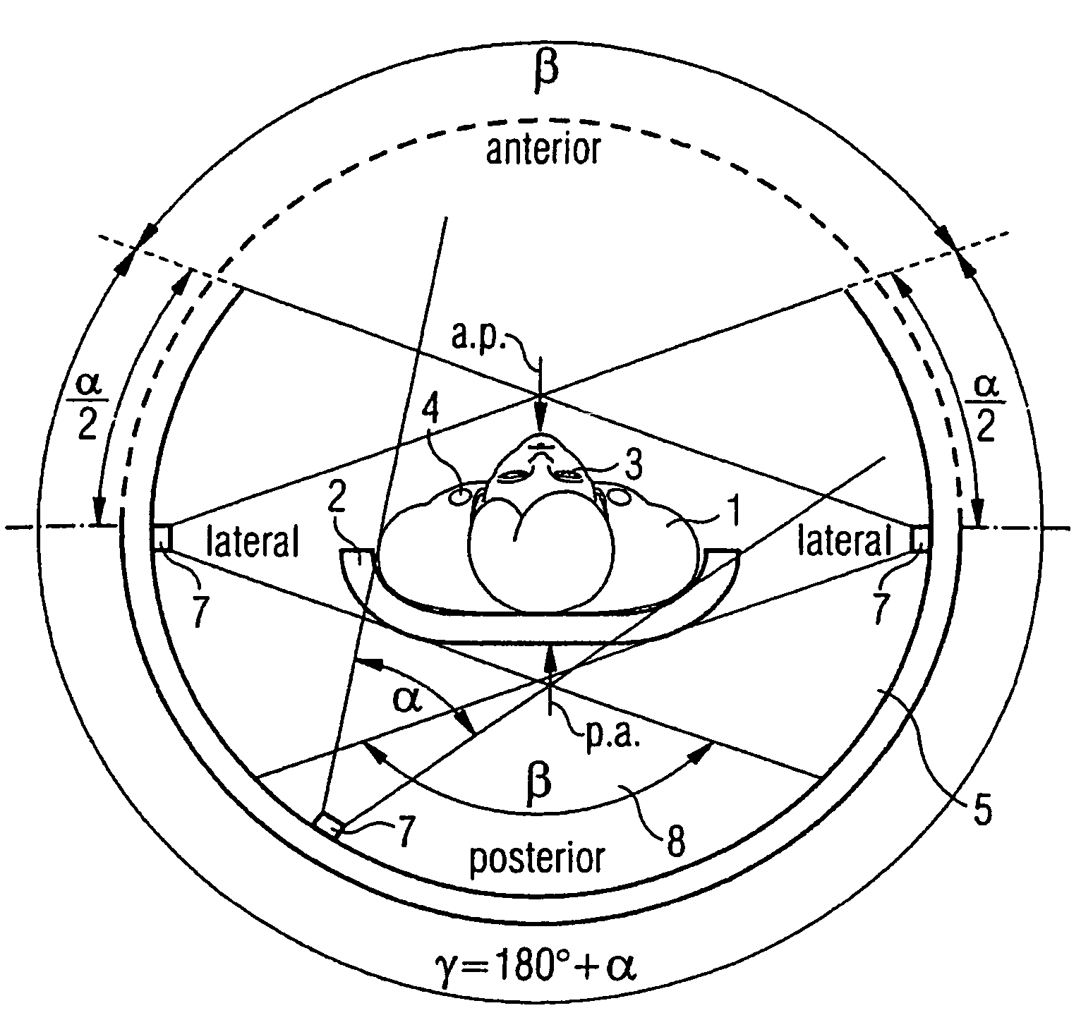
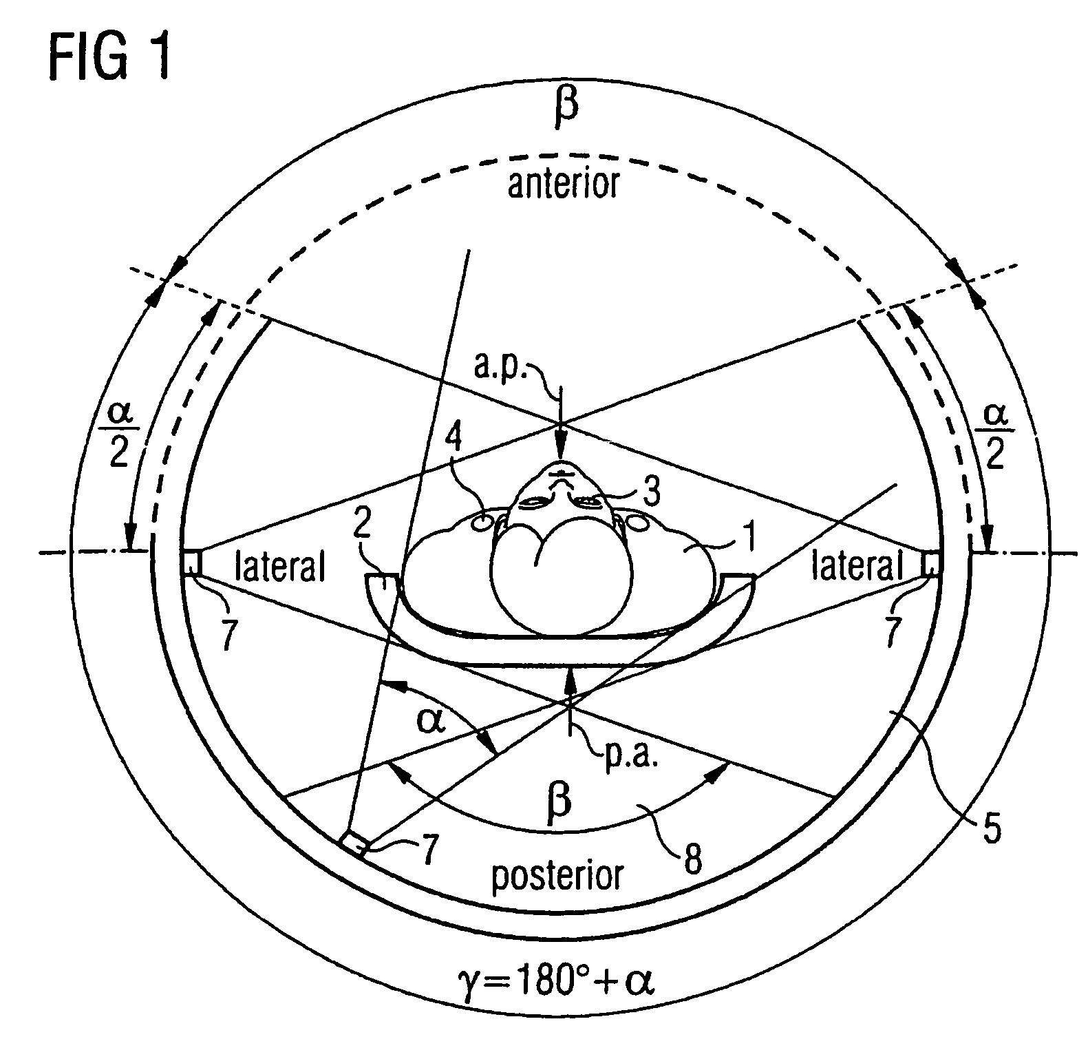
[0039]FIG. 1 schematically shows, in a front view, a patient 1 on a patient bed 2. The patient 1 lies on his or her back so that radiation-sensitive organs (such as the mammary glands 4 and the eye lens 3) are facing the upper area (anterior) of the gantry opening 5. The scanning takes place such that the X-ray tube detector unit (gantry) rotates in a circular manner around the patient 1 while the patient 1 is moved at a uniform speed (constant table feed d) along the patient's longitudinal axis z. The combination of the gantry rotation and the patient displacement results in, as shown in FIG. 2, a spiral-shaped or helix-shaped scanning trajectory 6 as is conventional in spiral CT.

[0040]An object of the present invention is, within the context of spiral scanning, to protect the aforementioned radiation-sensitive organs, or to expose them to the lowest possible radiation dose for the best possible image quality (minimal information loss). According to the invention, this is achieved ...


PUM


Abstract

In a method and CT apparatus and software product for reconstructing incomplete projection data sets for dose-reduced sectional spiral scanning along a z axis of a patient on a table that is movable in the z direction in the CT apparatus that has a radiation source supplied with tube current so as to emit a beam that strikes a detector system to obtain scanning data from which in an image computing device generates two-dimensional and three-dimensional scanning images of the examination subject, at a pitch greater or equal to one. For z positions having an incomplete projection data set for reconstruction of the image, in a preprocessing step data from another row of this projection and / or data for a previous or subsequent 360° rotation are used to compute a complete projection data set therefor.

Description

BACKGROUND OF THE INVENTION[0001]1. Field of the Invention[0002]The present invention relates in general to computed tomography as used in medicine to examine patients, particularly to a method for image reconstruction for sectional dose-reduced spiral scanning.[0003]2. Description of the Prior Art[0004]Using modern medical diagnostic techniques such as X-ray computed tomography, it is possible to acquire image data for an object to be examined. The examined object is generally a patient.[0005]X-ray computed tomography (CT for short) is an X-ray imaging technique that differs fundamentally in terms of the image formation from classic X-ray tomographic imaging techniques. With CT images, transverse image slices are obtained, i.e., images of body slices that are oriented essentially perpendicularly to the body axis. The tissue-specific physical quantity represented in the image is the distribution of the attenuation values of X-ray radiation μ(x, y) in the slice plane. The CT image is...

Claims


Application Information

Patent Timeline
Patent Type & Authority Patents(United States)
IPC IPC(8): H05G1/60A61B6/03H05G1/64A61B6/06G01T1/29G01T1/36G06T17/00G21K1/12
CPCA61B6/032A61B6/542A61B6/544G06T11/006G01T1/2985A61B6/027G06T2210/41G06T2211/432
Inventor KALENDER, WILLISCHALLER, STEFANSCHMIDT, BERNHARD
Owner SIEMENS HEALTHCARE GMBH
Why Eureka
Try Eureka