Automatic abnormal tissue detection in MRI images

a tissue detection and automatic technology, applied in image analysis, image enhancement, instruments, etc., can solve the problems of inconsistent and properly interpreting a series, difficult coordination of multiple images with respect to each other, and poor delineation of anatomy

Inactive Publication Date: 2008-01-08
CARESTREAM HEALTH INC
View PDF3 Cites 57 Cited by
  • Summary
  • Abstract
  • Description
  • Claims
  • Application Information

AI Technical Summary

Problems solved by technology

Positron emission tomography (PET) and single photon emission computed tomography (SPECT) scans display aspects of brain function and allow metabolic measurements but poorly delineate anatomy.
Unfortunately, such a coordination of multiple images with respect to each other is extremely difficult and even highly trained medical personnel, such as experienced radiologists, have difficulty in consistently and properly interpreting a series of medical images so that a treatment regime can be instituted which best fits the patient's current medical condition.
Another problem encountered by medical personnel today is the large amount of data and numerous images that are obtained from current medical imaging devices.
In order for medical personnel to properly review each image takes a great deal of time and, with the many images that current medical technology provides, a great amount of time is required to thoroughly examine all the data.
However, Daw's system fails to provide a function of automatically detect and differentiate image areas corresponding to materials (tissues) being imaged that have different time response to contrast enhancing agent.
It should be recognized that if the patient moves even slightly between the two scans, the shape or size of the image may be distorted—a big loss of information.

Method used


Image

  • Automatic abnormal tissue detection in MRI images
  • Automatic abnormal tissue detection in MRI images
  • Automatic abnormal tissue detection in MRI images

Examples


Embodiment Construction

[0032]The following is a detailed description of the preferred embodiments of the invention, reference being made to the drawings in which the same reference numerals identify the same elements of structure in each of the several figures.

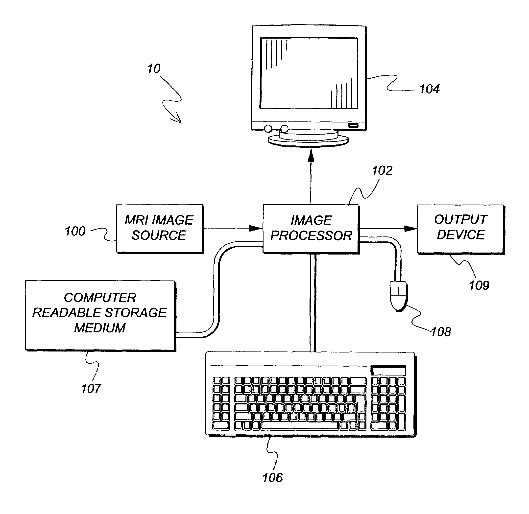
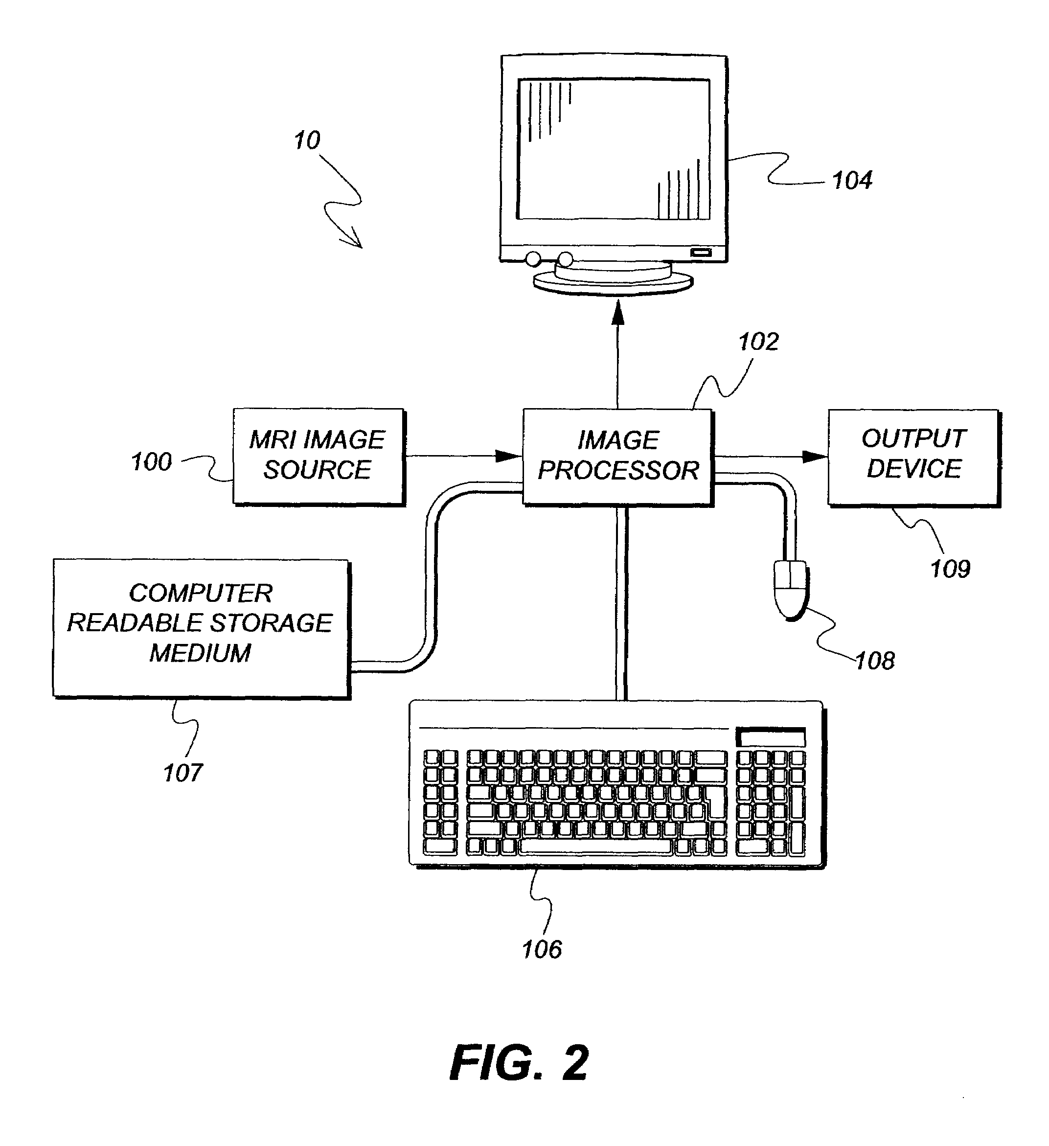
[0033]FIG. 2 shows an image processing system 10 useful in practicing the method in accordance with the present invention. System 10 includes a digital MRI image source 100, for example, an MRI scanner, a digital image storage device (such as a compact disk drive), or the like. The digital image from digital MRI image source 100 is provided to an image processor 102, for example, a programmable personal computer, or digital image processing work station such as a Sun Sparc workstation. Image processor 102 can be connected to a display 104 (such as a CRT display or other monitor), an operator interface such as a keyboard 106, and a mouse 108 or other known input device. Image processor 102 is also connected to computer readable storage medium 107. Im...


PUM


Abstract

A pattern recognition method for automatic abnormal tissue detection and differentiation using contrast enhanced MRI images. The method includes the steps of acquiring a plurality of MRI breast image sets; aligning the plurality of MRI breast images with respect to spatial coordinates; differencing the plurality of MRI breast image sets with a reference MRI image set, producing a plurality of difference image sets; segmenting the plurality of difference image sets, producing a plurality of MRI breast images with segmented intensity pixels; applying dynamic system identification to the segmented intensity pixels, producing a plurality of dynamic system parameters; and classifying the plurality of system parameters augmented with other physical or non-physical factors into different classes.

Description

FIELD OF THE INVENTION[0001]The present invention relates to a digital image processing / computer vision method for medical image analysis and, in particular, to automatically detecting and differentiating breast abnormal tissues using MRI images, particularly, contrast enhanced MRI images augmented with other factors.BACKGROUND OF THE INVENTION[0002]Digital imaging techniques in medicine were implemented in the 1970's with the first clinical use and acceptance of the Computed Tomography or CT scanner. Later, extensive use of x-ray imaging (CT) and the advent of the digital computer and new imaging modalities like ultrasound and magnetic resonance imaging (MRI) have combined to create an explosion of diagnostic imaging techniques in the past three decades.[0003]There are benefit to using digital medical imaging technology in health care. For example, angiographic procedures for looking at the blood vessels in the brain, kidneys, arms and legs, and heart all have benefited from the ad...

Claims


Application Information

Patent Timeline
Patent Type & Authority Patents(United States)
IPC IPC(8): G06K9/00
CPCG06K9/00G06T7/0012G06K2209/053G06T2207/30068G06V2201/032G06F18/00
Inventor CHEN, SHOUPUHUO, ZHIMINRAY, LAWRENCE A.
Owner CARESTREAM HEALTH INC
Why Eureka
Try Eureka