Tissue punch and tissue sample labeling methods and devices for microarray preparation, archiving and documentation

a tissue punch and tissue sample technology, applied in the field of tissue punch and tissue sample labeling methods and devices for microarray preparation, archiving and documentation, can solve the problems of inability of current available methods to accurately prepare tissue slides, time-consuming, laborious, etc., and achieves the effects of avoiding errors, avoiding waste of tissue or time, and being prepared efficiently

Inactive Publication Date: 2008-07-29
EXB TECH
View PDF26 Cites 23 Cited by
  • Summary
  • Abstract
  • Description
  • Claims
  • Application Information

AI Technical Summary

Benefits of technology

[0011]The present invention generally provides improved methods, devices, and systems for handling and preparation of tissues for biological analysis. In many embodiments, the improved techniques provided by the present invention make use of an array or set of punch tubes, with the punch tubes often being supported by a punch tube platform which allows each tube to slide along its axis. The punch tubes can extend from the platform into a tissue specimen that has been removed from a patient so as to capture a tissue sample within a receptacle of the punch tube. The punch tubes and / or platform may have a label (such as a barcode or radiofrequency identification device (“RFID”)) indicating one or more tube or sample identifiers. By labeling the specimen removed from the patient, the punch tubes, a microarray block containing a plurality of the samples, and each microarray slide separated from the microarray block, and by recording the relationships therebetween, a continuous tissue identification chain can be established between a test result at a specific location of a microarray slide and a tissue sample location within a tissue specimen of a particular patient. Such a tissue identification chain can help avoid errors when testing large numbers of biological tissues from different tissue specimens and different patients. Exemplary techniques for punching tissue samples from tissue specimens while recording the tissue sample locations are also provided, and the punch tubes (with their associated tissue samples) may be archived and / or reassembled into new tissue arrays by selectively removing punch tubes from one platform and / or selectively assembling punch tubes into another platform. In some embodiments, cooling of the punch tubes, tissue specimen, and / or tissue samples can be provided using cryogenic and / or other cooling techniques, and microarray blocks may be prepared efficiently and with little waste of tissue or time by aligning the tissue samples from the punch tubes both axially and laterally within the microarray block using the punch tube platform.
[0014]In many embodiments, at least some of the tissue specimens will be extruded from the punch tubes to form a tissue microarray. Typically, at least four punch tubes are extended by sliding the tubes from the platform, and the punch tubes may slide serially and / or sequentially. The platform can hold the punch tubes in a two-dimensional punch tube array so that the punch tubes slide along parallel longitudinal axes. The sample receptacle of each punch tube may punch the associated sample with a sample cross section suitable for use in a tissue microarray, with the tube receptacle typically having a cross sectional dimension of between about 0.4 mm and 0.5 mm. Conveniently, the platform may support each punch tube with a separation distance from an adjacent punch tube that is also appropriate for the tissue microarrays, with the sample separation distance between adjacent tissue samples typically being in a range of up to about 5.0 mm (measured between tissue sample centers). This can facilitate extruding the tissue samples from the array of punch tubes into the microarray while the punch tubes are supported by the platform. The platform will often support between about 1 and about 1,000 punch tubes.
[0021]After using the first punch tube to capture the first tissue sample, a second tissue sample may be manually input to an input device with reference to a slide tissue. The slide tissue may correspond to and be separated from the tissue specimen. The first motion stage may move the slide tissue and tissue specimen while maintaining registration therebetween. A system operator may view a display showing an image of the slide tissue, along with a target marker identifying a candidate location for a subsequent tissue sample. The target marker may be projected onto the slide tissue, or may be superimposed on the image using image processing techniques or the like. The first motion stage may effect lateral movement of the target marker to an alternative candidate location by moving the slide tissue laterally relative to a field of view of an image capture device coupled to the display. The arbor can remain aligned with the image capture device during movement of the first motion stage. In some embodiments, a plurality of prior punch tube sample locations may be displayed on the slide tissue image. This can help the user to select appropriate tissue samples, as the slide the user is viewing may, unlike the tissue specimen itself, may undergo no physical change so as to indicate where samples have already been removed.
[0024]Each punch tube may have a chamfer disposed radially outwardly about the sample receptacle so as to form a sharpened distal edge at the distal end of the punch tube. A cross section of the punch tube receptacle and / or sample may remain substantially constant proximal the sharpened edge. This can facilitate proximal entry of the tissue sample into and distal extrusion of the sample from the sample receptacle. In some embodiments, a friction-inhibiting coating may be disposed along an outer surface of the punch tube and / or an inner surface of the punch tube.
[0036]In another aspect, the invention provides a tissue punch microarray system for identifying specimens from a plurality of patients. The system comprises a plurality of tissue specimens including a first specimen and a second specimen. A plurality of tissue specimen identifiers help identify tissue samples taken from the first and second specimens. A first microarray block identifier helps identify a first microarray block. The first microarray comprises a first group of the samples. A second microarray block identifier identifies a second microarray block comprising a second group of the samples. A plurality of microarray slides are separated from the first microarray block, each having a microarray slide label indicating a microarray slide identifier. A tangible media stores each microarray slide identifier. The tangible media indicates, for each microarray slide: a microarray block identifier of an associated microarray block from which the microarray slide was separated; the tissue sample identifiers of associated tissue samples included in the associated microarray block; at least one tissue specimen identifier of an associated tissue specimen from which the associated tissue samples were taken; and at least one patient identifier of the patient from which the associated specimen was taken.

Problems solved by technology

The expanding range of analytical techniques that can be applied to tissue slides and the increasing use of these techniques for studying, diagnosing, and monitoring disease states may challenge the current capacity of laboratories to accurately prepare tissue slides, to reliably handle large numbers of test tissues, and to document the sources of these test tissues.
While known tissue microarray preparation techniques have achieved varying levels of success in allowing labs to handle tissues for testing, the currently available methods may generally suffer from deficiencies that have limited their use.
For example, many involve time consuming, labor intensive, and highly repetitious processing steps that can introduce errors and inaccuracies.
At least some of these methods could lead to cross contamination between samples, and / or degradation of tissues between specimen removal and the biological tissue testing.
Some or all of these methods may lack adequate flexibility and security for selectively handling tissues from a variety of sources and preparing them for any of a number of different tests, some of which tests (for example) need not be performed until long after the associated specimens have been removed from the patient and results from initial tests on tissue samples from the specimen have been obtained.

Method used


Image

  • Tissue punch and tissue sample labeling methods and devices for microarray preparation, archiving and documentation
  • Tissue punch and tissue sample labeling methods and devices for microarray preparation, archiving and documentation
  • Tissue punch and tissue sample labeling methods and devices for microarray preparation, archiving and documentation

Examples


Embodiment Construction

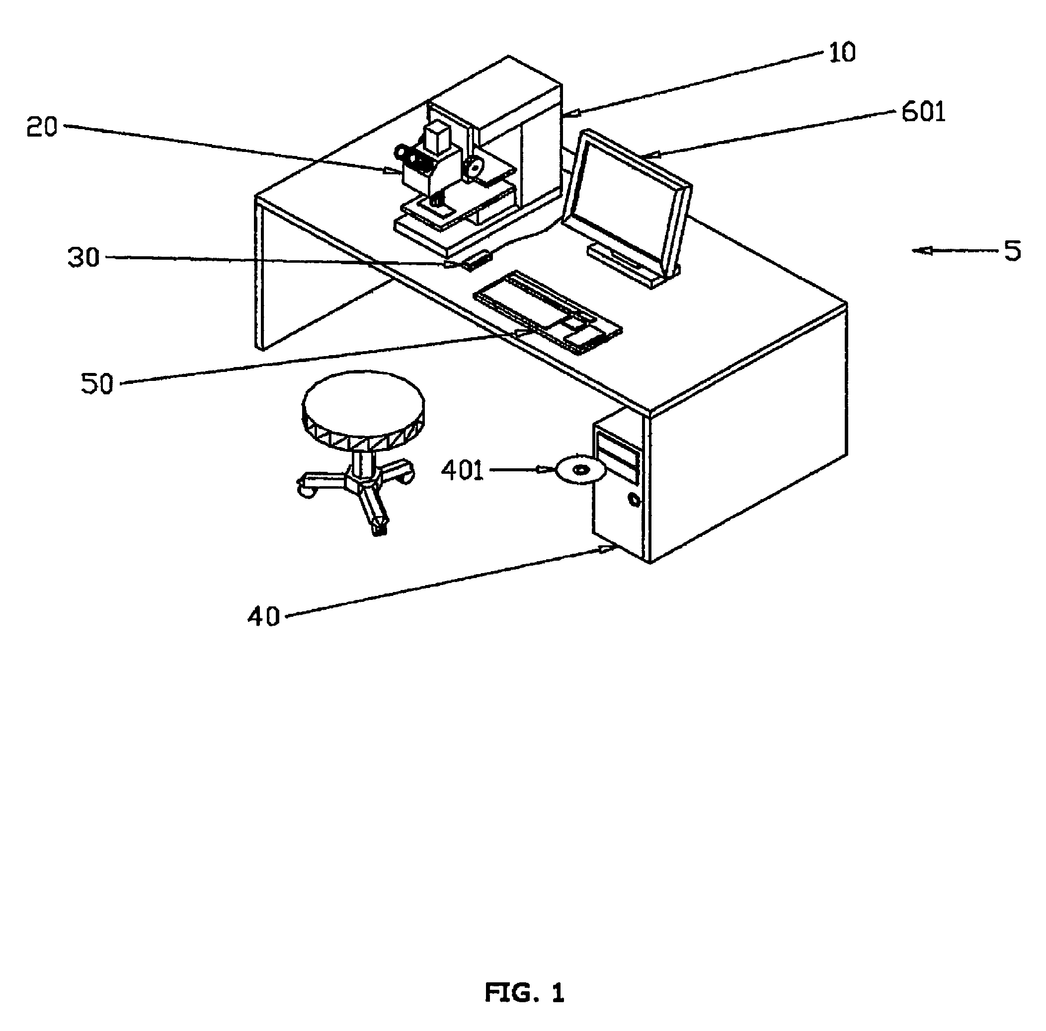
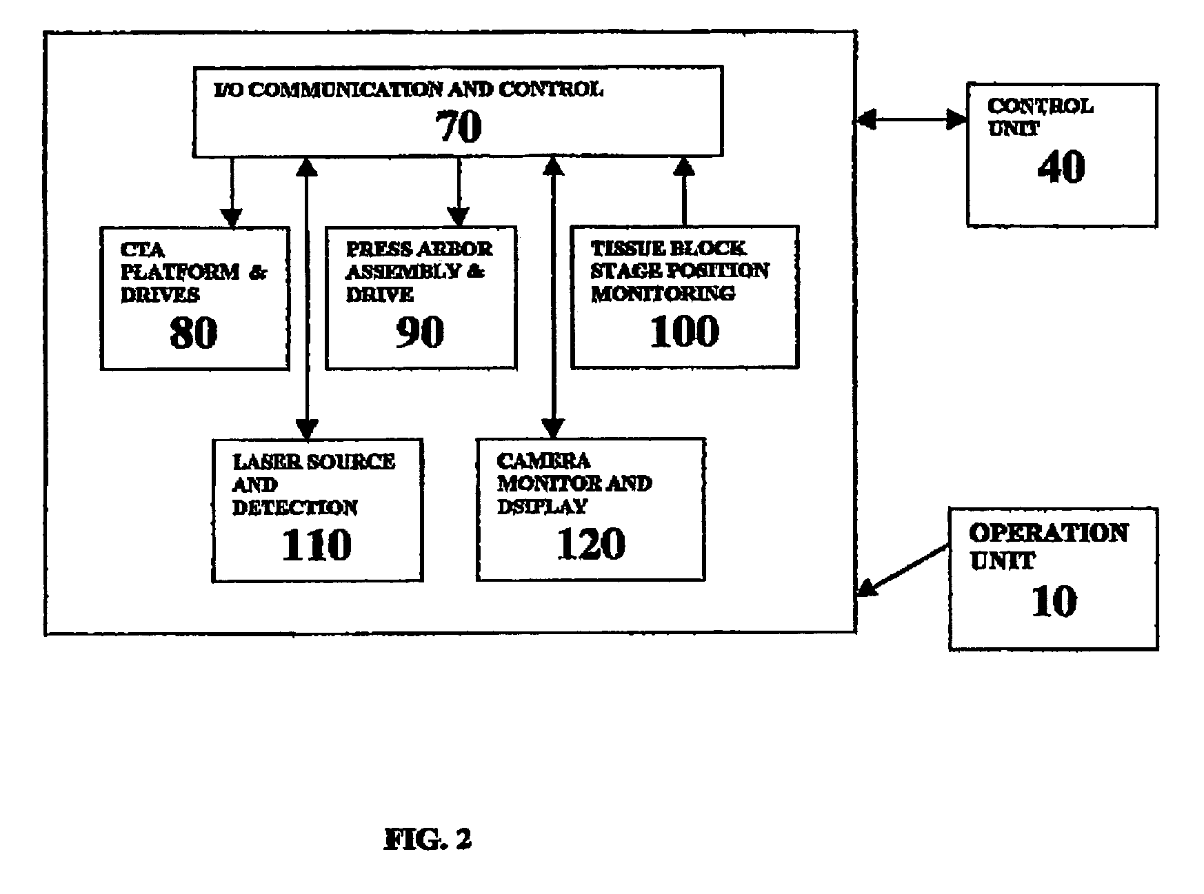
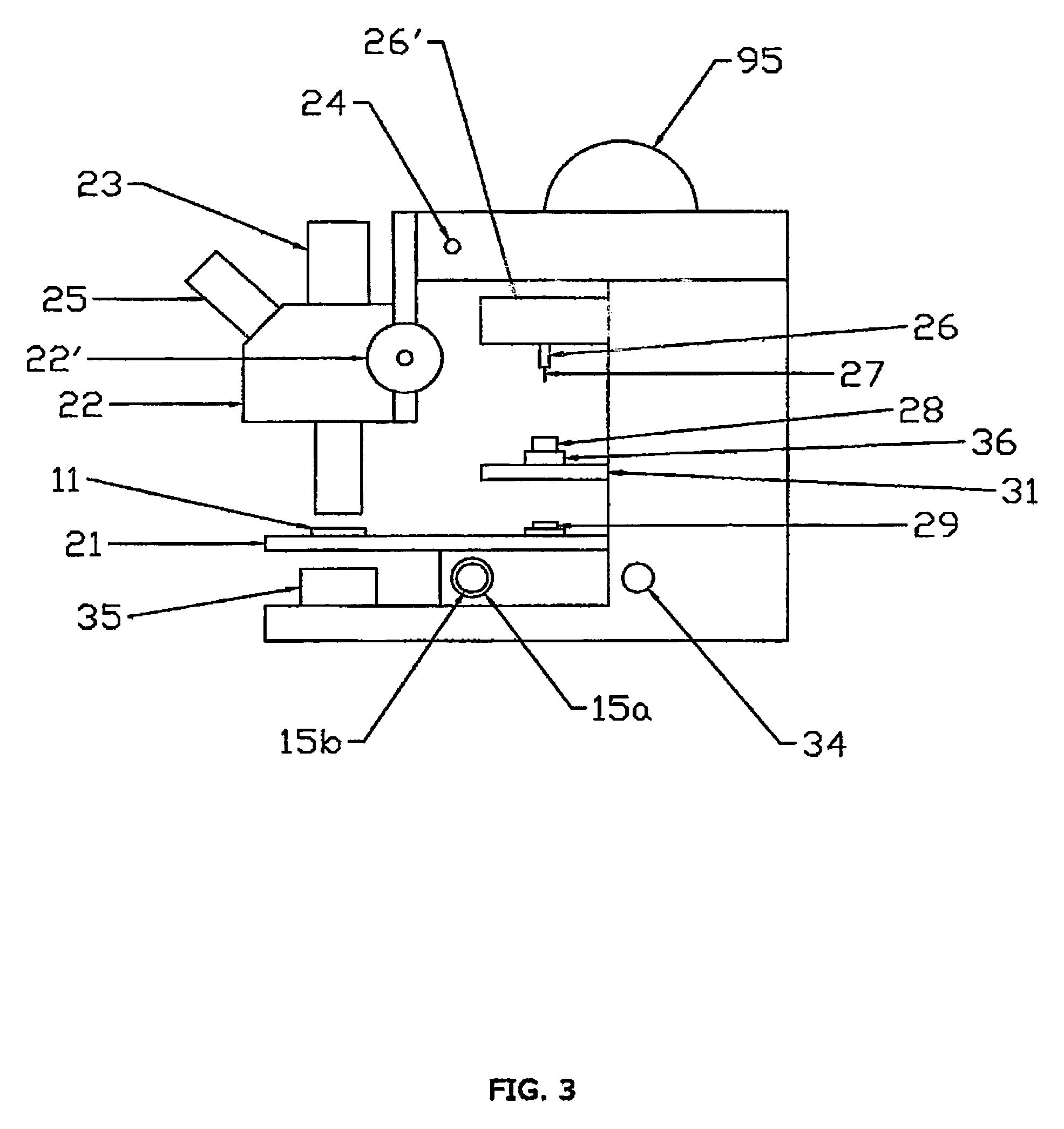
[0052]The present invention generally provides improved devices, systems, and methods for handling of tissues for biological analysis. Exemplary embodiments of the invention provide improved devices, systems, and methods for preparing microarray blocks and slides from tissue samples taken from one or more tissue specimens. Punch tubes will often be used to separate the tissue samples from the tissue specimens, and each punch tube will often include a label indicating a punch tube identifier that can be used to identify a tissue sample contained within a receptacle of the punch tube. The labels may comprise bar codes, radiofrequency identification (“RFID”) devices, or the like, and similar labels may be placed on the tissue specimen block, a microarray block formed from the samples, the microarray slides taken from a microarray block, and the like. The punch tubes may be releasably mounted to a platform that allows the punch tubes to slide for collection of the samples, and the platf...


PUM

PropertyMeasurementUnit
thickaaaaaaaaaa
depthaaaaaaaaaa
sizeaaaaaaaaaa

Abstract

A workstation that provides an efficient method to collect biological tissues in a column tissue array format from blocks of embedded, frozen tissues, or fresh tissues. The workstation has a control unit for directing operations of the workstation and the operation unit for performing the production of the tissue column array. The operation unit comprises an array of vertical tubes in a platform, an arbor which engages and presses down the designated tube in the array, the embedded tissue block which is mounted directly below the designated tube, assemblies of motors responsive to the control unit for driving the platform and the tissue block, a light source block for generating an alignment signal, and a light detector block which measures the signal from the light source to determine the degree of alignment between the arbor, punch tubes, and the specimen block.

Description

CROSS-REFERENCES TO RELATED APPLICATIONS[0001]Not ApplicableSTATEMENT AS TO RIGHTS TO INVENTIONS MADE UNDER FEDERALLY SPONSORED RESEARCH OR DEVELOPMENT[0002]Not ApplicableREFERENCE TO A “SEQUENCE LISTING,” A TABLE, OR A COMPUTER PROGRAM LISTING APPENDIX SUBMITTED ON A COMPACT DISK[0003]Not ApplicableBACKGROUND OF THE INVENTION[0004]This invention generally relates to tissue handling and preparation of tissue for biological analysis, and in particular embodiments provides methods, devices, and systems using labeled punch tubes in the preparation of tissue microarrays and microarray slides, for tissue sample archiving, for correlating pathology, histology, and other biological test results with specific source tissues through intermediary tissue configurations, and the like.[0005]Punch tubes are often used to obtain samples of tissue specimens for biological testing. The tissue specimens are often removed surgically from patients, and punch tubes can help to separate a desired sample ...

Claims


Application Information

Patent Timeline
Patent Type & Authority Patents(United States)
IPC IPC(8): G01N33/48A61B10/00
CPCG01N1/286A61B17/32053G01N2001/2873A61B2010/0208
Inventor LAM, EDWARDLAM, RONALD
Owner EXB TECH
Why Eureka
Try Eureka