Ischemia detection based on cardiac conduction time

a technology of cardiac conduction time and ischemia detection, applied in the field of cardiac health, can solve the problems of unreliable use of the st segment as an indicator of ischemia, myocardial ischemia, and myocardial infarction, so as to avoid false indication of ischemia events, increase the specificity of ischemia detection, and reliable indication of an ischemic episode

Active Publication Date: 2008-08-19
MEDTRONIC INC
View PDF27 Cites 36 Cited by
  • Summary
  • Abstract
  • Description
  • Claims
  • Application Information

AI Technical Summary

Benefits of technology

[0014]The invention may provide a number of advantages. In accordance with the invention, detection of changes in electrical conduction velocity within the heart, e.g., via measurement of conduction time, may provide a more reliable indication of an ischemic episode. In particular, the invention may be useful in increasing the specificity of ischemia detection, generally avoiding false indication of ischemic events due to axis shifts, electrical noise, cardiac pacing stimuli, high sin

Problems solved by technology

Myocardial ischemia, a leading cause of mortality, involves oxygen starvation of the myocardium.
Myocardial ischemia can lead to myocardial infarction if left untreated.
Unfortunately, many episodes of myocardial ischemia do not cause excessive pain or other noticeable warning signs, and often go undetected.
Unf

Method used


Image

  • Ischemia detection based on cardiac conduction time
  • Ischemia detection based on cardiac conduction time
  • Ischemia detection based on cardiac conduction time

Examples


Embodiment Construction

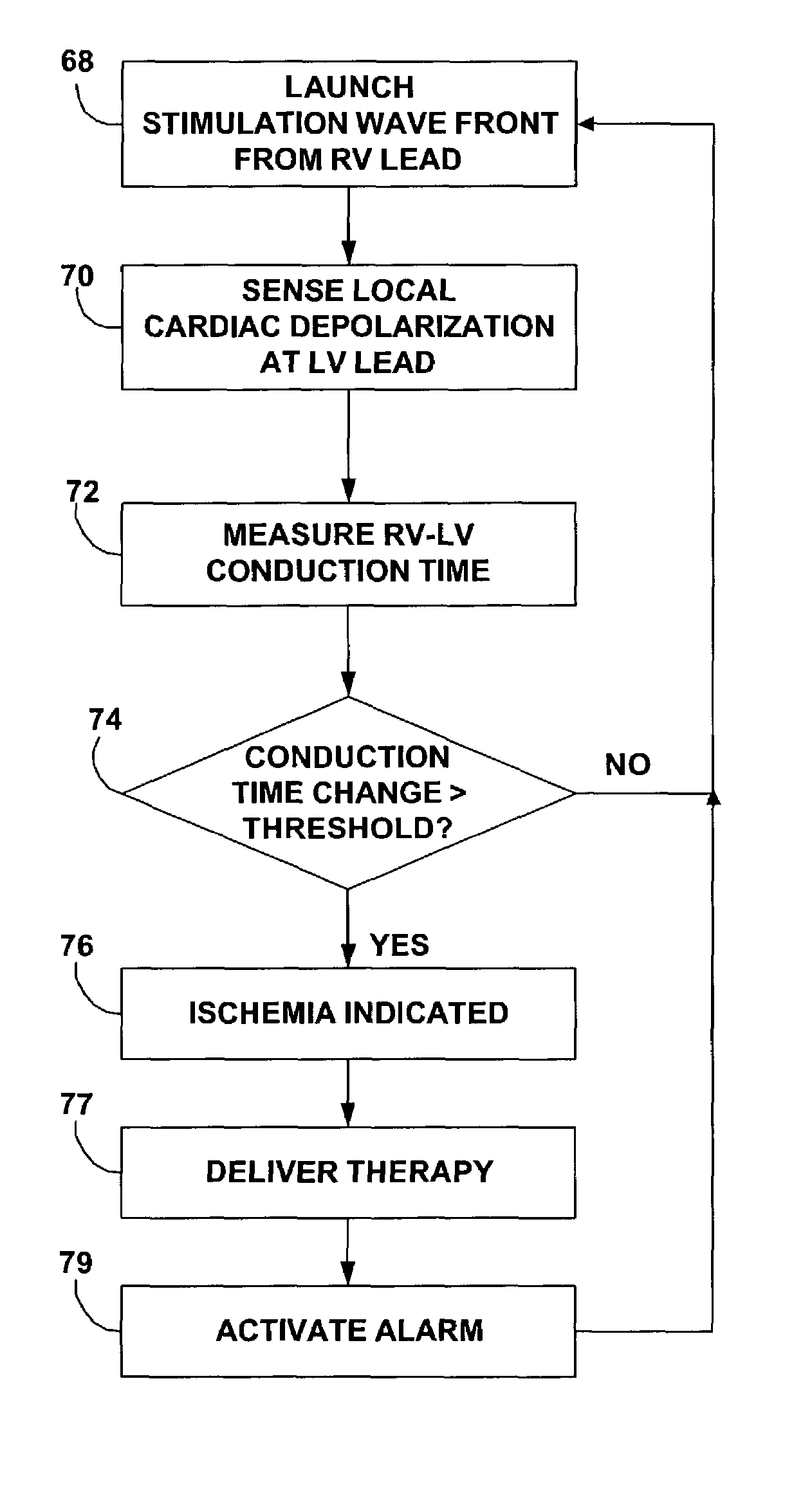
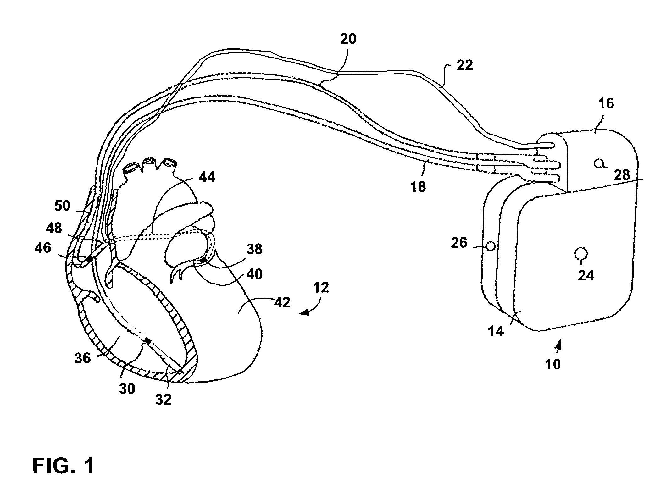
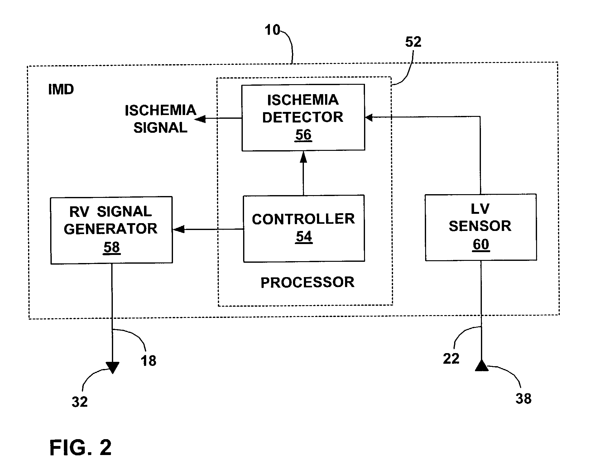
[0027]FIG. 1 is a diagram illustrating an exemplary implantable medical device (IMD) 10 in association with a human heart 12. IMD 10 may be dedicated to monitoring of heart 12, or integrate both monitoring and therapy features, as will be described. In accordance with the invention, IMD 10 is configured to detect cardiac conduction velocity, via measurement of conduction time, and detect myocardial ischemia based on the detected conduction time. Using conduction time, IMD 10 detects changes in the state of heart 12, and thereby obtains an indication of heart tissue conditions suggestive of myocardial ischemia within the heart 12.

[0028]When a change in cardiac conduction time reveals ischemic conditions, IMD 10 indicates an ischemic episode. Conduction velocity across cardiac muscle tends to decrease significantly when oxygen supply to the heart is reduced. At the same time, conduction time tends to increase. Consequently, the conduction time between two fixed electrodes can provide ...


PUM


Abstract

Methods and process for detection of myocardial ischemia involve detection and analysis of changes in electrical conduction velocity within the heart to monitor changes in the condition of the cardiac muscle and indicate possible ischemia. Conduction velocity slows considerably when oxygen supply to the heart is reduced. Analysis of electrical conduction velocity can be used to verify the occurrence of myocardial ischemia in a more reliable manner. Changes in conduction velocity may be monitored based on conduction time between electrodes positioned in the left and right ventricles of the heart. The electrodes may be endocardial or epicardial electrodes. In general, the techniques may involve launching a stimulation waveform at one electrode and sensing a local cardiac depolarization at another electrode to assess conduction time.

Description

FIELD[0001]The invention relates to cardiac health and, more particularly, to techniques for detection of myocardial ischemia.BACKGROUND[0002]Myocardial ischemia, a leading cause of mortality, involves oxygen starvation of the myocardium. Myocardial ischemia can lead to myocardial infarction if left untreated. Early detection of myocardial ischemia provides the opportunity for a wide range of effective therapies such as revascularization, neural stimulation, and drug delivery to reduce cardiac workload or improve cardiac circulation. Unfortunately, many episodes of myocardial ischemia do not cause excessive pain or other noticeable warning signs, and often go undetected.[0003]An electrocardiogram (ECG) or electrogram (EGM) presents a PQRST waveform sequence that characterizes the cyclical cardiac activity of a patient. The ST segment, also associated with the repolarization of the ventricles, is typically close in amplitude to the baseline, i.e., isoelectric amplitude of the signal ...

Claims


Application Information

Patent Timeline
IPC IPC(8): A61N1/00A61B5/05A61N1/362
CPCA61B5/05A61B5/7203A61N1/362A61N1/3629
Inventor SHARMA, VINODOLSON, WALTER H.
Owner MEDTRONIC INC
Why Eureka
Try Eureka