Direct imaging of neural currents using ultra-low field magnetic resonance techniques

a magnetic resonance and ultra-low field technology, applied in the field of medical and biological imaging systems and methodologies, can solve the problems of ill-posed inverse problem of electromagnetism, poor temporal resolution of the applicator, and inability to directly measure neural activity,

Inactive Publication Date: 2009-08-11
TRIAD NAT SECURITY LLC +1
View PDF12 Cites 22 Cited by
  • Summary
  • Abstract
  • Description
  • Claims
  • Application Information

AI Technical Summary

Benefits of technology

[0017]It is also a feature of the present invention to provide a system to provide imaging in accordance with the methods described herein, the system including a single SQUID sensor adapted for operating at measurement fields of 2-25 uT and is further adapted with a pre-polarizing field from 4-30 uT generated by at least one room-temperature, wire-wound coil that is turned off during measurement operations. The system can have geometry with samples located outside of a cryostat at room temperature, removing constraints on sample size and enabling the capture of signals from living tissue.
[0018]The present inventors have found that neuronal currents (both intra- and extra-cellular) will interact with the proton spins in tissue resulting in a measurable change in the NMR signal that can be localized using ULF MRI techniques. The present inventors have enabled development of new models demonstrating an entirely new signature of neural (and any bioelectric) activity that can be tomographically imaged. The inventors demonstrate that nuclear magnetic resonance (NMR) techniques at ultra-low fields (<100 microtesla) can be used to unambiguously detect and ultimately directly image neural activity. NMR at ULF provides several benefits, based on the basic properties of the process, which are not realized at high fields.

Problems solved by technology

The approaches all suffer from poor temporal resolution, poor spatial localization, or indirectly measuring neural activity.
While these modalities yield detailed temporal information (milliseconds or better), the spatial localization must be inferred from spatial modeling priors resulting in the classic ill posed inverse problem of electromagnetism.
A common problem with all of these studies was the use of effective DC currents while human brain activity is more accurately characterized by waveforms that contain a broad distribution of frequencies with roughly “zero-mean” amplitude over typical MR measurement time windows.
A serious confound for this, or any HF measurement, is the “susceptibility artifact” caused by the different magnetic properties of oxy- and de-oxy hemoglobin—an effect that is orders of magnitude larger than the expected direct neural effect on the NMR signal.

Method used


Image

  • Direct imaging of neural currents using ultra-low field magnetic resonance techniques
  • Direct imaging of neural currents using ultra-low field magnetic resonance techniques
  • Direct imaging of neural currents using ultra-low field magnetic resonance techniques

Examples


Embodiment Construction

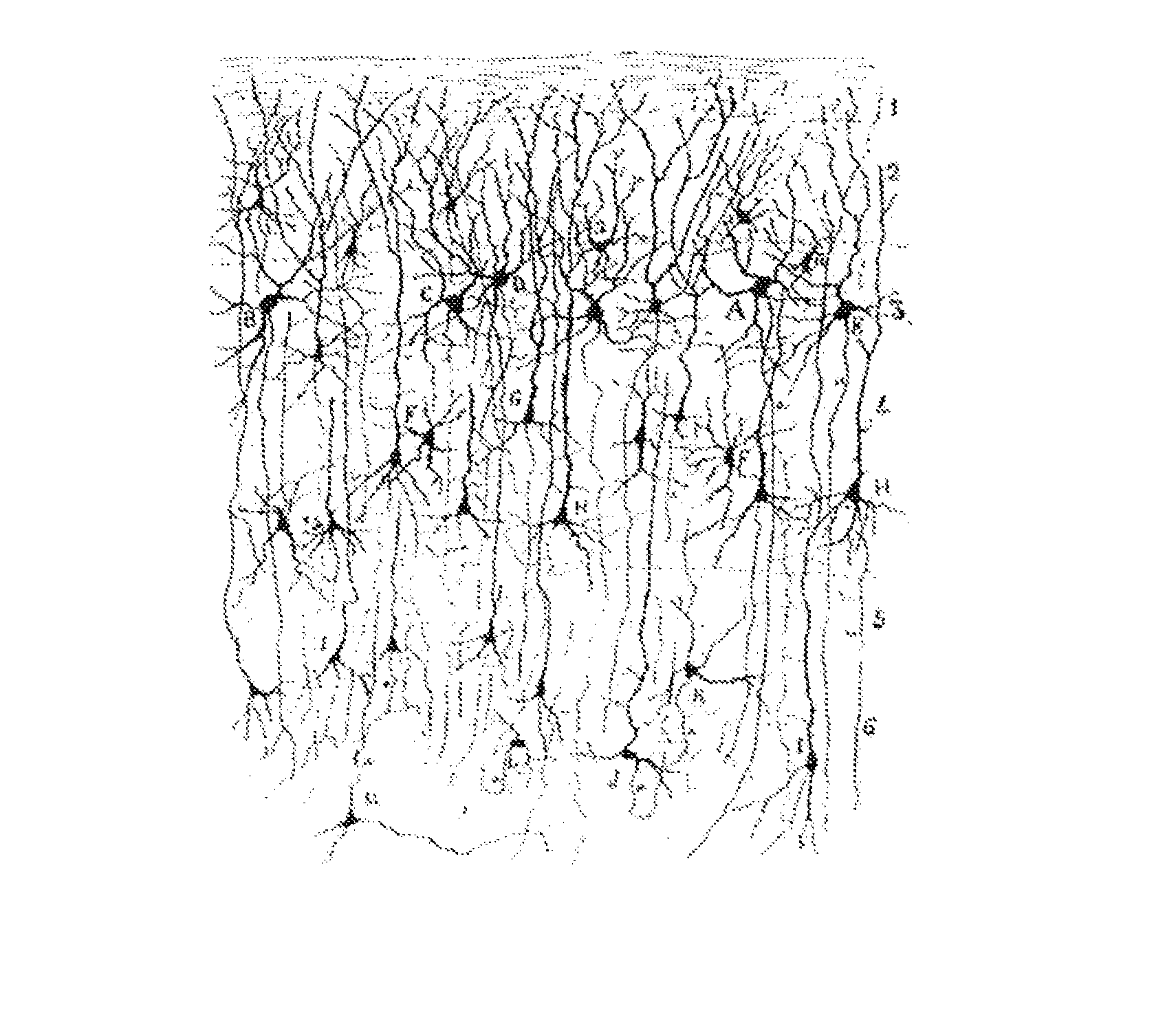
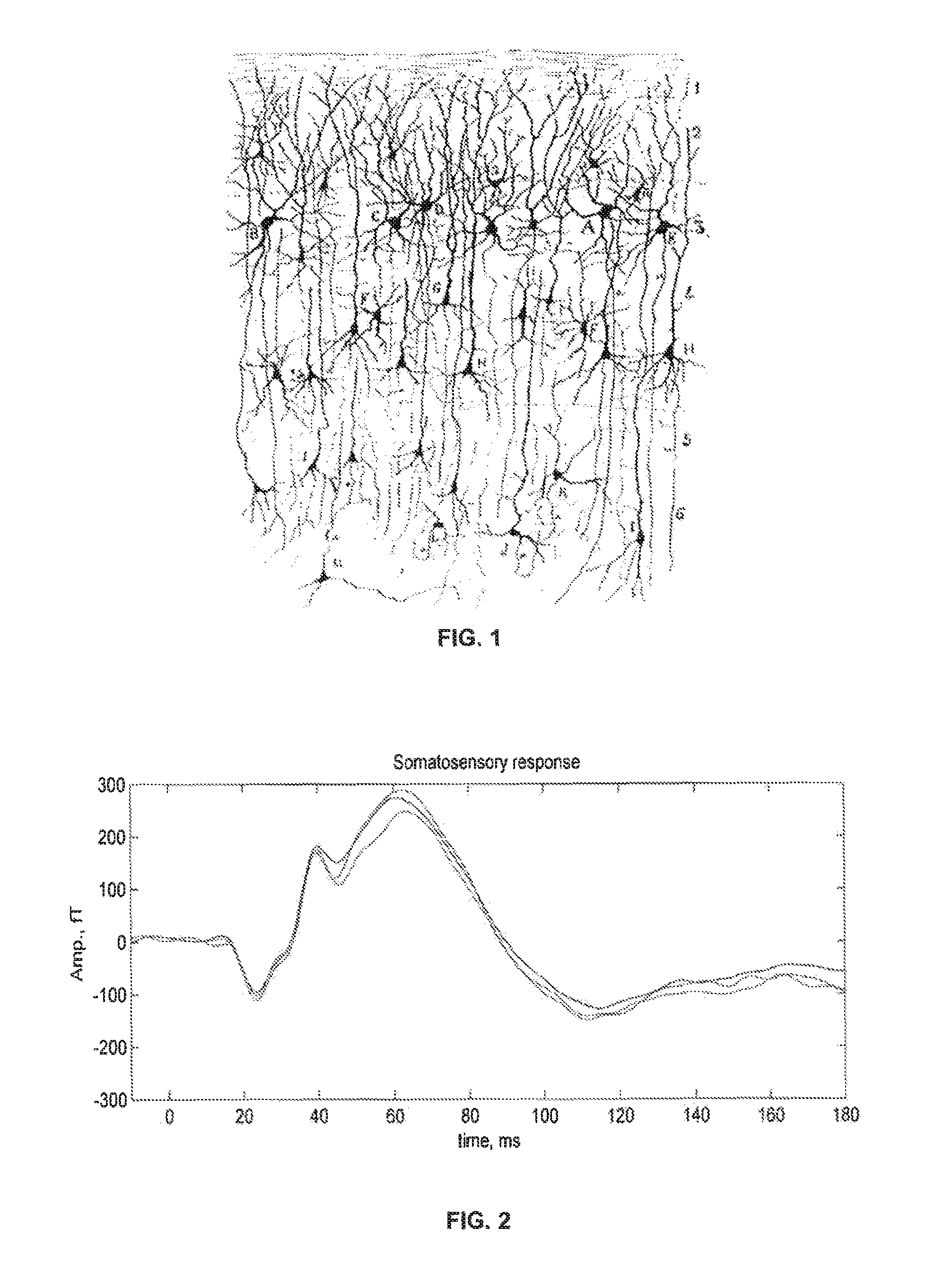
[0044]Electric currents associated with neuronal activity generate magnetic fields that affect the precession of the spins in cortical tissue. This effect can be used to detect (and image) neuronal activity. The currents result from a large number of fast microscopic ion transport processes such as postsynaptic currents and action potential currents from many individual neurons comprising cortical tissue. FIG. 1 illustrates the complex spatial organization of neurons in cortex. The average duration of a composite postsynaptic potential is on the order of 10 ms; the average duration of an action potential is on the order of 1 ms. From a distance, postsynaptic currents appear to be a current dipole with average strength of roughly 20 fA-m per neuron. In contrast, currents associated with action potentials can be approximated by two oppositely oriented current dipoles with a separation of about 1 mm and magnitude of each dipole about 100 fA-m. Since the two dipoles are in opposite dire...


PUM


Abstract

Using resonant interactions to directly and tomographically image neural activity in the human brain using magnetic resonance imaging (MRI) techniques at ultra-low field (ULF), the present inventors have established an approach that is sensitive to magnetic field distributions local to the spin population in cortex at the Larmor frequency of the measurement field. Because the Larmor frequency can be readily manipulated (through varying Bm), one can also envision using ULF-DNI to image the frequency distribution of the local fields in cortex. Such information, taken together with simultaneous acquisition of MEG and ULF-NMR signals, enables non-invasive exploration of the correlation between local fields induced by neural activity in cortex and more ‘distant’ measures of brain activity such as MEG and EEG.

Description

CROSS-REFERENCE TO RELATED APPLICATIONS[0001]This application claims the benefit of U.S. Provisional Application No. 60 / 775,905 filed Feb. 22, 2006.STATEMENT REGARDING FEDERAL RIGHTS[0002]This invention was made with government support under U.S. Department of Energy Contract No. W-7405-ENG-36. Accordingly, the United States Government has certain rights in the invention.TECHNICAL FIELD OF THE INVENTION[0003]The present invention is generally related to the field of medical and biological imaging systems and methodologies. More particularly, the present invention is related to methods of imaging neurological currents in the brain and other biological currents in tissue. The present invention is also related to methods using ultra-low magnetic fields and highly sensitive magnetometers to measure biological currents including neural currents.BACKGROUND[0004]A variety of techniques have been developed to non-invasively image human brain function that are central to understanding how th...

Claims


Application Information

Patent Timeline
Patent Type & Authority Patents(United States)
IPC IPC(8): G01V3/00
CPCA61B5/055
Inventor VOLEGOV, PETR L.MATLASHOV, ANDREI N.MOSHER, JOHN C.ESPY, MICHELLE A.KRAUS, JR., ROBERT H.
Owner TRIAD NAT SECURITY LLC
Why Eureka
Try Eureka