
Indicator for in-situ detecting of lysozyme

a lysozyme and insitu detection technology, applied in the direction of chemical indicators, instruments, food testing, etc., can solve the problems of inability to provide an indication of wound condition, inconvenient operation, and inability to detect insitu, so as to achieve convenient determination and use the effect of working li

Inactive Publication Date: 2009-11-03
CAMBRIDGE MEDITECH LTD
View PDF22 Cites 7 Cited by
  • Summary
  • Abstract
  • Description
  • Claims
  • Application Information

AI Technical Summary

Benefits of technology

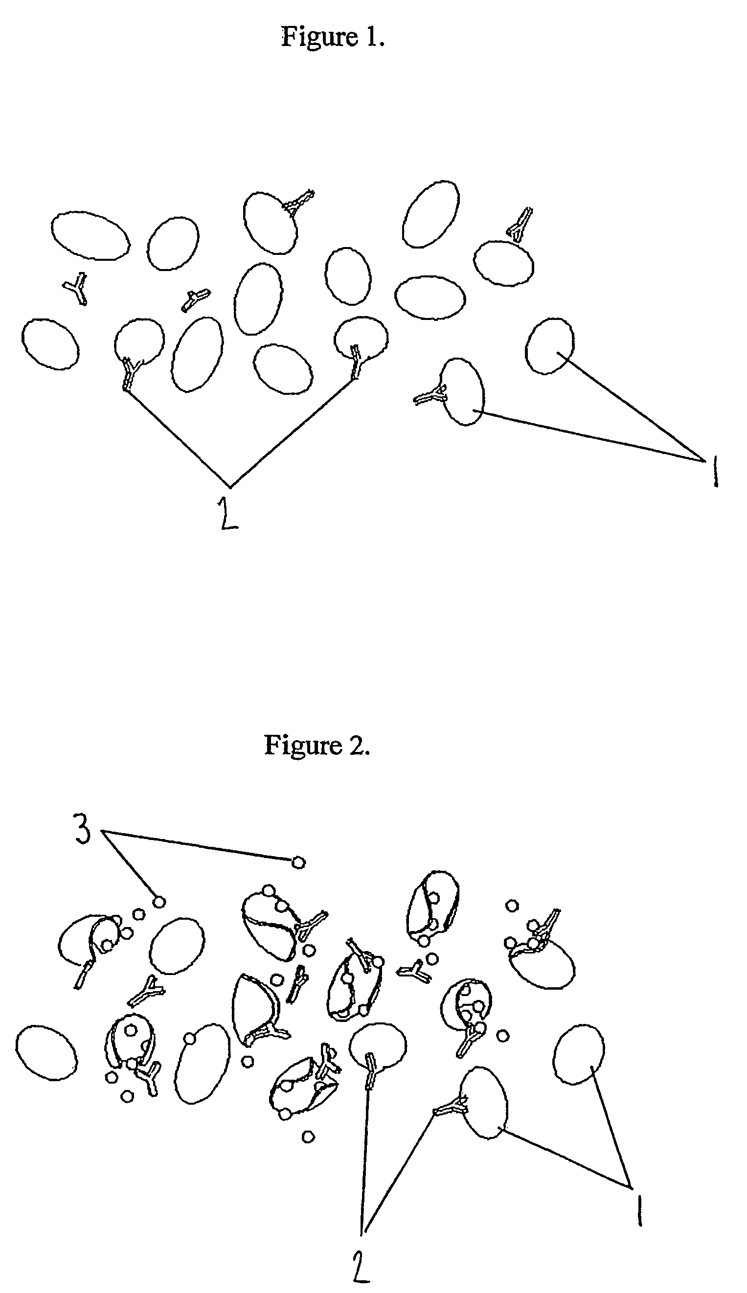
[0017]One advantage of the invention is that it can give an indication of when the amount of a substance or microbe at a particular location has reached a predetermined amount, for example a dangerous level or, in the case of a microbe where the location is a wound in a human or animal, a level at which infection might develop. For example, where the location is a foodstuff, the amount of the microbe to be detected could be an amount at which the foodstuff is dangerous to eat.
[0018]A further advantage of the invention is that it can measure the body's response to the presence of microbes rather than the microbes themselves. The invention therefore provides a more relevant indication of a clinical problem.
[0019]A further advantage of the invention is that whilst traditional methods of microbiological detection tend to be selective and may miss certain species of microbe, the current invention has the advantage over these traditional methods in that it gives a measure of general not specific microbial contamination.
[0020]Another advantage of the invention is that a background level of a first substance which may be present at a location will enable a positive signal in the indicator regardless of whether a contaminating substance or a microbe is present. The time required for this reaction to occur is significantly longer than the time required for a reaction to occur when a contaminating substance or microbe is present and therefore can provide the user with an indication of when the product has reached the end of its working life.
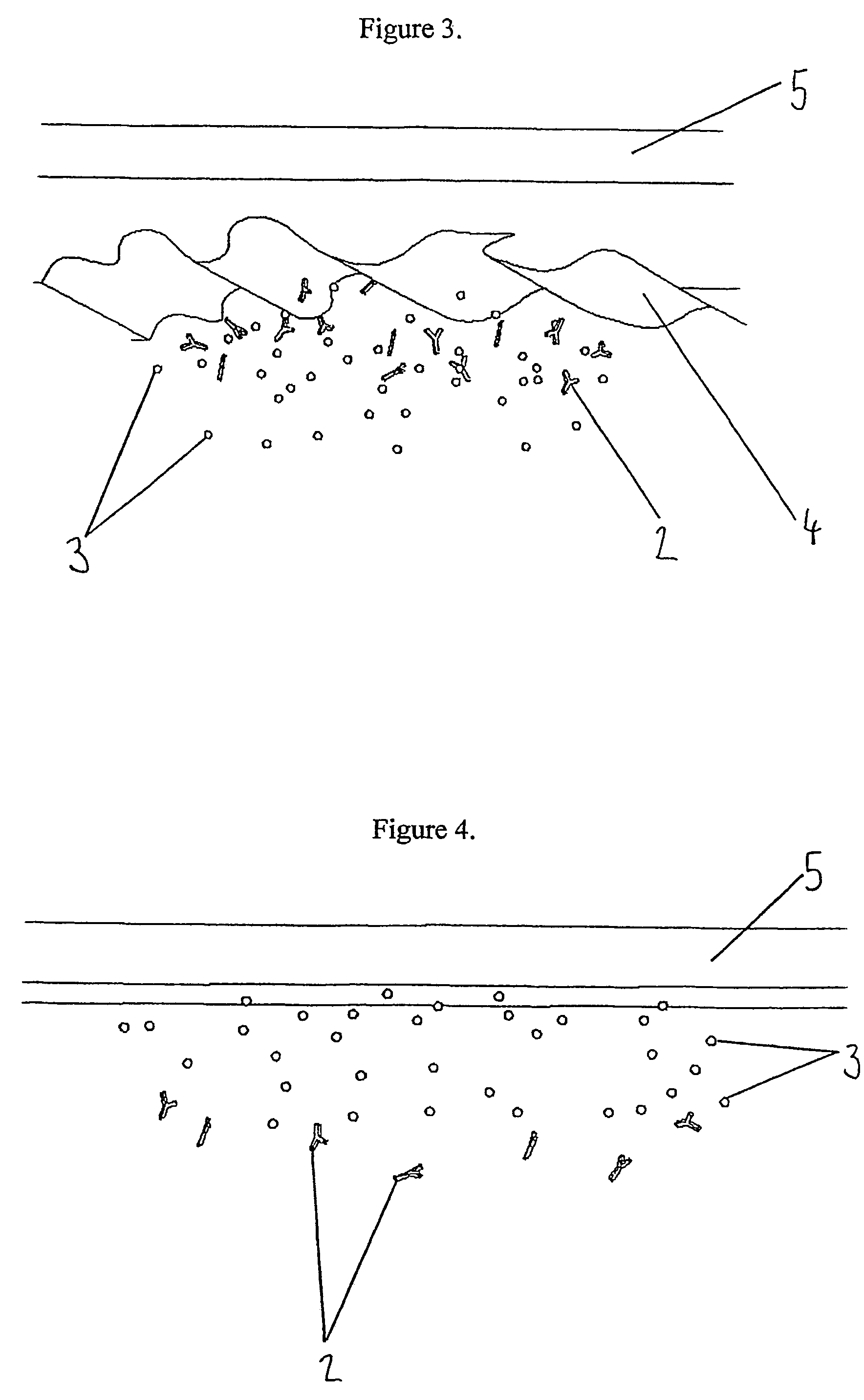
[0021]The sensitivity of the indicator according to the invention is determined by the nature of layer (a). It can be seen that the thicker layer (a) is, the higher the amount of the substance, microbe or the first substance associated with the microbe, or the longer the period of time that the layer (a) is exposed to the substance or microbe or the first substance associated with the microbe, has to be before the layer (a) is degraded sufficiently to allow the substance or microbe or the second substance associated with the microbe or the further substance which is at substantially the same location as the substance or microbe to contact the indicator.
[0022]The thickness of layer (a) and the material used to make it is preferably chosen according to the location at which the indicator is to be used such that when the indicator is designed to detect the presence of a microbe or of substance(s) associated with a microbe, the layer (a) is structured such that the signalling layer produces a detectable signal before the concentration of the microbe reaches a dangerous level. On the other hand, the layer (a) should be sufficiently robust that the time required before it is degraded by a background level of a microbe or of a first substance associated with a microbe is long enough such that the indicator has a useful working life. The thickness of layer (a) and the material used to make it can easily be determined by trial and error by a person of skill in the art when the location at which the indicator is to be used and the type of microbe which is likely to be present are known.

Problems solved by technology

However, these methods generally require a multistep assay to be performed in a laboratory or other clinical environment and not directly at the location where microbial contamination may exist.
During the process of wound healing there is often a serious danger of microbes entering a wound site, multiplying and resulting in the wound becoming infected.
This is usually at a point at which infection has taken hold and is considered to be seriously detrimental to the patient.
In extreme cases septicaemia will result.
None of the systems proposed above provide an indication of wound condition or of the presence of microbial contamination at a wound site.
However, this approach has the disadvantage that these tests are performed on a small random selection of the total production batch, and are therefore helpful only in indicating gross contamination problems at the point of manufacture.
Furthermore, once the packaging has been breached, for example by the action of opening and subsequent reuse of the product, opportunistic organisms can contaminate the product.
The presence of these micro-organisms can jeopardise the quality and organoleptic characteristics of the product, compromise performance, and shorten shelf-life.
Also traditional methods of microbiological detection tend to be selective and may miss certain species of organism.
Consumers are becoming more demanding and expecting products to contain more natural ingredients with fewer preservatives, together with the added requirements of having longer shelf-life and the ability to perform under difficult conditions.
These customer trends are powerful and can increase the risk of contamination both at the point of manufacture and whilst the product is in use in the hands of the customer.

Method used


Image

  • Indicator for in-situ detecting of lysozyme
  • Indicator for in-situ detecting of lysozyme
  • Indicator for in-situ detecting of lysozyme

Examples


example 1

Formation of Layer (a)

[0095]A chitosan (poly (D-glucosamine) of formula I) membrane

[0096]

Formula I

was formed for use as layer (a) using the method described below:[0097]A homogenous solution of 1% (w / w) purified 60% chitosan was prepared in 1% acetic acid by continuous stirring overnight at 22° C. (room temperature).[0098]20 g of the solution was transferred to a sterile polystyrene petri dish (120×120×17 mm) placed on a level surface at 30° C. for several days until the solution was dry and a uniform membrane was formed.[0099]The resulting membrane was removed carefully and neutralized by immersion for 1 hour in 100 ml 2% (w / v) sodium hydroxide solution.[0100]The film was then washed in distilled water several times over a period of 1 hour.[0101]The swollen film was then placed between glass plates and placed in a drying oven until dry.[0102]The film was then removed from the glass plate.

[0103]60% deacetylated chitosan was used as it had been determined experimentally that this lev...

example 2

Determination of Concentration of Signalling Substance

[0104]Meldola's blue (formula II) and nitro blue tetrazolium (formula III) were chosen for use as indicators.

[0105]

Formula II

[0106]

Formula III

[0107]Meldola's blue (8-dimethylamino-2,3-benzophenoxazine) acts as an electron donor to tetrazolium salts, such as nitro blue tetrazolium to form an insoluble formazan product. This product is coloured and precipitates out of solution, giving a visual indication of the presence of electron donors such as NADH. The reaction is shown below:

[0108]

[0109]Optimization experiments were carried out to determine the concentrations of meldola's blue and nitro blue tetrazolium in order that the formation of the formazan product (and therefore colour) was increased, and that allowed the meldola's blue to be continuously recycled. NADH was used as the electron donor. The concentrations of meldola's blue (blue crystals) and nitro blue tetrazolium (clear yellow solution) were kept as low as possible so t...

example 3

Signalling Presence of Substance

[0122]A simple cell with two chambers separated by a vertical wall of chitosan as produced in Example 1 was constructed to demonstrate the degradation of chitosan in a time-dependent manner in the presence of lysozyme. The left hand chamber was filled with a blue coloured dye to which lysozyme was added. The right hand chamber contained water. The chitosan between the two chambers prevented flow of liquid between them.

[0123]The diffusion of colour between the chambers was examined for up to 30 minutes. Appropriate controls were run where no lysozyme was present.

[0124]Two concentrations of lysozyme (25 and 50 μg per ml) were used, and the strength of colour visible in the second chamber up to 30 minutes was determined. A rough estimation of the relative rate of degradation of the chitosan by lysozyme was therefore given. The results are presented in the table below, the stronger the colour observed, the more positives indicated. Where no lysozyme was p...


PUM

PropertyMeasurementUnit
temperatureaaaaaaaaaa
absorbanceaaaaaaaaaa
concentrationsaaaaaaaaaa

Abstract

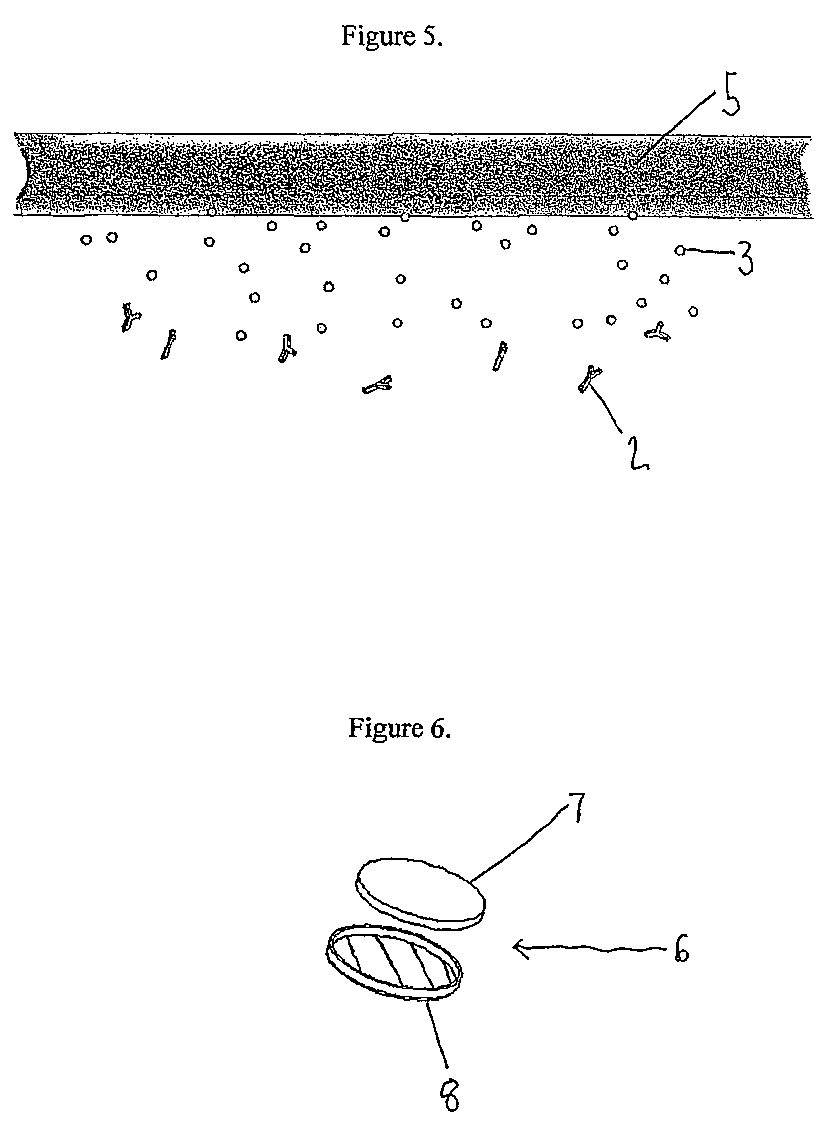
The present invention provides an indicator for the in-situ detection of the presence of a lysozyme or a microbe producing lysozyme at a location. The indicator comprises a layer (8) which is susceptible to degradation by lysozyme and a signaling layer (7) which is adapted to produce a detectable signal which indicates the presence of the lysozyme or the microbe producing lysozyme which is located at substantially the same location as the lysozyme or microbe. In use, the signaling layer is at least initially protected from contact with the lysozyme or microbe which is located at substantially the same location as the lysozyme or microbe by the degradable layer.

Description

[0001]This application is the US national phase of international application PCT / GB01 / 04588 filed 15 Oct. 2001 which designated the U.S.FIELD OF THE INVENTION[0002]The present invention provides a new indicator for the in situ detection of the presence of a substance or a microbe at a location, and methods for its use and construction.BACKGROUND OF THE INVENTION[0003]For a number of applications, it is important to be able to detect the presence of microbes at a given location, for example to determine whether foodstuffs are safe for consumption or whether a wound has become infected. There are currently a number of methods for the detection of the presence or absence of microbial contamination. However, these methods generally require a multistep assay to be performed in a laboratory or other clinical environment and not directly at the location where microbial contamination may exist.[0004]During the process of wound healing there is often a serious danger of microbes entering a w...

Claims


Application Information

Patent Timeline
Patent Type & Authority Patents(United States)
IPC IPC(8): C08L5/08C12Q1/34G01N33/53A61F13/00A61F13/42A61L15/56A61L17/00C12Q1/04G01N21/77G01N21/80G01N31/22G01N33/02G01N33/36G01N33/52
CPCA61F13/00055A61F13/42G01N31/22A61L15/56C12Q1/04A61F2013/00182A61F2013/00846
Inventor FERGUSON, DREW MMILAN, GUY D.DOW, CRAWFORD S.SWOBODA, UTHAYA
Owner CAMBRIDGE MEDITECH LTD
Learn More