Protease inhibitor sample collection system

a sample collection and protease inhibitor technology, applied in the field of protease inhibitor sample collection system, can solve the problems of degrading or fragmenting proteins, affecting and elapsed time between obtaining samples, etc., and achieves the effect of increasing the relevance of protein research and analysis

Inactive Publication Date: 2010-01-12
BECTON DICKINSON & CO
View PDF89 Cites 24 Cited by
  • Summary
  • Abstract
  • Description
  • Claims
  • Application Information

AI Technical Summary

Benefits of technology

[0012]Surprisingly, it has now been found that collecting a biological sample from a patient and introducing that sample into a container containing a stabilizing agent can be efficient in maintaining protein content and composition. Inclusion of the stabilizing agent within the collection device for immediate contact with the biological sample substantially prevents the degradation and / or fragmentation of proteins that otherwise occurs between the time the sample is collected and the stabilizing agent is added. As a result, the percentage of intact proteins is greatly increased.
[0013]The present invention, therefore, is directed to methods and devices for collecting a biological sample. More particularly, the invention is directed to a collection container and to a method of collecting a biological sample, which comprises immediately contacting the sample with a stabilizing additive to inhibit endogenous protease for the isolation and purification of proteins. The stabilizing agent of the invention is a suitable agent that is able to inhibit, prevent or reduce the occurrence of protein degradation and / or fragmentation during storage of the biological sample.
[0015]One aspect of the present invention is to prepare a biological sample that is stable at room temperature for extended periods of time with little or no degradation or fragmentation of proteins. Accordingly, a method is provided for producing a biological sample that is stable at room temperature for extended periods of time with little or no incidence of degradation or fragmentation of proteins during storage.
[0018]A further aspect of the present invention is to provide a method for stabilizing a biological sample, particularly whole blood or a component thereof, immediately upon collection from the patient to inhibit or prevent degradation or fragmentation of proteins when the sample is stored at various temperatures.
[0020]Still another aspect of the present invention is to provide a blood collection container for collecting an amount of blood and mixing the blood with a stabilizing agent at the point of collection to produce a blood sample that is stable by preventing degradation or fragmentation of proteins such that isolation and purification of proteins in the sample can be conducted at a later time.
[0024]The methods and collection devices of the present invention have several distinct advantages. One advantage of the collection devices is the offering of a system, preferably a closed system, that includes the stabilizing agent and that protects the sample from deleterious exposures. Another advantage is labor relief from manual fabrication of such collection systems. Still another advantage is routine line production of such collection devices, whereby quality control measures and procedures are applied to the product. Yet another advantage is the standardization of such collection devices where no industry standards currently exist and where the industry is calling for such standardization. Moreover, the relevance of protein research and analysis is increased by preserving and being able to characterize and study proteins in a state that is as close to the in vivo state as possible.

Problems solved by technology

Generally, a considerable amount of time elapses between obtaining the sample and analyzing it.
A common and recurring problem, therefore, is the maintenance of the biological sample in a manner that prevents degradation, alteration or destruction of essential materials during the manipulations and / or preparations preceding analysis of the biological sample as a test specimen.
Once the cells are frozen, broken up or disrupted, however, protease enzymes begin to react on cellular proteins unspecifically, thereby quickly degrading the cellular proteins.
In protein isolation, this can lead to degraded or fragmented proteins.
This also can lead to a decline in protein levels during storage and transport of biological samples, thereby limiting the sensitivity of testing methods.
The yield of the protein(s), therefore, can be drastically reduced and can endanger all further subsequent experiments such as, for example, protein quantification, 2-D gel plotting of proteins, drug development, Western blotting, reporter gene analysis, immunoprecipitations, epitope tagging, specific protein activity assays, etc.
Despite the development and commercial availability of stabilizing preparations and compositions, the overwhelming majority of these are quite limited as to their usefulness and efficacy and do not lend themselves without major modification and alterations to specific clinical problems or a broad variety of different clinical and analytical settings.
For example, many such compositions do not stabilize the proteases in the sample, thereby failing to preserve sensitive proteins in the biological samples.
This procedure, however, is dangerous because of the likelihood of needle stick injuries.
Moreover, the volumes of the protease inhibitor cocktails are subject to technician-to-technician variability because of the manual method of loading the protease inhibitor cocktails into the tubes.
The issue of industry standardization for a protease cocktail, i.e. a combination of protease inhibitors, is also significant.
The effectiveness and behavioral differences of the protease inhibitors and combinations thereof are, ostensibly, not well studied and, therefore, it is extremely difficult to correlate one researcher's analytical results to another researcher's results.
In addition, most protease inhibitors range in toxicity from mildly toxic to highly toxic.
Presently, there is no control and / or limit to potential exposure to these toxins, thereby jeopardizing the safety of healthcare and laboratory workers.

Method used


Image

  • Protease inhibitor sample collection system
  • Protease inhibitor sample collection system
  • Protease inhibitor sample collection system

Examples


example 1

Plastic Tubes with Mechanical Separator

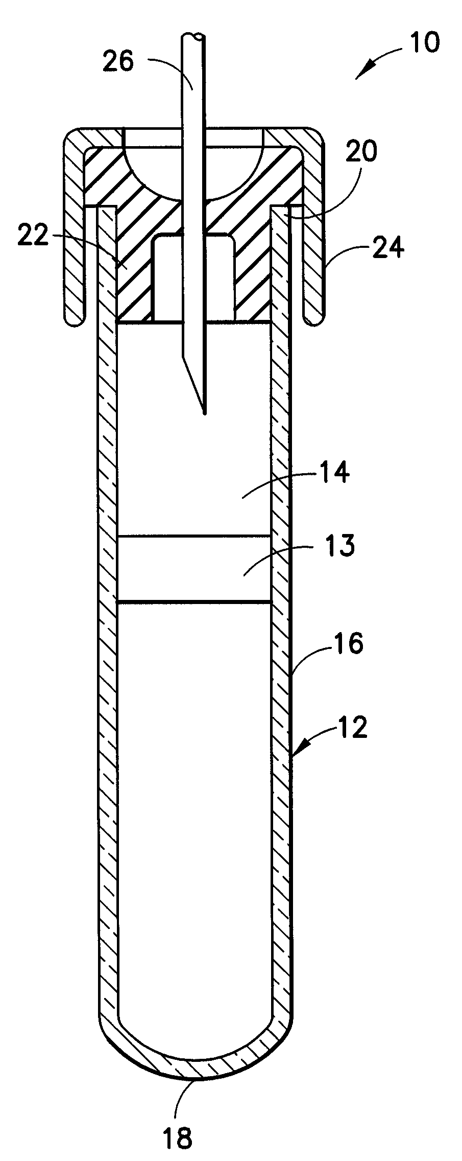
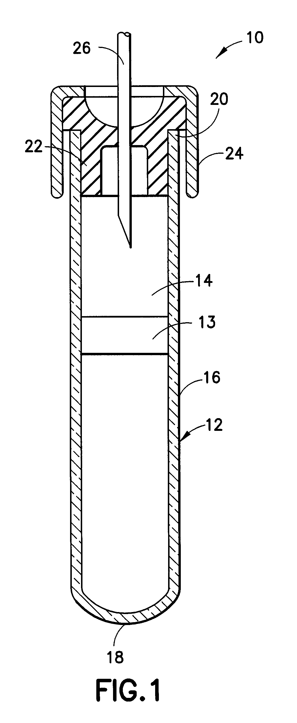
[0097]Fifteen hundred (1,500) plastic tubes were spray coated with a lithium heparin solution and dried. A stabilizing agent was prepared by dissolving SIGMA® protease inhibitor cocktail in deionized water using about 22 ml of deionized water per vial of protease inhibitor. Into each tube was placed approximately 300 μl of the stabilizing agent solution. A mechanical separator was assembled into the closures (HEMOGUARD® closures lubricated with silicone) of each tube and then the closure / separator assemblies were placed on the tubes. Next, the tubes were lyophilized and evacuated. The tubes were then placed into barrier packages, about five (5) tubes per pouch, with desiccant. The pouches were heat sealed, and the product was sterilized.

example 2

Glass Tubes with Mechanical Separator

[0098]The procedure of Example 1 was followed, this time using glass tubes.

example 3

Plastic Tubes with Mechanical Separator

[0099]The procedure of Example 1 is again followed, except that the stabilizing agent solution also includes about 0.5 to about 20% trehalose.

[0100]A suitable example of a formulation for a protease inhibitor cocktail is as follows: about 800 μg / ml benzamidine HCL, about 500 μg / ml phenanthroline, about 500 μg / ml aprotinin, about 500 μg / ml leupeptin, about 500 μg / ml pepstatin A and about 50 μg / ml PMSF. The cocktail is in the form of a lyophilized powder. Before use, about 1 ml of pure ethanol is added to the lyophilized powder to obtain a 50× protease inhibitor cocktail.


PUM


Abstract

A collection container and a method for collecting a biological sample, particularly whole blood, includes at least one stabilizing agent in an amount effective to stabilize and inhibit protein degradation and / or fragmentation. The stabilizing agent is able to stabilize proteases in the biological sample, particularly at the point of collection, by inhibiting protein degradation and / or fragmentation in the sample when the sample is stored. The stabilizing agent includes one or more protease inhibitors.

Description

CROSS-REFERENCE TO RELATED APPLICATIONS[0001]The present application is a continuation of U.S. Patent application Ser. No. 10 / 436,263 filed May 13, 2003, now U.S. Pat. No. 7,309,468 issued on Dec. 18, 2007, which claims priority to U.S. Provisional Patent Application Ser. No. 60 / 379,399, which was filed on May 13, 2002.FIELD OF THE INVENTION[0002]The present invention is directed to a method and device for collecting and stabilizing a biological sample, particularly a whole blood sample, directly from a patient. More specifically, the present invention relates to sample collection containers having a stabilizing additive contained therein for stabilizing proteases immediately on collection of a biological sample and for inhibiting protein degradation and / or fragmentation during storage thereof.BACKGROUND OF THE INVENTION[0003]In clinical diagnostic settings, it has often been necessary to collect biological samples such a whole blood, red blood cell concentrates, platelet concentrat...

Claims


Application Information

Patent Timeline
Patent Type & Authority Patents(United States)
IPC IPC(8): B01L3/00C12M1/34A61B5/15A61B5/154A61J1/12A61J1/14A61J7/00B01L3/02B01L3/14C12M1/26C12Q1/02G01N1/00
CPCA61B5/1411A61B5/417A61J1/05A61J7/00A61J7/0053B01L3/50B01L3/5082B01L3/02A61J1/12Y10T436/2525B01L2200/16B01L2300/0609G01N35/1095Y10T436/25375A61B5/15003A61B5/150221A61B5/150236A61B5/150244A61B5/150274A61B5/150351A61B5/150366A61B5/150389A61B5/150503A61B5/150755A61B5/150893A61B5/154C12M23/12C12M33/04A61B5/1405B01L3/00
Inventor HAYWOOD, BRUCEDASTANE, AJITLIN, FU-CHUNG
Owner BECTON DICKINSON & CO
Why Eureka
Try Eureka