Direct quantification of ribosome inactivating protein

a technology of inactivating protein and quantification method, which is applied in the field of detection of enzymes, can solve the problems of cellular destruction, loss of protein synthesis, and high cost, and achieve the effect of reducing the number of ribosomes

Inactive Publication Date: 2010-06-29
UNITED STATES OF AMERICA
View PDF0 Cites 6 Cited by
  • Summary
  • Abstract
  • Description
  • Claims
  • Application Information

AI Technical Summary

Problems solved by technology

RIPs exert their highly toxic action by catalytically inactivating ribosomes in eukaryotic cells that in turn inhibits protein synthesis leading to cellular destruction.
This depurination subsequently prevents elongation factor 2 from binding to the ribosome resulting in loss of protein synthesis.
Their extreme toxicity makes them potential candidates for use or deployment during warfare or acts of terrorism.
Currently available military systems for detecting and identifying RIPs are laboratory based and require sophisticated and expensive equipment.
Therefore, such systems are of limited practical use in the field.
The recent domestic attacks employing ricin via the postal system exposed several vulnerabilities of the current process that include their indirect identification of toxin and inability to directly quantitate the toxin's activity or distinguish between active and inactive toxin.
These are significant limitations because ricin is easily produced through a number of processes that yield toxin with various purity and stability and thus lethality at the time of dissemination.
Current processes to assess the N-glycosidase activity of type II RIPs are arduous and lack sensitivity.
Moreover, direct labeling processes of the excised purine consistently suffer from low sensitivity, and are thus not amenable to use as a field detection technique for identification and assessment of RIP activity.

Method used


Image

  • Direct quantification of ribosome inactivating protein
  • Direct quantification of ribosome inactivating protein
  • Direct quantification of ribosome inactivating protein

Examples


example 1

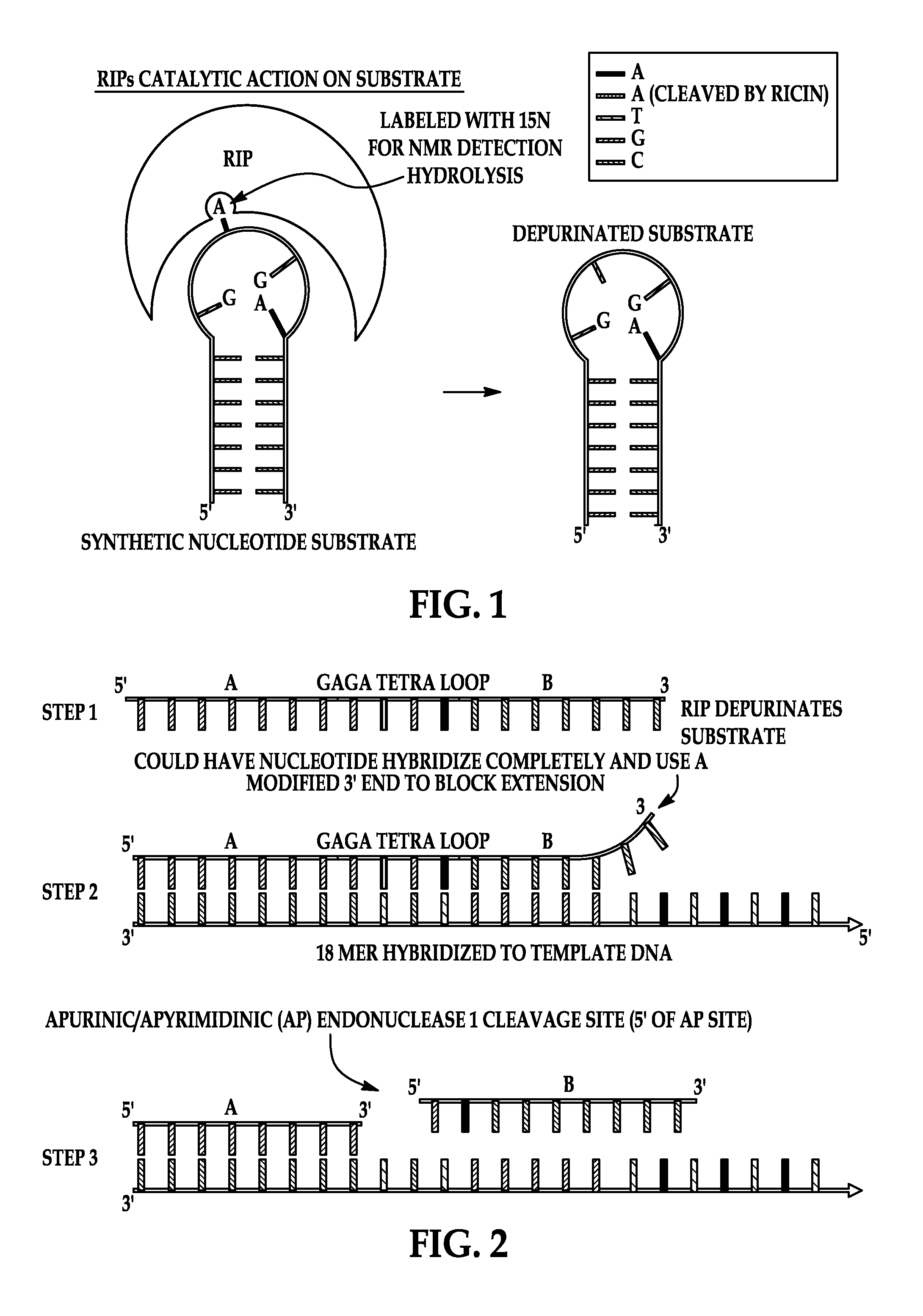
Depurination by Ricin A-Chain

[0076]100 μM of DNA oligonucleotide substrate (SEQ ID NO: 2) is reacted with 0.5 μM Ricin A chain (Sigma Chemical Co, St. Louis, Mo.) at 37° C. for 30 minutes in 10 mM potassium citrate and 1 mM EDTA (pH 4.0). Adenine release is verified with LC / MS.

example 2

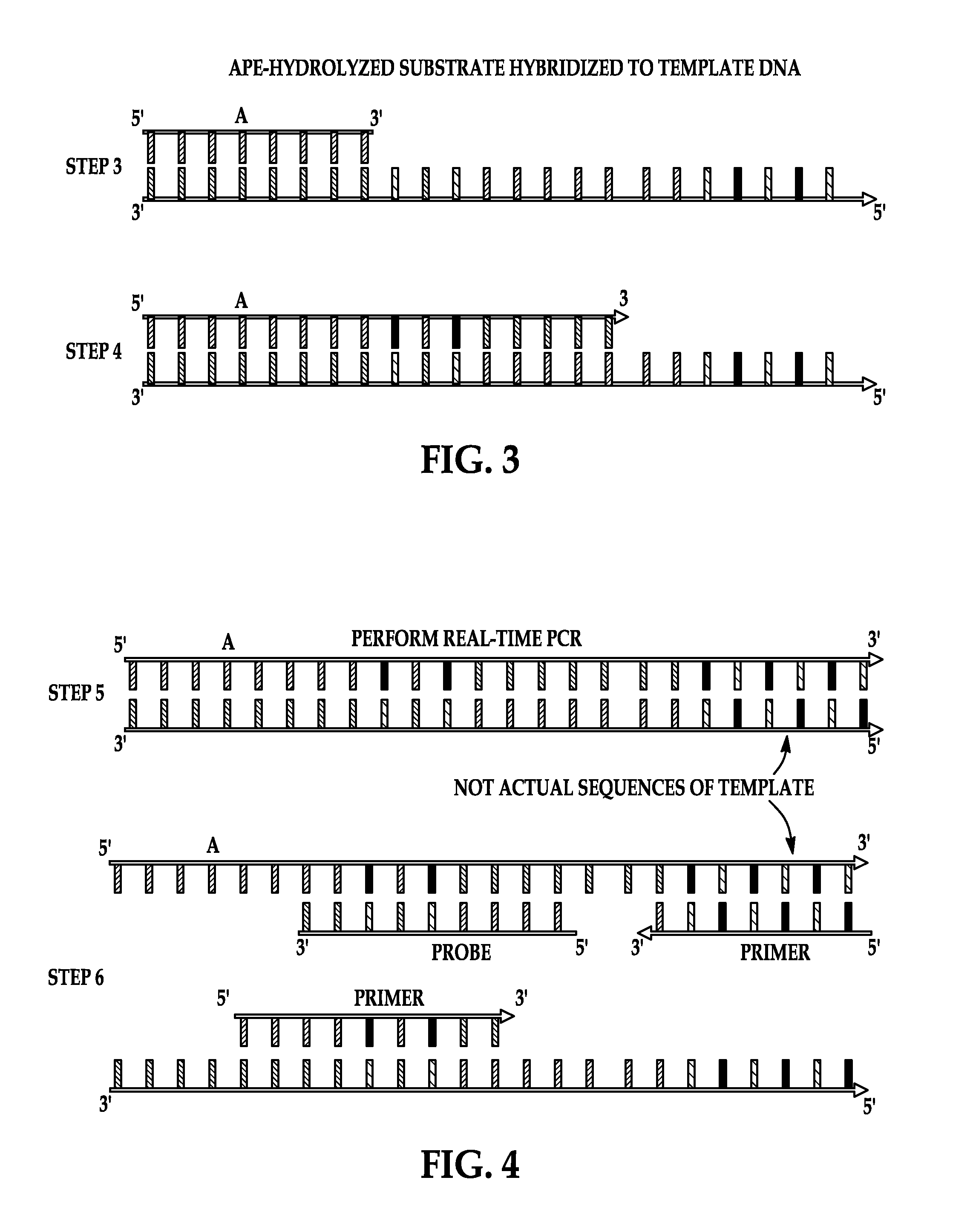
Hydrolysis of Product by APE

[0077]To perform the APE activity assay, 1 μl of the reaction product of Example 1 is hybridized to 1 μl of 100 μM template oligonucleotide (SEQ ID NO: 5) (Gene Link, Inc., Hawthorne, N.Y.) by mixing at room temperature. To this 1 unit of APE1, 1 μl of 10× NEBuffer4 (20 mM Tris-acetate, 50 mM potassium acetate, 10 mM Magnesium Acetate, 1 mM Dithiothreitol, pH 7.9 at 25° C.) (New England Biolabs) and 6 μl of molecular grade water is added. Recombinant human APE1 is purchased from New England Biolabs, Ipswich, Mass. (10,000 Units / ml) in 50% glycerol. One unit is defined as the amount of enzyme required to hydrolyze 20 μmol of a 34 by oligonucleotide duplex containing a single AP site in a reaction volume of 10 μl in one hour at 37° C. The reaction mixture is incubated for 1 hour at 37° C.

example 3

Detection by Real-Time PCR

[0078]Following incubation, 5 μl of the reaction mix of Example 2 is added to a PCR reaction mixture (Bio-Rad, Hercules, Calif.) consisting of 12.5 μl Bio-Rad master mix, 1 μl forward primer (SEQ ID NO: 4), 1 μl reverse primer (SEQ ID NO: 3), 1 μl probe (SEQ ID NO: 7), and 4.5 μl water. Real-time PCR is performed on a Bio-Rad iCycler using Taq Polymerase (Bio-Rad). Cycle times are 95° C. for 10 minutes followed by an amplification program of 50 cycles of 3 seconds at 95° C., 5 seconds at 61° C., and 20 seconds at 72° C. with fluorescence acquisition at the end of each extension.

[0079]The ability of a DNA substrate that was simulated to have been depurinated by a RIP and subsequently hydrolyzed by APE was examined to determine its ability to hybridize to the template DNA, followed by extension with Taq polymerase and amplification through the polymerase chain reaction. Oligonucleotide representing RIP depurinated and APE cleaved product is amplified when com...


PUM

PropertyMeasurementUnit
pHaaaaaaaaaa
pHaaaaaaaaaa
pHaaaaaaaaaa

Abstract

A method for detecting ribosome inactivating protein (RIP) activity in a sample is provided that involves contacting a sample that contains an RIP with an inventive substrate that is depurinated to form product. The product is hybridized to a template and cleaved by an apurinic / apyrimidinic endonuclease such that releasing the blocked 3′ end only in a sample that contains RIP activity. The 5′ end of the product serves as a primer for extension by a polymerase reaction. The newly synthesized strand complementary to the template is detected by RT-PCR processes. A kit is provided suitable for field or laboratory use.

Description

GOVERNMENT INTEREST[0001]The invention described herein may be manufactured, used, and licensed by or for the United States Government.FIELD OF THE INVENTION[0002]The invention relates generally to a process of detecting enzymes in a fluid or solid sample; and in particular, to the detection of ribosome inactivating proteins in fluid mediaBACKGROUND OF THE INVENTION[0003]Ribosome inactivating proteins (RIPS, also known as ribotoxins) are toxic to animals and humans. RIPs exert their highly toxic action by catalytically inactivating ribosomes in eukaryotic cells that in turn inhibits protein synthesis leading to cellular destruction.[0004]RIPs are generally divided into two classes, the Type I RIP and the Type II RIP. There is significant amino acid sequence homology between members of Type I and Type II RIPs, and bacterial Shiga and Shiga-like toxins. However, the Type II RIPs and the bacterial shiga and shiga-like toxins have similar mechanisms of action.[0005]Abrin, ricin and shig...

Claims


Application Information

Patent Timeline
Patent Type & Authority Patents(United States)
IPC IPC(8): C12Q1/68
CPCC12Q1/6804C12Q2525/119C12Q2525/301C12Q2521/301
Inventor DORSEY, RUSSELL M.
Owner UNITED STATES OF AMERICA
Why Eureka
Try Eureka