
Tumor markers for ovarian cancer diagnosis

a tumor marker and ovarian cancer technology, applied in the field of tumor markers for ovarian cancer diagnosis, can solve the problems of not being recommended as a diagnostic tool or target, yielding many false positives, and difficult to detect early ovarian cancer

Inactive Publication Date: 2010-06-29
NAT TAIWAN UNIV
View PDF0 Cites 7 Cited by
  • Summary
  • Abstract
  • Description
  • Claims
  • Application Information

AI Technical Summary

Benefits of technology

The present invention provides an ovarian cancer marker and a method for detecting ovarian cancer at an early stage. The tumor markers include galectin-1, cathepsin B, MHC class I antigen, heat shock protein 27, ubiquitin carboxy-terminal esterase L1, plasma retinol-binding protein, transthyretin, SH3 binding glutamate-rich protein, tubulin-specific chaperone A, RNA binding protein regulatory subunit, γ-actin, tropomyosin, and calcium / calmodulin-stimulated cyclic nucleotide phosphatase. The expression levels of these tumor markers are significantly different between ovarian cancer tissues and non-cancerous tissues, indicating their potential use as diagnostic tools for ovarian cancer. The detection method involves obtaining ovarian tissue samples from an individual to be diagnosed and a non-cancerous ovarian tissue, determining the expression levels of the tumor markers, and comparing them to determine if the individual is affected with ovarian cancer or not.

Problems solved by technology

The lack of reliable tumor marker has made the early detection of ovarian cancer difficult.
It yields many false positive results.
Therefore it is not recommended as a diagnostic tool or target for ovarian cancer in early stage cancers.

Method used


Image

  • Tumor markers for ovarian cancer diagnosis

Examples


example 1

Screening of Tumor Markers

[0029]The ovarian tissues collected in the present invention comprised of 36 epithelial ovarian cancers, 10 borderline malignancies and 18 normal ovaries. Clinical and histological characteristics of these 36 ovarian cancer tissue samples are summarized in Table 1.

[0030]The histologic subtypes of ovarian cancer include clear cell, endometrioid, mucinous, serous and others as shown in Table 1. Among these 36 ovarian cancer tissue samples, 10 were of clinical stage I, 6 of clinical stage II, 18 of clinical stage III, and 2 of clinical stage IV.

[0031]Protein extracts from the normal ovarian tissues and the ovarian cancer tissues were separated on SDS-PAGE followed by 2D-polyacrylamide gel electrophoresis.

[0032]2D-polyacrylamide gel electrophoresis was performed on a 130 mm, linear immobilized pH 4-7 Immobiline DryStrip (Amersham Pharmacia Biotech, Piscataway, N.J., USA) using MULTIPHOR II Electrophoresis system. The ovarian tissues were frozen in liquid nitrog...

example 2

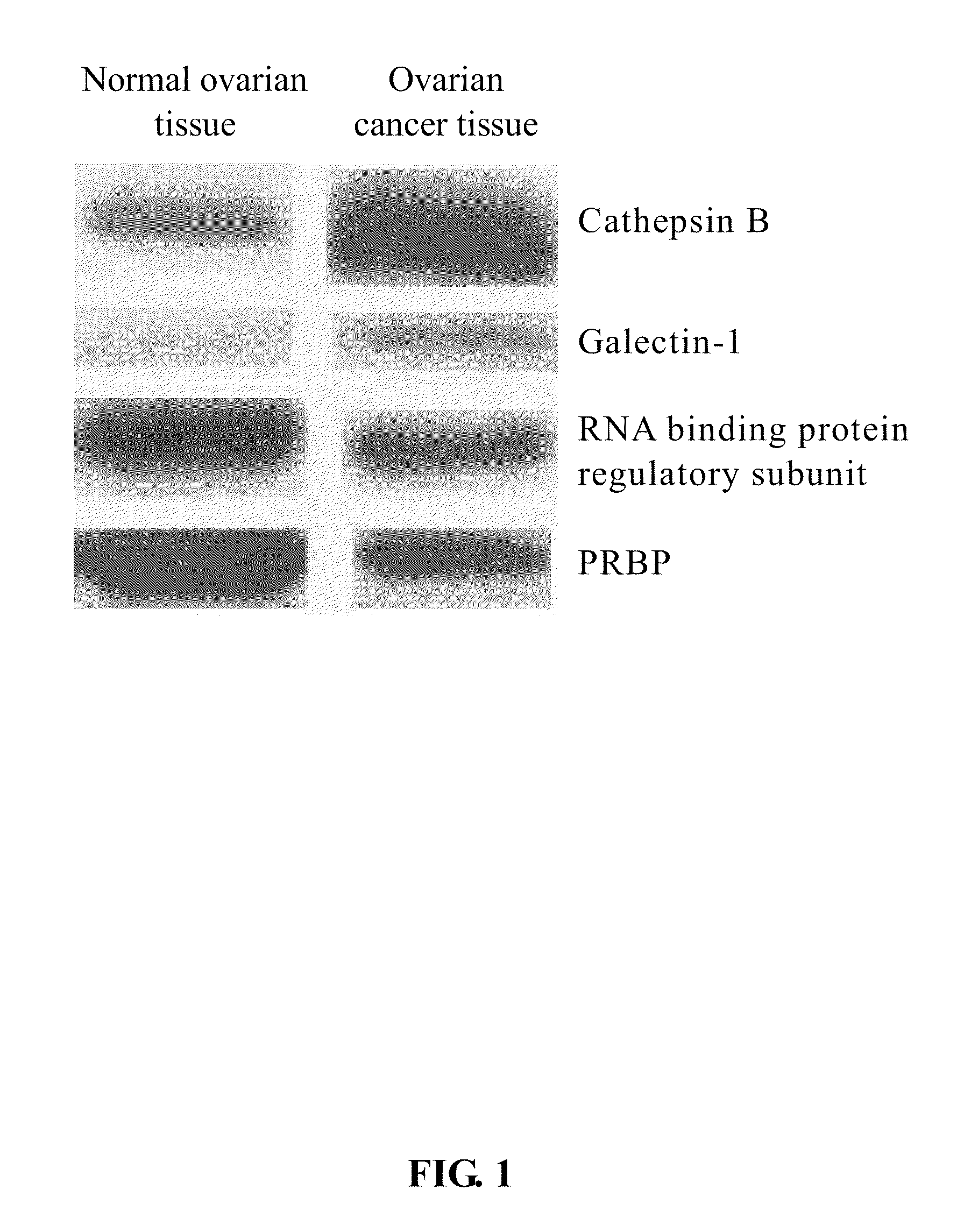
[0040]Four of the protein spots detected in 2D-gel electrophoresis images, which included: cathepsin B, galectin-1, RNA-binding protein regulatory subunit, and plasma retinol-binding protein (PRBP), were further identified through Dot blot, or SDS-PAGE followed by Western blotting analyses.

[0041]The tissues from normal or cancer ovaries were grinded with plastic pestles in ¼ PBS buffer containing protease inhibitor cocktail (Calbiochem). After centrifugation 15,000×g for 10 min at 4° C., the supernatant was transferred to an eppendorf tube and subjected to Dot blot, or SDS-PAGE followed by Western blotting analyses. The protein concentration was determined by the absorption of A280.

[0042]For SDS-PAGE analysis, 30 μg of protein sample was applied to each lane. All samples were heated for 5 min at 95° C. before loading into the 15% polyacrylamide gel. After electrophoresis, proteins were electroblotted onto polyvinylidene difluoride (PVDF) membrane. For Dot blot analysis, 5 μg of prot...


PUM

PropertyMeasurementUnit
pHaaaaaaaaaa
pHaaaaaaaaaa
accelerating voltageaaaaaaaaaa

Abstract

The present invention relates to a tumor marker for diagnosis of ovarian cancer, which is selected from the group consisting of galectin-1, cathepsin B, MHC class I antigen, heat shock protein (HSP) 27, ubiquitin carboxy-termal esterase L1, plasma retinol-binding protein (PRBP), transthyretin, SH3 binding glutamate-rich protein, tubulin-specific chaperone A, RNA binding protein regulatory subunit, γ-actin, tropomyosin and calcium / calmodulin-stimulated cyclic nucleotide phosphatase. The ovarian cancer is diagnosed effectively and efficiently based on detecting the expression levels of the tumor markers in the invention from the ovarian tissue sample of an individual to be diagnosed.

Description

BACKGROUND OF THE INVENTION[0001]1. Field of the Invention[0002]The present invention relates to a tumor marker for diagnosis of cancer, especially for diagnosis of ovarian cancer, which can be applied in early diagnosis of ovarian cancer.[0003]2. The Prior Arts[0004]Human ovarian cancer is one of the common gynecological malignancies. In the developed country, it is one of the leading causes of death of the gynecological cancers, and the five-year survival rate is only about 30%. Overall about one woman in 70 will get ovarian cancer, and estimated one woman in 100 will die from this cancer in USA. This is because the illness is often diagnosed during late stage of the cancer. The cancer has often spread beyond the ovaries at that time, and therefore related to the low survival rate. Though Taiwanese women do not have a high incidence of ovarian cancer, but the incidence has increased over past two decades.[0005]The five-year survival rate will up to 90-95 percent if the ovarian can...

Claims


Application Information

Patent Timeline
Patent Type & Authority Patents(United States)
IPC IPC(8): G01N33/53
CPCG01N33/57449
Inventor CHOW, SONG-NAN
Owner NAT TAIWAN UNIV
Learn More