Extendible stent apparatus

a stent and stent technology, applied in the field of stent apparatus, can solve the problems of limiting the ability to insert a second stent into the side branch, compromising the degree of patency of the primary vessel and/or its branches, and generally produced in a straight tubular configuration

Inactive Publication Date: 2010-08-03
BOSTON SCI SCIMED INC
View PDF550 Cites 30 Cited by
  • Summary
  • Abstract
  • Description
  • Claims
  • Application Information

AI Technical Summary

Benefits of technology

[0016]The stent apparatuses of the invention offers significant and novel advantages over prior art stents in that the stents of the invention 1) can completely cover the bifurcation point of a branched vessel; 2) can accomodate main and branch stents of differing sizes, thus providing a better fit where the main and branch vessels are of different sizes or where the main and branch vessels are occluded to different degrees; 3) can fit branched vessels where the branch extends laterally from the side of the main vessel; 4) may be used to treat lesions in one branch of a bifurcation while preserving complete access to the other branch for future treatment; 5) may be delivered intraluminally by catheter; and 6) are marked with, or at least partly constructed of, material which is imageable by commonly used intraluminal catheterization visualization techniques including but not limited to ultrasound or x-ray, but not endoscopy.

Problems solved by technology

One of the drawbacks of conventional stents is that they are generally produced in a straight tubular configuration.
The use of such stents to treat diseased vessels at or near a bifurcation (branch point) of a vessel may create a risk of compromising the degree of patency of the primary vessel and / or its branches, or the bifurcation point and also limits the ability to insert a second stent into the side branch if the result of treatment of the primary, or main, vessel is suboptimal.
The risk of branch compromise is increased generally in two anatomical situations.
First, a side branch may be compromised when there is a stenosis in the origin of the side branch.
Second, when there is an eccentric lesion at the bifurcation site, asymmetric expansion can cause either plaque shifting or dissection at the side branch origin.
There are reports of attempts to solve this problem by inserting a balloon into the side branch through the struts of a stent deployed in the main branch spanning the bifurcation point; however, this technique carries the risk of balloon entrapment and other major complications (Nakamura, S. et al., Catheterization and Cardiovascular Diagnosis 34: 353-361 (1995)).
Moreover, adequate dilation of the side branch is limited by elastic recoil of the origin of the side branch.
In addition, insertion of a traditional stent into a main vessel spanning a the bifurcation point may pose a limitation to blood flow and access to the side branch vessel.
In this regard, the tubular slotted hinged design of the Palmaz-Schatz intracoronary stent, in particular, is felt to be unfavorable for lesions with a large side branch and is generally believed to pose a higher risk of side branch vessel entrapment where the stent prevents or limits access to the side branch.
The first removal of the guidewire poses the risk of occlusion of the side branch during the deployment of the stent in the main branch.
However, none of these patents relate to stents which are structurally adapted for the treatment of bifurcation lesions, or disclose a bifurcating stent apparatus.
However, bifurcated stents are not taught or disclosed, and the purpose of the stent as used in these inventions is simply to anchor the graft into the vessel wall.
It does not reinforce the vessel wall, treat a lesion, or prevent restenosis after angioplasty.
In addition, studies with hinge-containing stents have shown that there is a high incidence of restenosis (tissue growth) at the hinge point that may cause narrowing or total occlusion of the vessel and thus compromise blood flow.
Furthermore, this design has a relatively large size as compared to the present invention, which makes insertion into many smaller vessels difficult and poses a greatly increased risk of complications.
Also, by having the two additional smaller stents connected to an end of the trunk stent, tracking into a wide-angle lateral side branch may be difficult and may carry the risk of dissection of the vessel wall.
Furthermore, once the device of the '071 patent is implanted, it is impossible to exchange a branch stent should the need for a different stent size or repair of a branch stent arise.
In general, when treating a bifurcation lesion using commercially available stents, it is important to cover the origin of the branch because if left uncovered, this area is prone to restenosis.
Another frequent complication experienced when stenting bifurcated vessels is the narrowing or occlusion of the origin of a side branch spanned by a stent placed in the main branch.
Additionally, placement of a stent into a main vessel where the stent partially or completely extends across the opening of a branch makes future access into such branch vessels difficult if not impossible.
Lastly, conventional stents are difficult to visualize during and after deployment, and in general are not readily imaged by using low-cost and easy methods such as x-ray or ultrasound imaging.

Method used


Image

  • Extendible stent apparatus
  • Extendible stent apparatus
  • Extendible stent apparatus

Examples


Embodiment Construction

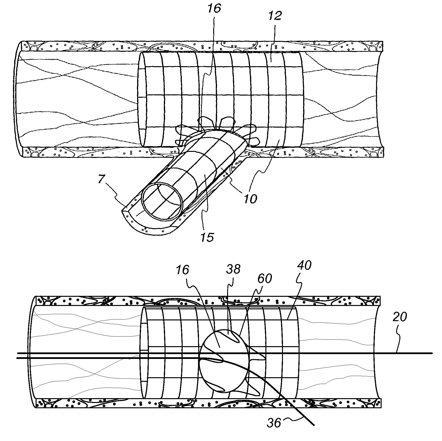
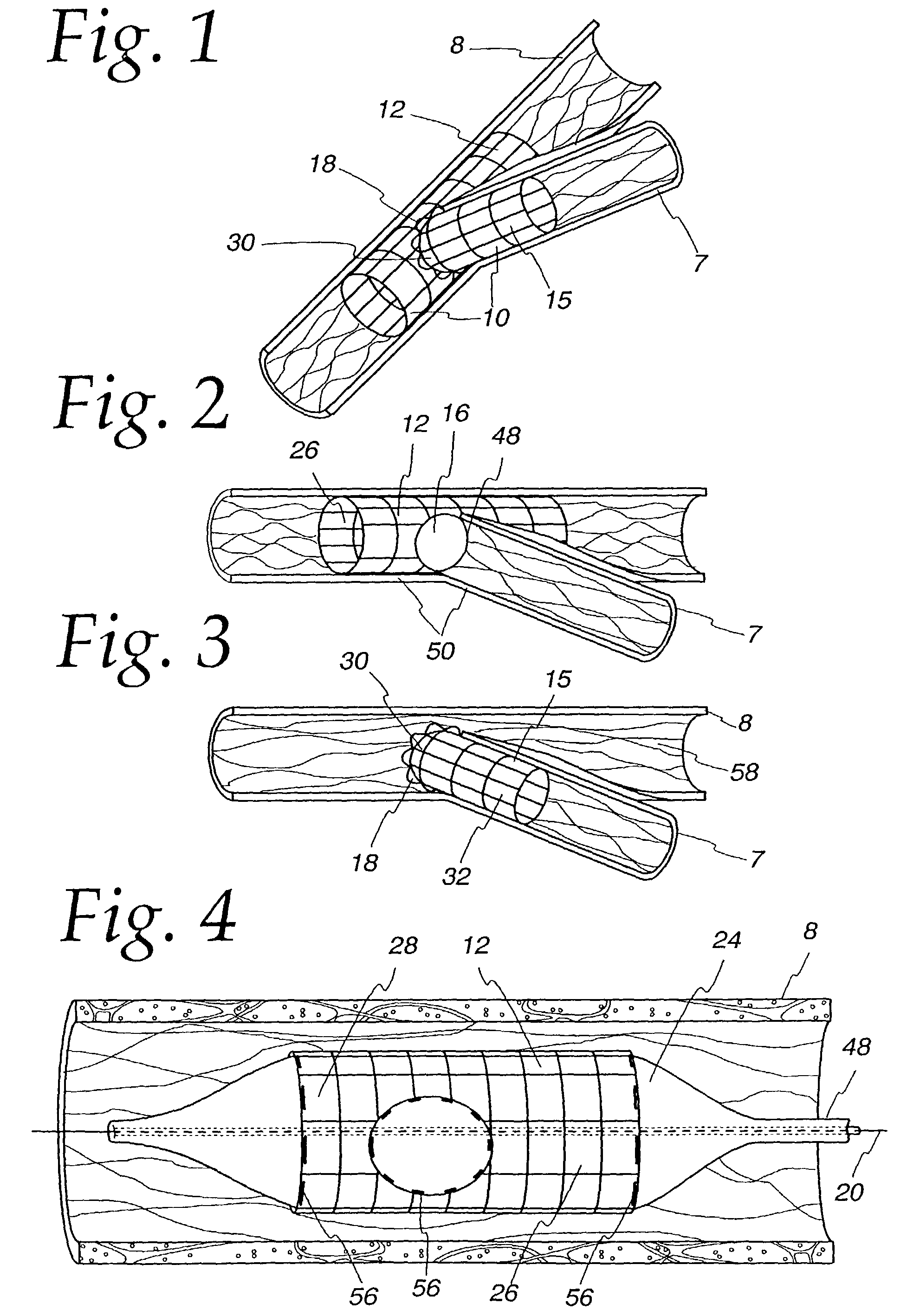
[0039]The bifurcating double-stent apparatus 10 of the present invention comprises a generally cylindrical main stent 12 and a generally cylindrical branch stent 15, which are shown as fully dilated in a subject main vessel 8 and a subject branch vessel 7, as illustrated in FIG. 1.

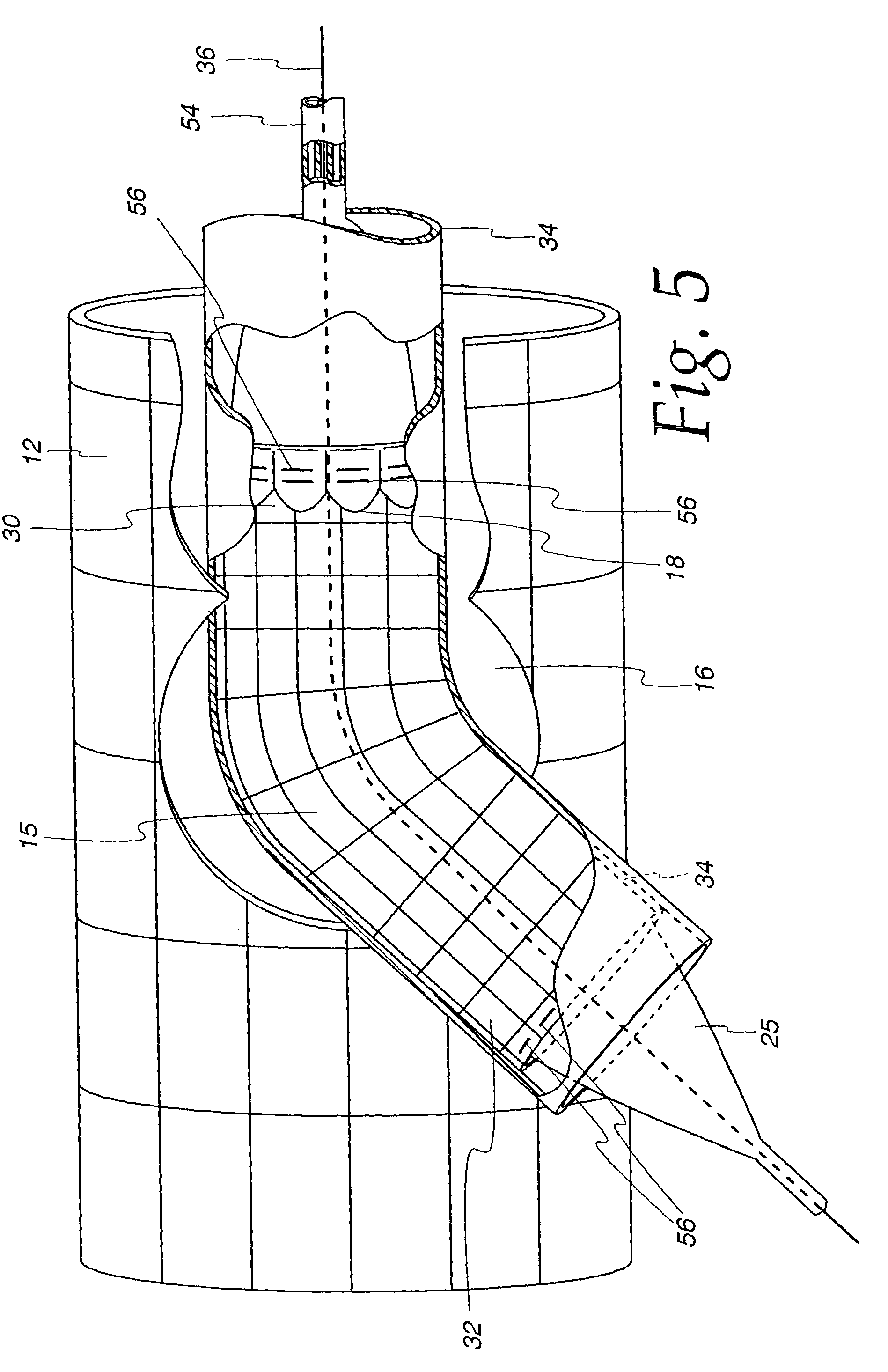
[0040]The main stent 12 contains at least one generally circular side opening 16 located between the proximal end 26 and the distal end 28 of the main stent 12 (FIG. 2), which opening is positioned over and in registry with the opening 48 of a branch vessel in a vessel bifurcation 50, as shown in FIG. 2. The stent 12 and the side opening are imaged during imaging procedures either by constructing the stent of imageable materials or by placing markers 56 at appropriate locations, such as around the perimeter of the side opening 16 in the main stent 12, and at the proximal end 26 and distal end 28 of the main stent, as illustrated in FIG. 4.

[0041]As shown in the embodiment of the invention illustrated in FIG...


PUM


Abstract

The present invention concerns novel stent apparatuses for use in treating lesions at or near the bifurcation point in bifurcated cardiac, coronary, renal, peripheral vascular, gastrointestinal, pulmonary, urinary and neurovascular vessels and brain vessels. More particularly, the invention concerns a stent apparatus with at least one side opening which may further comprise an extendable stent portion laterally extending from the side opening and at least partly in registry with the wall of the side opening. Devices constructed in accordance with the invention include, singularly or in combination, a main expandable stent comprising at least one substantially circular side opening located between its proximal and distal end openings, which side opening may further comprise an expandable portion extending radially outward from the edges of the side opening; and a branch stent comprising proximal and distal end openings and which may further comprise a contacting portion at its proximal end, and which may optionally be constructed to form either a perpendicular branch or a non-perpendicular branch when inserted through a side opening of the main stent. The stents of the invention are marked with, or at least partially constructed of, a material which is imageable during intraluminal catheterization techniques, most preferably but not limited to ultrasound and x-ray.

Description

RELATED APPLICATIONS[0001]This Application is a continuation of U.S. patent application Ser. No. 09 / 750,372 filed Dec. 27, 2000, now U.S. patent application Ser. No. 6,599,316; which is a continuation-in-part of application Ser. No. 09 / 007,265 filed on Jan. 14, 1998, now U.S. Patent No. 6,210,429; which is a continuation-in-part of U.S. Patent application Ser. No. 08 / 744,002, filed on Nov. 4, 1996, now abandoned, the disclosures of which are hereby incorporated by reference herein in their entirety.BACKGROUND[0002]A type of endoprosthesis device, commonly referred to as a stent, may be placed or implanted within a vein, artery or other tubular body organ for treating occlusions, stenoses, or aneurysms of a vessel by reinforcing the wall of the vessel or by expanding the vessel. Stents have been used to treat dissections in blood vessel walls caused by balloon angioplasty of the coronary arteries as well as peripheral arteries and to improve angioplasty results by preventing elastic ...

Claims


Application Information

Patent Timeline
Patent Type & Authority Patents(United States)
IPC IPC(8): A61F2/06A61F2/00A61F2/07A61F2/82A61F2/856A61F2/91A61F2/915A61F2/954A61F2/958
CPCA61F2/82A61F2/856A61F2/91A61F2/915A61F2/954A61F2/958A61F2/064A61F2/07A61F2/852A61F2002/067A61F2002/821A61F2002/91508A61F2002/91516A61F2002/91525A61F2002/91533A61F2002/9155A61F2002/91558A61F2002/91575A61F2002/91583A61F2250/006A61F2250/0098A61F2230/005
Inventor VARDI, GILDAVIDSON, CHARLES J.
Owner BOSTON SCI SCIMED INC
Why Eureka
Try Eureka