System and method for semi-automatic aortic aneurysm analysis

a semi-automatic, aneurysm technology, applied in the field of computed tomography angiography, can solve the problems of requiring a great deal of user interaction, time-consuming, and death in 90% of cases

Inactive Publication Date: 2008-02-07
SIEMENS MEDICAL SOLUTIONS USA INC +1
View PDF5 Cites 33 Cited by
  • Summary
  • Abstract
  • Description
  • Claims
  • Application Information

AI Technical Summary

Benefits of technology

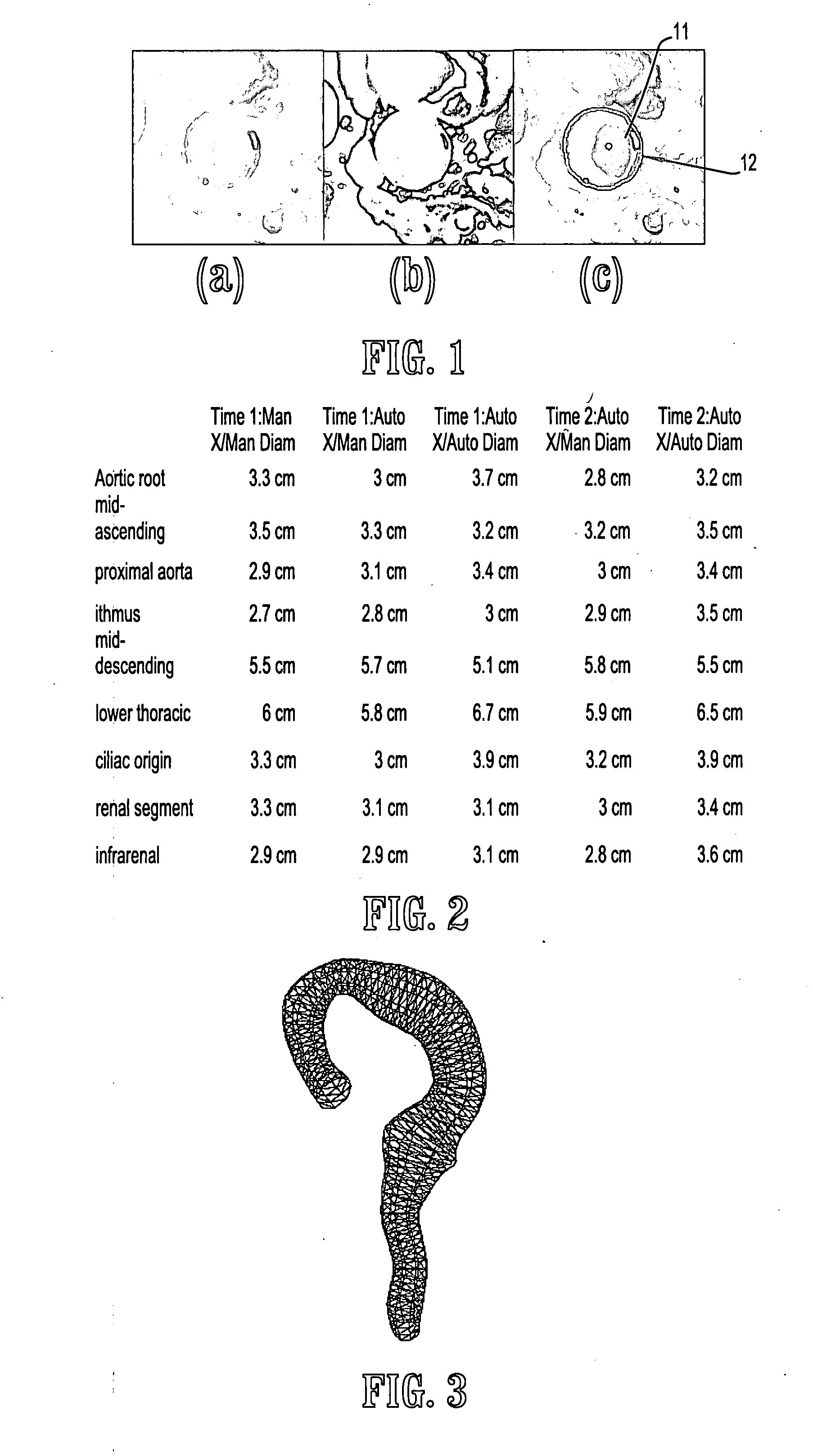
[0011] According to an embodiment of the invention, it is possible to obtain complete coverage of the aorta. It is not unusual for a practicing clinician to be constrained to only a few minutes to examine a study. Using system according to an embodiment of the invention, within seconds the clinician has available a series of optimal, reproducible, orthogonal cross-sections of the entire vessel which may be visually inspected for any irregularities. Further, the user can segment these cross-sections and have the guaranteed maximal diameter automatically computed. With the creation of a 3D model, it becomes possible to evaluate the efficacy of wall stress, wall stiffness, etc., as well as volumetric features. After the 3D model is constructed, stent planning is possible and the stage is set to compute rupture risk indicators such as wall stress (in conjunction with blood pressure readings). Finally, via registration, side by side comparison of the same aorta at different time points becomes straightforward. Since the monitoring of both AAs at risk as well as repaired aortas is commonplace, this feature can be valuable.
[0019] According to a further aspect of the invention, minimizing said isoperimetric ratio comprises representing said lumen intensities by a Laplacian matrix L whose entries are defined by voxels i, j by representing said lumen intensities by a Laplacian matrix L whose entries are defined by voxels i, j by Li, ⁢j={diif⁢ ⁢i=j,-w⁡(eij)if⁢ ⁢eij∈E,0otherwise. wherein eij represents an edge connecting neighboring voxels i,j, w(eij) is a weight for edge eij defined by w(eij)=e−(D<sub2>L< / sub2>(i)+D<sub2>T< / sub2>(i)−D<sub2>L< / sub2>(j)=D<sub2>T< / sub2>(j))<sup2>2 < / sup2>where DL is an estimated lumen distribution, DT is the estimated thrombus distribution, di is a degree of voxel i defined by summing the weights of edges connecting said voxel, and minimizing a cost function g⁡(x)=xT⁡(L+γ⁢ ⁢U)⁢xxT⁡(d+γ⁢ ⁢u), wherein d is a vector of voxel degrees, x is a partition indicator function defined by xi={0if⁢ ⁢voxel⁢ ⁢i∈S_1if⁢ ⁢voxel⁢ ⁢i∈S, U indicates a Laplacian matrix with uniform weights, u represents the vector of degrees for a graph with uniform weights, and γ is a circularity parameter.
[0020] According to a further aspect of the invention, minimizing said cost function comprises selecting a node corresponding to the intersection of the centerline with the MPR as a ground voxel, vg, eliminating the row / column corresponding to vg to form a reduced Laplacian and degree vector L0, d0, solving L0x0=d0 for x0 allowing x to take on any real value, and thresholding the partition indicator x at a value that yields a partition corresponding to a lowest isoperimetric ratio.

Problems solved by technology

An aortic aneurysm (AA) is a permanent and irreversible localized dilation of this vessel, and if left untreated, will gradually expand until rupture, resulting in death in 90% of cases.
Current clinical tools for acquiring these measurements requires a great deal of user interaction and can be quite time consuming.
Treatment for this disorder, such as open repair, runs significant risk including infection, pseudoaneurysm formation, and secondary impotence.
Endovascular stent repair is gaining popularity but the long term outcome of this procedure is not yet known and not all AAs are candidates for stents.
The drawback of this approach is that it is time consuming and as a result the aorta may be sparsely sampled due to practical limits on the duration of the analysis.
In addition, when performed manually, the orthogonal plane may not be correct, introducing error.
Reproducing the same orthogonal cross-section position in a longitudinal study may also prove difficult.
Finally, the manual measurement made may not be correct as it relies on the user subjectively determining which points are connected to form the maximal diameter.
The focus is on the abdominal aorta where the central axis of the vessel is approximately perpendicular to the image stack and thus does not require a centerline calculation, but does require a training set and runs the risk of degeneracy of the modes of variation since the aortic cross-sections are frequently circular.
In this domain, however, the challenges come in the morphology of the vessel, the brain vascular being more detailed and complex as compared to the aorta, and the issue of thrombi is not addressed.

Method used


Image

  • System and method for semi-automatic aortic aneurysm analysis
  • System and method for semi-automatic aortic aneurysm analysis
  • System and method for semi-automatic aortic aneurysm analysis

Examples


Embodiment Construction

[0031] Exemplary embodiments of the invention as described herein generally include systems and methods for automatically segmenting aortic cross-sections on computed tomography angiography acquisitions. Accordingly, while the invention is susceptible to various modifications and alternative forms, specific embodiments thereof are shown by way of example in the drawings and will herein be described in detail. It should be understood, however, that there is no intent to limit the invention to the particular forms disclosed, but on the contrary, the invention is to cover all modifications, equivalents, and alternatives falling within the spirit and scope of the invention.

[0032] As used herein, the term “image” refers to multi-dimensional data composed of discrete image elements (e.g., pixels for 2-D images and voxels for 3-D images). The image may be, for example, a medical image of a subject collected by computer tomography, magnetic resonance imaging, ultrasound, or any other medic...


PUM


Abstract

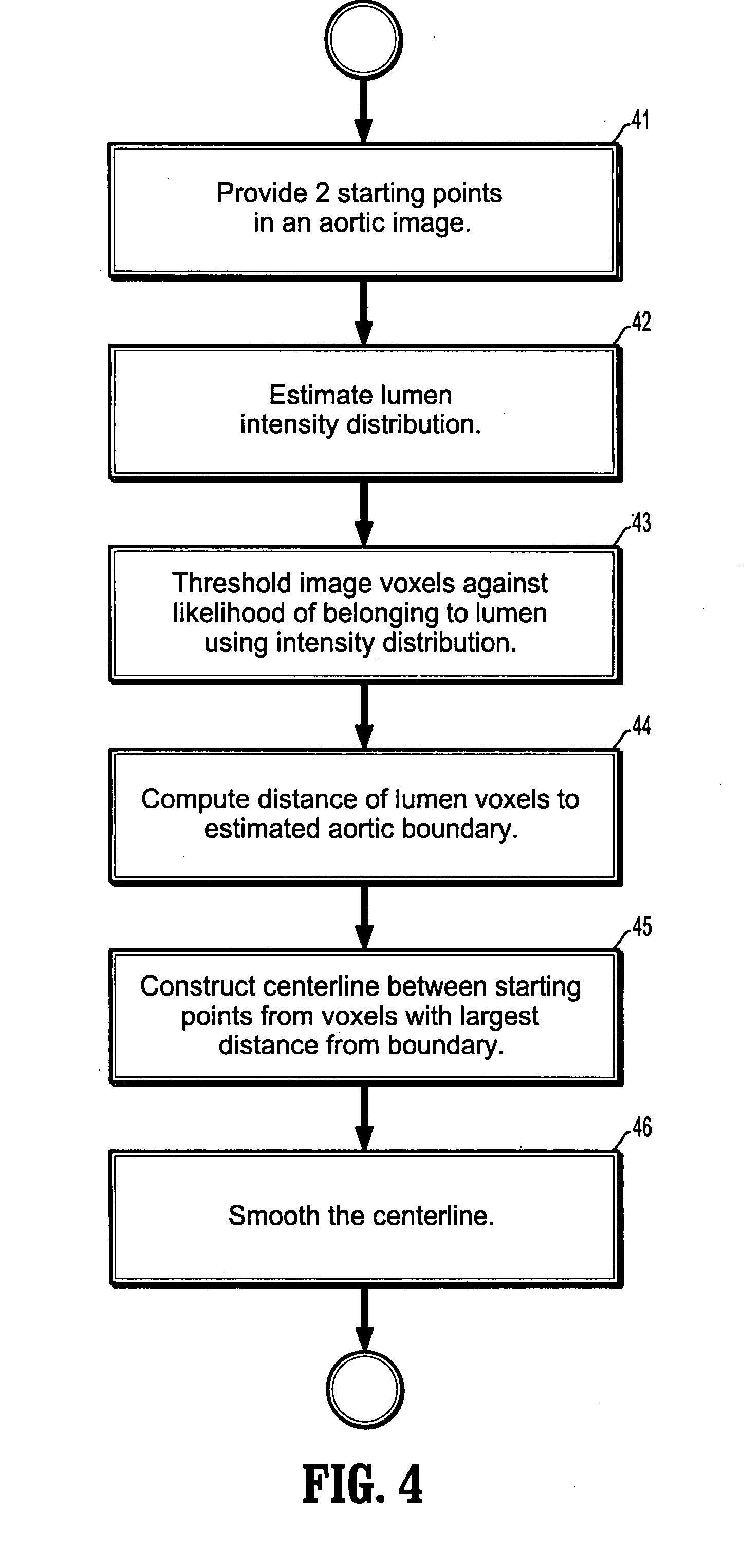
A method for automatically analyzing an aortic aneurysm includes providing a digitized 3-dimensional image volume of an aorta, determining which voxels in said image are likely to be lumen voxels, determining a distance of said lumen voxels from an aortic boundary, finding a centerline of the aorta in said image volume based on said lumen voxel distances, constructing a series of 2-dimensional multiplanar reformatted (MFR) image planes orthogonal to this centerline, segmenting aortic cross sections in each said MPR image plane wherein an aortic wall is located in each MPR image, and constructing from said aortic wall locations a 3D model of the aorta.

Description

CROSS REFERENCE TO RELATED UNITED STATES APPLICATIONS [0001] This application claims priority from: “Semi-Automatic Aortic Aneurysm Analysis”, U.S. Provisional Application No. 60 / 793,866 of O'Donnell, et al., filed Apr. 21, 2006, the contents of which are herein incorporated by reference.TECHNICAL FIELD [0002] This disclosure is directed to methods for automatically segmenting aortic cross-sections on computed tomography angiography acquisitions. DISCUSSION OF THE RELATED ART [0003] The aorta is the largest artery in the body and is the primary conduit of oxygenated blood. An aortic aneurysm (AA) is a permanent and irreversible localized dilation of this vessel, and if left untreated, will gradually expand until rupture, resulting in death in 90% of cases. AAs are the 13th leading cause of death in the United States. Standard procedure assesses the risk of aneurysm rupture based on maximal aortic diameter. Current clinical tools for acquiring these measurements requires a great deal...

Claims


Application Information

Patent Timeline
Patent Type & Authority Applications(United States)
IPC IPC(8): A61B5/02G06V10/26
CPCG06K9/34G06K2009/00932G06T2207/30101G06T7/0012G06T7/606G06K2209/05G06T7/66G06V40/14G06V10/26G06V2201/03
Inventor GRADY, LEOBODUR, OSMANSETSER, RANDOLPH M.STILLMAN, ARTHUR E.O'DONNELL, THOMASFUNKA-LEA, GARETH
Owner SIEMENS MEDICAL SOLUTIONS USA INC
Why Eureka
Try Eureka