Colorimetric sensor using polydiacetylene supramolecule
a colorimetric sensor and supramolecule technology, applied in the field of colorimetric sensors, can solve problems such as long time, difficulty in detecting color, and difficulty in detecting color, and achieve the effects of improving accuracy, reducing labor intensity, and improving accuracy
- Summary
- Abstract
- Description
- Claims
- Application Information
AI Technical Summary
Benefits of technology
Problems solved by technology
Method used
Image
Examples
example 1
Preparation of a Liposome
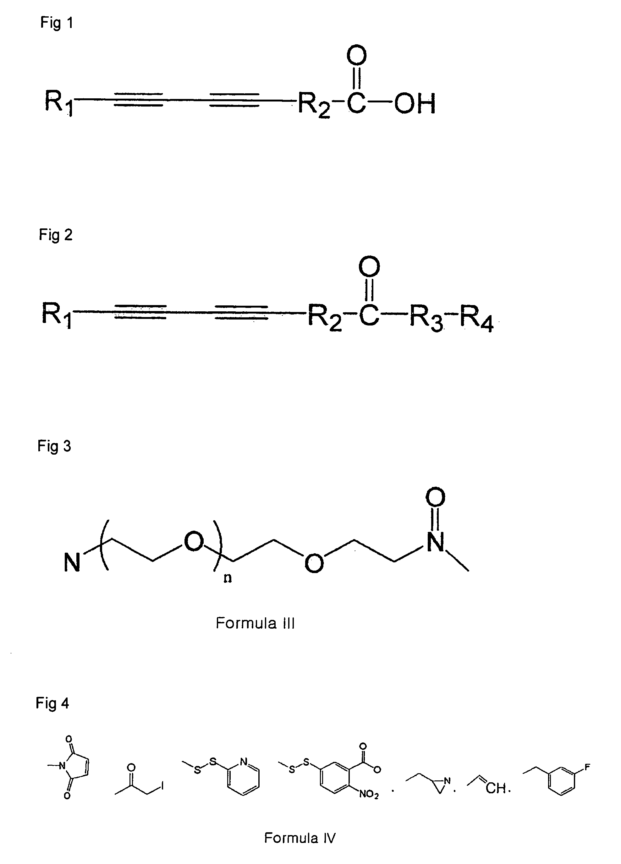
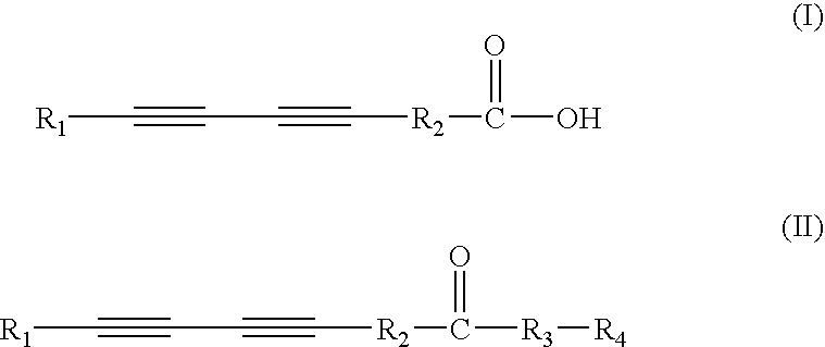
[0043]PCDA and PCDA-maleimide diacetylene monomer were mixed in ratios of 1:9, 2:8 or 3:7 in chloroform in a vitreous bottle, and then chloroform was evaporated with nitrogen gas or by rotary evaporation to form a thin film consisting of the two components. Then, the film was soaked in 10 mM HEPES buffer (pH=7.4) and they are shaken slowly for 15 minutes at high temperature of above 80° C., and then the film was treated by probe sonication for 20 minutes at 30 W. Thus prepared liposome was stored for about 4 hours in a refrigerator, and then was used in next experiment.
example 2
Reduction and Immobilization of an Antibody
[0044]A monoclonal antibody (Waterborne Inc., USA) against 0.3 mg / ml of Cryptosporidium parvum was mixed with 20 mM TCEP (Tris 2-carboxyethyl phosphine) at ratio of 1:1, and then the mixture was reacted for 1 hour at room temperature. Buffer exchange was performed with HEPES buffer containing 0.02 mM TCEP by employing 30 kDa MWCO (molecular weight of fraction) filter available at Amicon. The antibody treated according to such method was mixed with the liposome solution of the Example 1 to final concentration of 0.1 mg / ml and then the mixture was reacted for 3 hours at room temperature. Then, L-cysteine was added to final concentration of 1 mM and then the remaining maleimide functional group of the liposome was capped.
example 3
Detection of a Pathogenic Bacteria Employing an Antibody-immobilized Liposome
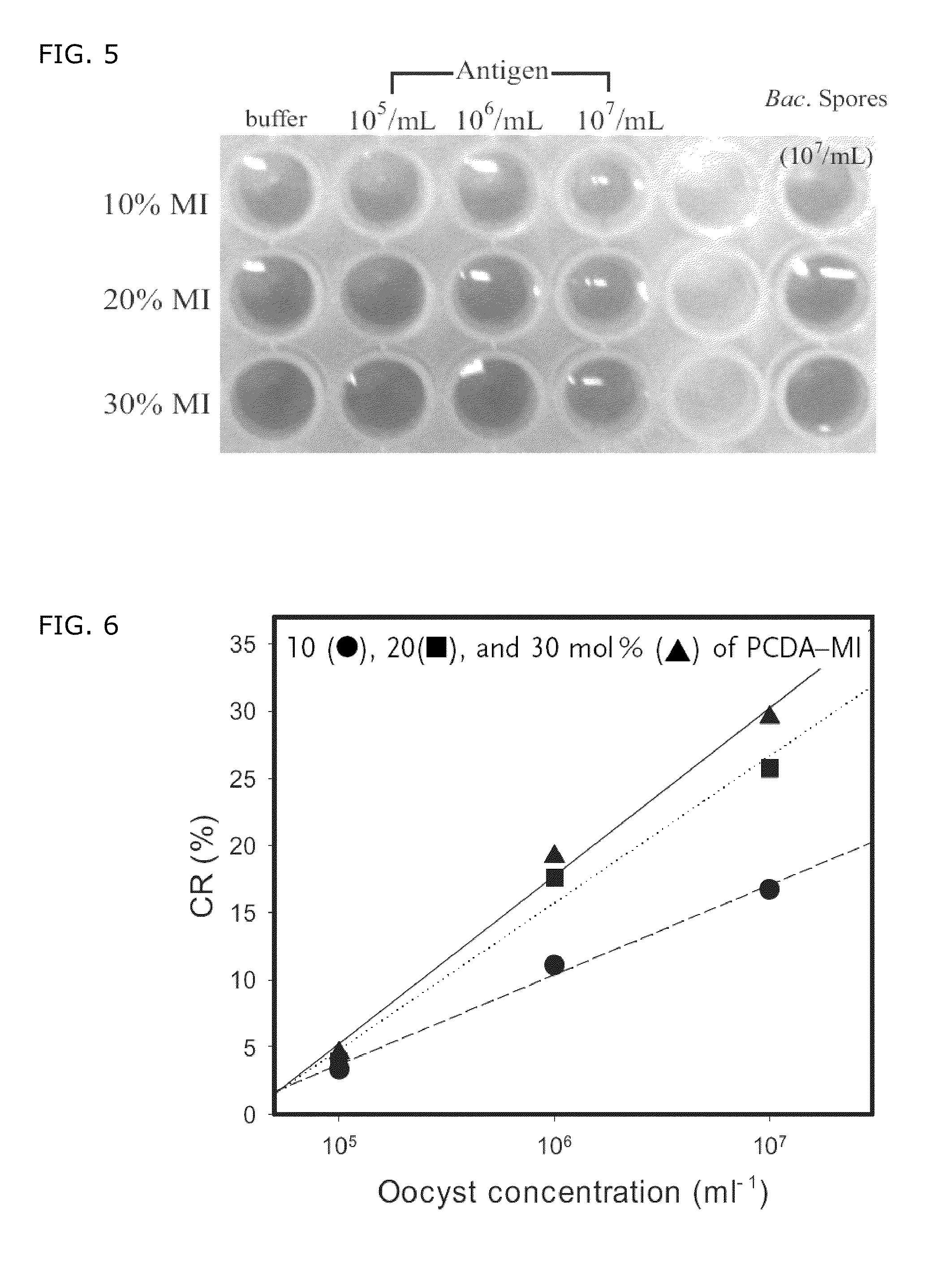
[0045]When an antibody-polydiacetylene liposome was exposed to UV light at 254 nm, it was confirmed that a blue solution was generated. When 50 μl of the solution and 100 μl of HEPES buffer containing C. parvum were mixed and reacted for 2.5 hours at 37° C., it was observed that the color of the antibody-polydiacetylene liposome was changed from blue to violet or red depending on the concentration of C. parvum (FIG. 5). Columns 1˜3 in FIG. 5 represent liposome sensors containing 10, 20 and 30 mol % of PCDA-maleimide, respectively. Row 1 represents color reaction of the liposome against a buffer, rows 2˜4 represent color reaction of the liposome against a buffer containing 105, 106 and 107 / ml of C. parvum, respectively, and row 6 represents color reaction of the liposome against a buffer containing 107 / ml of Bacillus spore.
[0046]FIG. 6 shows standard graph about the concentration of the ligand, expressed num...
PUM
| Property | Measurement | Unit |
|---|---|---|
| mol % | aaaaa | aaaaa |
| distance | aaaaa | aaaaa |
| temperature | aaaaa | aaaaa |
Abstract
Description
Claims
Application Information
 - R&D
- Intellectual Property
- Life Sciences
- Materials
- Tech Scout
- Unparalleled Data Quality
- Higher Quality Content
- 60% Fewer Hallucinations
Browse by: Latest US Patents, China's latest patents, Technical Efficacy Thesaurus, Application Domain, Technology Topic, Popular Technical Reports.
© 2025 PatSnap. All rights reserved.Legal|Privacy policy|Modern Slavery Act Transparency Statement|Sitemap|About US| Contact US: help@patsnap.com


