
Transmission spectroscopy system for use in the determination of analytes in body fluid

a technology of total transmission spectroscopy and body fluid, which is applied in the field of spectroscopy, can solve the problems of large angle loss of light exiting the sample, the cost of integrating spheres and photomultipliers, and the cost of these devices, so as to achieve the effect of cost-prohibition in the production of existing total transmission spectroscopy systems for use by patients

Inactive Publication Date: 2010-08-31
ASCENSIA DIABETES CARE HLDG AG
View PDF20 Cites 31 Cited by
  • Summary
  • Abstract
  • Description
  • Claims
  • Application Information

AI Technical Summary

Problems solved by technology

A drawback associated with existing total transmission spectroscopy systems that use an integrating sphere is a low signal level that requires using a photomultiplier tube for reading the reflected light from a small portion of the inside surface of the integrating sphere.
Another drawback associated with conventional total transmission spectroscopy systems is the cost of an integrating sphere and photomultiplier tube.
The cost of these devices makes it cost-prohibitive to produce existing total transmission spectroscopy systems for use by a patient needing to self-test, for example, the patient's blood-glucose concentration level.
As discussed above, only the light emerging from the sample at small angles is collected using existing regular transmission spectroscopy measurements, often resulting in losing light exiting the sample at large angles.
A significant portion of light scattered by the red blood cells is not collected with existing systems using regular transmission measurements, which can lead to significant loss of light resulting in very low transmission levels through whole blood.
The addition of a lysing reagent and subsequent lysis of the red blood cells is time consuming relative to the overall measurement process.
This problem is not present in existing total transmission spectroscopy methods because the scattered transmitted light and regular transmitted light is collected by the optics.
Another drawback associated with existing systems using regular transmission spectroscopy is a transmission bias at wavelengths of light where the chromatic reaction occurs.
This transmission bias causes inaccuracies in determining the analyte (e.g., glucose) concentration.
The hematocrit level of whole blood may also cause a total transmission bias due to differences in the amount of scattered light at different hematocrit levels.
Existing systems using regular transmission or total transmission spectroscopy are not capable of detecting the difference in hematocrit levels because of poor transmission level and poor separation between hematocrit levels at certain wavelengths of light.
Another drawback to existing systems using regular transmission spectroscopy is accuracy errors that result from the sample path length.
A 10% variation in the path length of the sample cell area results in a 10% error in the concentration measurement for both regular and total transmission methods.
Existing systems using regular transmission methods, however, require a shorter path length to make up for transmission losses due to red blood cell scatter.
Thus, the mechanical tolerance at a shorter pathlength results in higher concentration errors.

Method used


Image

  • Transmission spectroscopy system for use in the determination of analytes in body fluid
  • Transmission spectroscopy system for use in the determination of analytes in body fluid
  • Transmission spectroscopy system for use in the determination of analytes in body fluid

Examples


examples

[0063]Referring now to FIG. 3a, one embodiment of the present invention (transmission spectroscopy system 10) measured the total transmission levels of whole blood samples having hematocrit levels of 20% reacted with reagents, and each had a different glucose concentration level—54, 104, 210, and 422 milligrams of glucose per deciliter of blood (“mg / dL glucose”). The transmission spectroscopy system 10 will be referred to in the examples as the “inventive system.” White light from the light source 12 (FIG. 1a) was transmitted through the sample. The total transmission level measured from 500 nm to 940 nm was plotted in FIG. 3a for each of the glucose concentration levels. The transmission was lower from 500 to 600 nm due to the absorption of hemoglobin. Transmission loss caused by light scattered by red blood cells (hematocrit) affects the transmission from 500 nm to 940 nm. The indicator in the glucose reaction absorbs between 500 and 750 nm, so there was separation between the glu...

embodiment b

Alternate Embodiment B

[0084]The system of Alternate Embodiment A wherein the fluid sample is blood and wherein the sample cell receiving area includes a dried reagent in the absence of a hemolyzing agent is adapted to produce a chromatic reaction when reconstituted with blood.

embodiment c

Alternate Embodiment C

[0085]The system of Alternate Embodiment A wherein each of the first and second lenses is a half-ball lens.


PUM

PropertyMeasurementUnit
angle of acceptanceaaaaaaaaaa
acceptance angleaaaaaaaaaa
angle of divergenceaaaaaaaaaa

Abstract

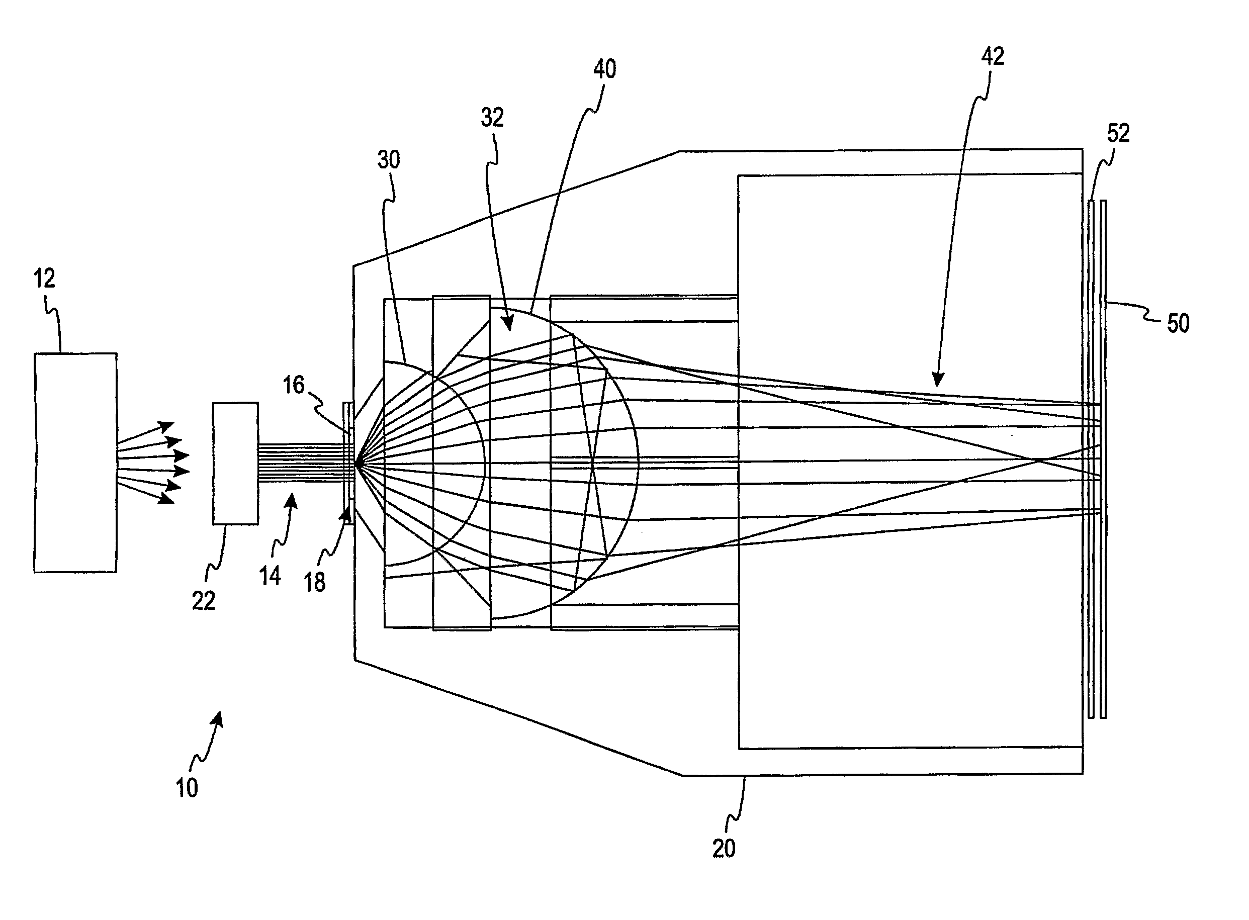
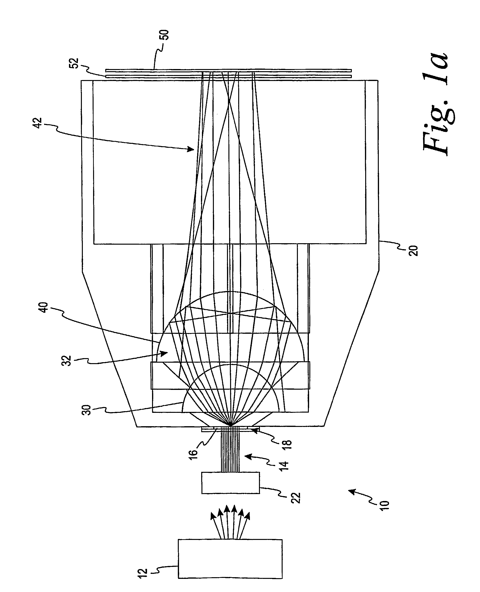
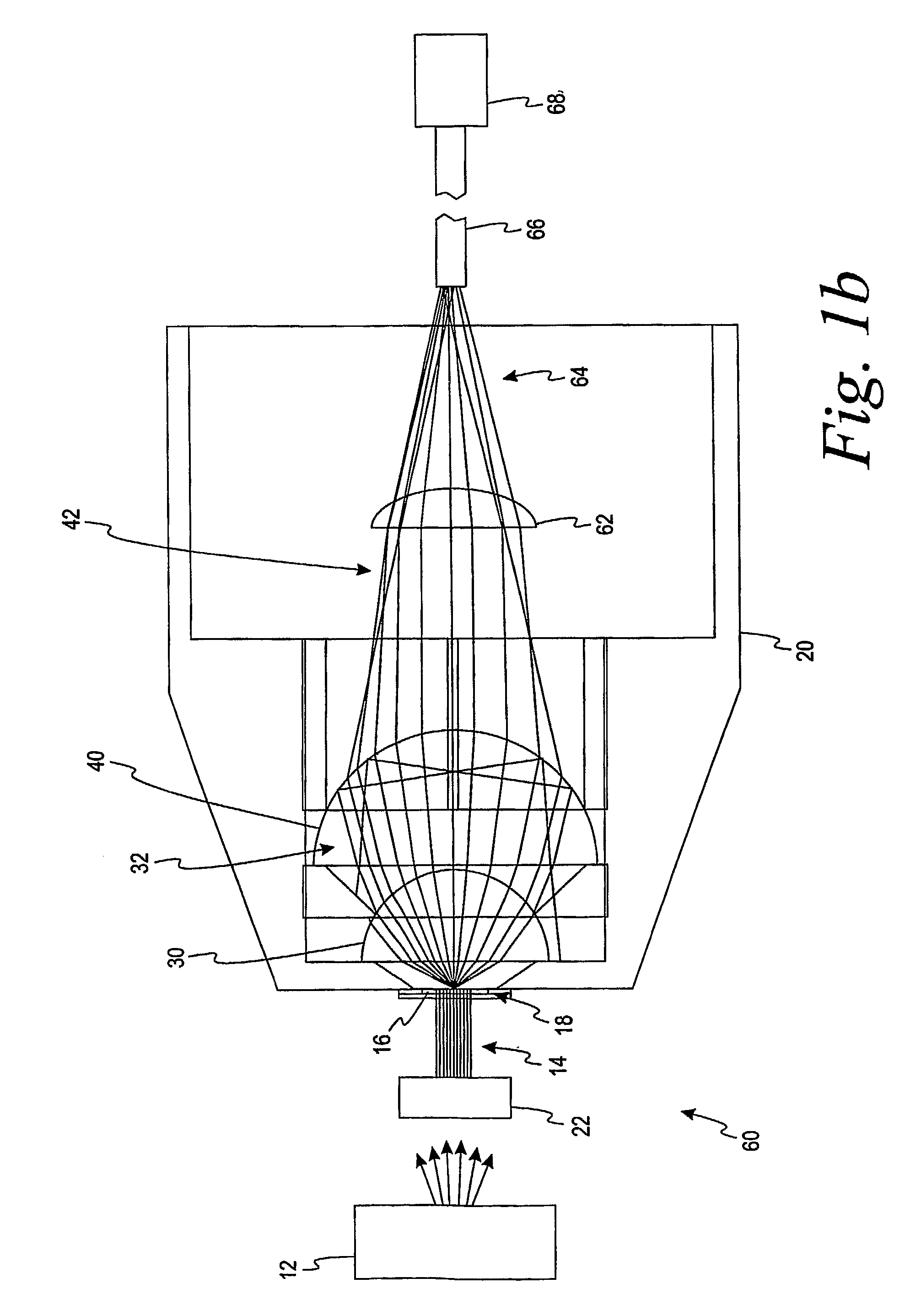
A total transmission spectroscopy system for use in determining the analyte concentration in a fluid sample comprises a sample cell receiving area, a light source, a collimating lens, a first lens, a second lens, and a detector. The sample cell receiving area is adapted to receive a sample to be analyzed. The sample cell receiving area is constructed of a substantially optically clear material. The collimating lens is adapted to receive light from the light source and adapted to illuminate the sample cell receiving area with a substantially collimated beam of light. The first lens is adapted to receive regular and scattered light transmitted through the sample at a first angle of divergence. The first lens receives light having a first angle of acceptance. The first lens outputs light having a second angle of divergence. The second angle of divergence is less than the first angle of divergence. The second lens is adapted to receive light from the first lens and adapted to output a substantially collimated beam of light. The detector is adapted to measure the light output by the second lens.

Description

CROSS-REFERENCE TO RELATED APPLICATIONS[0001]This application claims priority to Application No. 60 / 635,666 filed on Dec. 13, 2004, which is incorporated by reference in its entirety.FIELD OF THE INVENTION[0002]The invention generally relates to spectroscopy and, more particularly, to the use of total transmission spectroscopy for determining the concentration of an analyte in body fluid.BACKGROUND OF THE INVENTION[0003]Transmission spectroscopy is used to perform quantitative analysis of a sample based on the transmission of a light beam through a sample contained by a sample cell. Different frequency components of the light beam are absorbed by components of the sample, whereby a frequency analysis of light transmitted through the sample permits analysis of the sample itself. Dry chemical reagents are dissolved by the sample and react with the analyte of interest to produce a chromaphoric response at certain wavelengths of light ranging from about 450 nanometers (“nm”) to about 95...

Claims


Application Information

Patent Timeline
Patent Type & Authority Patents(United States)
IPC IPC(8): G01N33/48
CPCG01N21/255G01N21/31G01N21/78G01N21/274G01N21/314G01N21/3151G01N21/3577G01N21/39
Inventor DOSMANN, ANDREW J.NELSON, CHRISTINE D.WARCHAL-WINDHAM, MARY ELLEN
Owner ASCENSIA DIABETES CARE HLDG AG
Learn More