System and method for quantitative reconstruction of Zernike phase-contrast images

a phase contrast image and quantitative reconstruction technology, applied in the field of system and method for quantitative reconstruction can solve the problems of complex image interpretation, complex image processing, and inability to accurately represent features, so as to achieve the effect of reducing the artifacts of zernike phase contrast images

Active Publication Date: 2010-08-31
CARL ZEISS X RAY MICROSCOPY
View PDF3 Cites 76 Cited by
  • Summary
  • Abstract
  • Description
  • Claims
  • Application Information

AI Technical Summary

Benefits of technology

[0006]The principle of reciprocity states that full-field and scanning microscopes can produce equivalent images by interchanging the roles of condenser and detector. Thus, the contrast transfer function inversion previously used for images from scanning systems can be appl...

Problems solved by technology

One drawback of Zernike phase contrast imaging is the mixing of absorption and phase contrast signals and the resulting halo-like artifacts that occur at features' edges.
These artifacts can make image interpretation difficult.
With three dimensional (3D) imaging, e.g. computed tomography (...

Method used


Image

  • System and method for quantitative reconstruction of Zernike phase-contrast images
  • System and method for quantitative reconstruction of Zernike phase-contrast images
  • System and method for quantitative reconstruction of Zernike phase-contrast images

Examples


Embodiment Construction

[0021]Many types of specimens exhibit higher phase contrast than absorption contrast. For example, biological samples studied with visible light, electron, or x-ray microscopy and light metals and ceramic materials examined using x-ray microscopy can often be better analyzed using phase contrast.

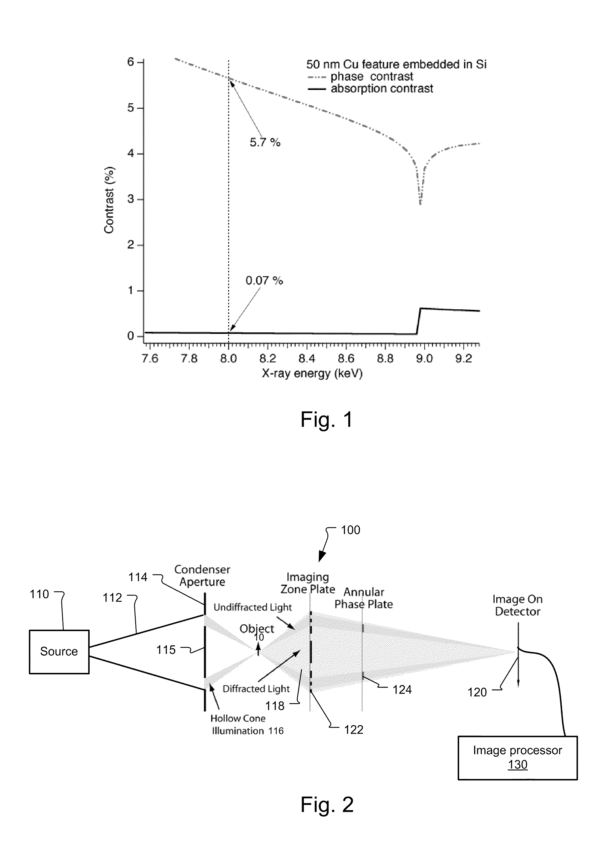
[0022]FIG. 1 shows the phase and absorption contrast of a 50 nanometer (nm) sized copper feature in a silicon substrate for a range of different x-ray energies. The contrast is defined here as:

[0023]C=Ipeak-IvalleyIpeak+Ivalley.

[0024]Note that absorption generates relatively weak contrast except at copper's absorption edge at 9 keV. In comparison, phase contrast dominates with sometimes several orders of magnitude higher contrast. This advantage in intrinsic contrast makes phase contrast better suited for imaging specimens by bringing benefits of lower dose, shorter exposure time, and better image quality.

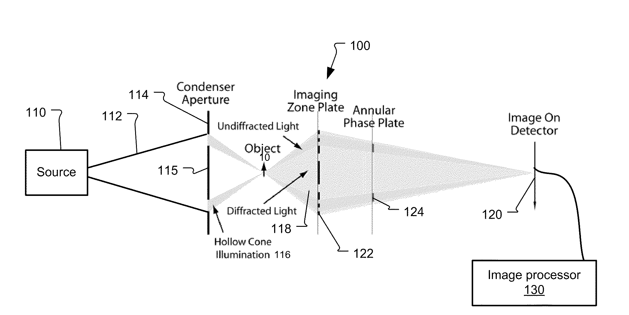
[0025]FIG. 2 shows a typical full-field imaging microscope 100.

[0026]Radiation 112 is g...


PUM


Abstract

The principle of reciprocity states that full-field and scanning microscopes can produce equivalent images by interchanging the roles of condenser and detector. Thus, the contrast transfer function inversion previously used for images from scanning systems can be applied to Zernike phase contrast images. In more detail, a full-field x-ray imaging system for quantitatively reconstructing the phase shift through a specimen comprises a source that generates x-ray radiation, a condenser x-ray lens for projecting the x-ray radiation onto the specimen, an objective x-ray lens for imaging the x-ray radiation transmitted through the specimen, a phase-shifting device to shift the phase of portions of x-ray radiation by a determined amount, and an x-ray detector that detects the x-ray radiation transmitted through the specimen to generate a detected image. An image processor then determines a Fourier filtering function and reconstructs the quantitative phase shift through the specimen by application of the Fourier filtering function to the detected image. As a result, artifacts due to absorption contrast can be removed from the detecting image. This corrected image can then be used in generating three dimensional (3D) images using computed tomography.

Description

RELATED APPLICATIONS[0001]This application claims the benefit under 35 USC 119(e) of U.S. Provisional Application Nos. 61 / 082,326, filed on Jul. 21, 2008 and 61 / 083,669, filed on Jul. 25, 2008, both of which are incorporated herein by reference in their entirety.BACKGROUND OF THE INVENTION[0002]Many imaging methods have been developed to take advantage of phase contrast. The most widely used technique with lens-based far-field microscopes is the Zernike method developed by Frits Zernike. Features that are difficult or impossible to observe in absorption contrast can be effectively studied in phase contrast mode, such as biological samples.[0003]The set up for Zernike phase contrast imaging is similar to microscopes for absorption contrast, but a phase plate is used. The phase plate is usually placed at or near the back focal plane of the objective lens to shift the phase of the unscattered beam by π / 2 or 3π / 2. The unscattered light then interferes with the diffracted light to produc...

Claims


Application Information

Patent Timeline
IPC IPC(8): G21K7/00G01N23/00
CPCG01N23/046G21K7/00G01N2223/419
Inventor YUN, WENBINGFESER, MICHAELHORNBERGER, BENJAMIN
Owner CARL ZEISS X RAY MICROSCOPY
Why Eureka
Try Eureka