Antibody production method

a production method and antibody technology, applied in the direction of peptides, prosthesis, drug compositions, etc., can solve the problems of infection such as mastitis, time-consuming injections, limited antibody yield,

Inactive Publication Date: 2011-01-04
AGRI BIOTECH PTY LTD
View PDF1 Cites 2 Cited by
  • Summary
  • Abstract
  • Description
  • Claims
  • Application Information

AI Technical Summary

Benefits of technology

[0010]Other objects, features, and advantages of the instant invention, in its details as seen from the above, and from the following description of the preferred embodiment will become apparent to those skilled in the art.

Problems solved by technology

Hence, the yield of antibodies is limited (approximately 3 to 30 mg per month) due to the finite amount of blood that can be obtained.
Such injections are time consuming as they must be repeated to re-stimulate antibody production.
Furthermore, injection directly into the teat of a mammal frequently results in infections such as mastitis.
Intramammary immunization techniques have generally not been preferred as a route for vaccination under field conditions due to the high chance of mammary infection (R. F. Sheldrake (1987) Australian Journal of Dairy Technology, 42:30-32) and often requires application by highly skilled practitioners.

Method used


Image

  • Antibody production method
  • Antibody production method
  • Antibody production method

Examples


example 1

Preparation of Antigen Releasing Device Minus Adjuvant

Materials

[0062](i) 0.15 g mannitol-D (Sigma-Aldrich)[0063](ii) 0.15 g sodium citrate (Proanalys)[0064](iii) 11.25 mg lipase from Pseudomonas fluorescens (Sigma-Aldrich)[0065](iv) 0.75 ml part A Silastic (Dow Corning Q74850)[0066](v) 0.75 ml part B Silastic (Dow Corning Q7-4850)[0067](vi) 2×2.5 ml disposable syringes (Terumo)[0068](vii) 2×1 ml syringe (Terumo)[0069](viii) 2×12G×4 inch stainless steel hypodermic needles[0070](ix) 37° C. incubator[0071](x) sterile petri dish[0072](xi) sterile scalpel[0073](xii) sterile spachella[0074](xiii) 32 ml glass McCartney bottle

Methods[0075](i) Remove pistons from all syringes[0076](ii) Part A silastic placed into 1 ml syringe using spatula. Piston replaced into syringe and 0.75 ml quantity dispensed into one 2.5 ml syringe. Procedure repeated for Part B.[0077](iii) Lipase, mannitol and sodium citrate are combined and mixed in a small glass McCartney then placed into the 2.5 ml syringe contai...

example 2

Preparation of Antigen Releasing Device Including Adjuvant

Materials

[0078](i) 0.3 g mannitol-D (Sigma-Aldrich)[0079](ii) 0.3 g sodium citrate (Proanalys)[0080](iii) 22.50 mg lipase from Pseudomonas fluorescens (Sigma-Aldrich)[0081](iv) 1.2 mg IL1-B Fragment 163-171 synthetic human (Sigma)[0082](v) 1.5 ml part A Silastic (Dow Corning Q7-4850)[0083](vi) 1.5 ml part B Silastic (Dow Corning Q7-4850)[0084](vii) 2×2.5 ml disposable syringes (Terumo)[0085](viii) 2×1 ml syringe (Terumo)[0086](ix) 2×12G×4 inch stainless steel hypodermic needles[0087](x) 37° C. incubator[0088](xi) sterile petri dish[0089](xii) sterile scalpel[0090](xiii) sterile spachella[0091](xiv) 32 ml glass McCartney bottle

Methods[0092](i) Remove pistons from all syringes[0093](ii) Part A silastic placed into 1 ml syringe using spatula. Piston replaced into syringe and 0.75 ml quantity dispensed into one 2.5 ml syringe. Procedure repeated for Part B.

[0094]Lipase, mannitol and sodium citrate mixed in a small glass McCartney...

example 3

Delivery of ARD

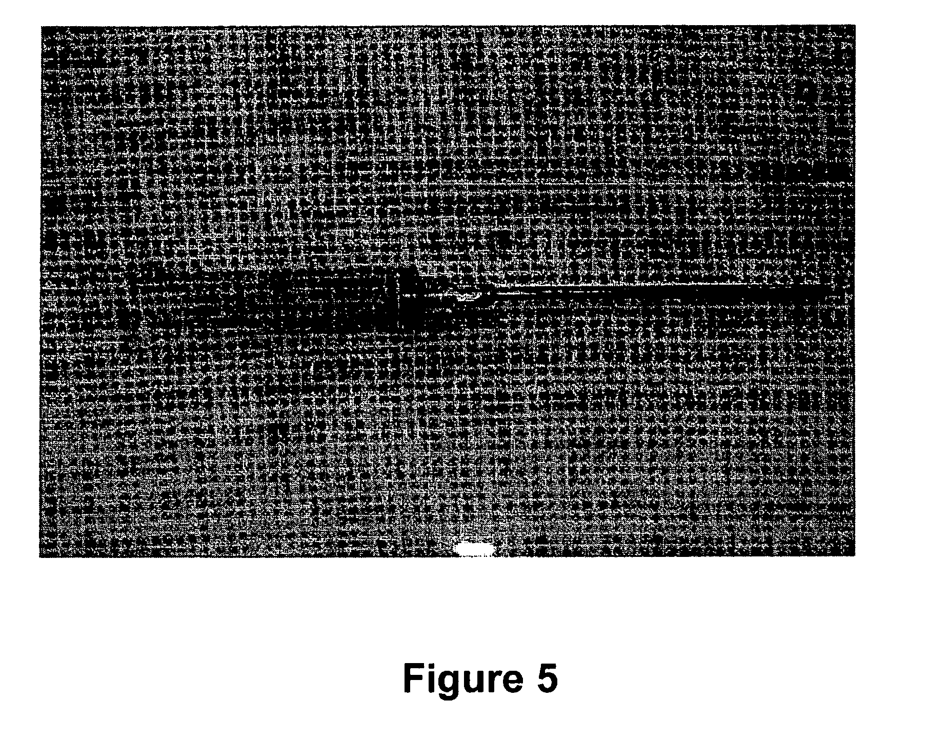
[0095]A device was purpose designed and built comprising 10 ml disposable luer lock syringe, 10G×4 inch stainless steel hypodermic needle and a 90 mm×10G stainless steel welding rod (see FIG. 5 below). The device was assembled with the rod attached to the piston of the syringe and passing through the needle. The piston was withdrawn about 3 cm allowing for free space near the tip of the needle in which to insert the antigen releasing device. Thus, when the piston is depressed, the antigen releasing device is expelled from the needle by the rod (see FIG. 5 below).

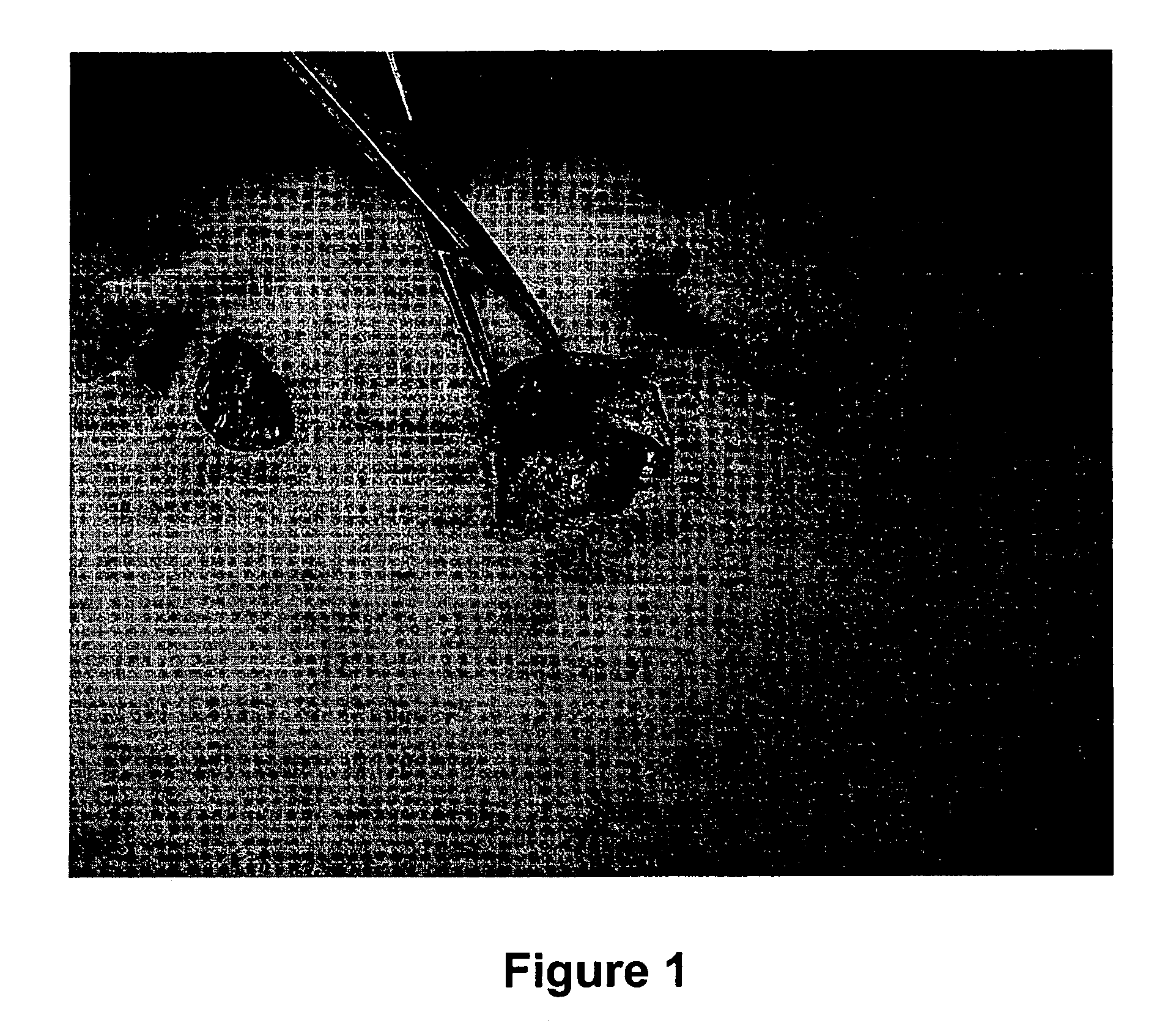
[0096]The animal was placed on the floor on its back and restrained by animal handlers. An area approximately 3 cm×10 cm right lateral and adjacent to the udder was swabbed with iodine. 2 ml of 2% lignocaine infiltrated the cutaneous tissue through a 26G hypodermic needle as a local anaesthetic. The 10G needle housing the ARD was inserted in the posterior end of this area and pushed to the anterior end subcuta...


PUM

PropertyMeasurementUnit
distanceaaaaaaaaaa
distanceaaaaaaaaaa
distanceaaaaaaaaaa

Abstract

A method of inducing the sustained release of antibodies in milk comprising the step of: a) implanting at least one antigen releasing device adjacent to, within close proximity of or within at least one supramammary lymph node, wherein in use the antigen releasing device releases an antigen into the tissue area around the supramammary lymph node which stimulates antibody secretion into a mammary gland.

Description

[0001]This application is a Continuation of International Application No. PCT / AU2005 / 001540, filed Oct. 6, 2005, which claims priority to Australian Application No. 2004905762, filed Oct. 6, 2004. The entire contents of the foregoing applications are incorporated herein in their entirety.FIELD OF THE INVENTION[0002]The present invention relates to a method for inducing a sustained production of antibodies or immunoglobulins. More particularly, it relates to a method of inducing a sustained production of antibodies in the milk of a mammal.BACKGROUND ART[0003]The advantage of producing antibodies in milk over the traditional source of antibodies from blood lies in the constancy of supply through the daily access to milk. After blood products are harvested, the production animal is allowed to recover for extended periods (for example, up to one month). Hence, the yield of antibodies is limited (approximately 3 to 30 mg per month) due to the finite amount of blood that can be obtained. ...

Claims


Application Information

Patent Timeline
Patent Type & Authority Patents(United States)
IPC IPC(8): A61K49/00A61K39/38
CPCA23C9/1585C07K16/40C07K16/04A23C2230/15A61P31/04A61P43/00
Inventor TAY, KWANG G.PENHALE, WILLIAM J.GEERLINGS, PETER M.
Owner AGRI BIOTECH PTY LTD
Why Eureka
Try Eureka