Lesion assessment by pacing
a technology of lesion assessment and pacing, which is applied in the field of lesion assessment by pacing, validating and monitoring percutaneous cardiac ablation procedures, can solve the problems of difficult to determine the proper dosage of energy, arrhythmias may persist or return, and dangerous damage to the tissue at and around the ablation site, so as to mitigate the danger of excessive ablation
- Summary
- Abstract
- Description
- Claims
- Application Information
AI Technical Summary
Benefits of technology
Problems solved by technology
Method used
Image
Examples
embodiment 1
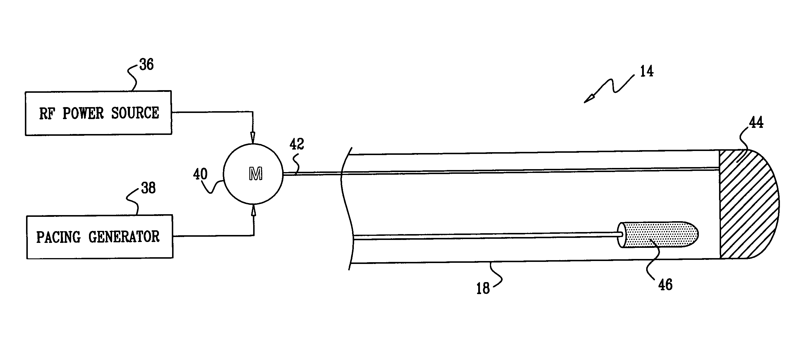
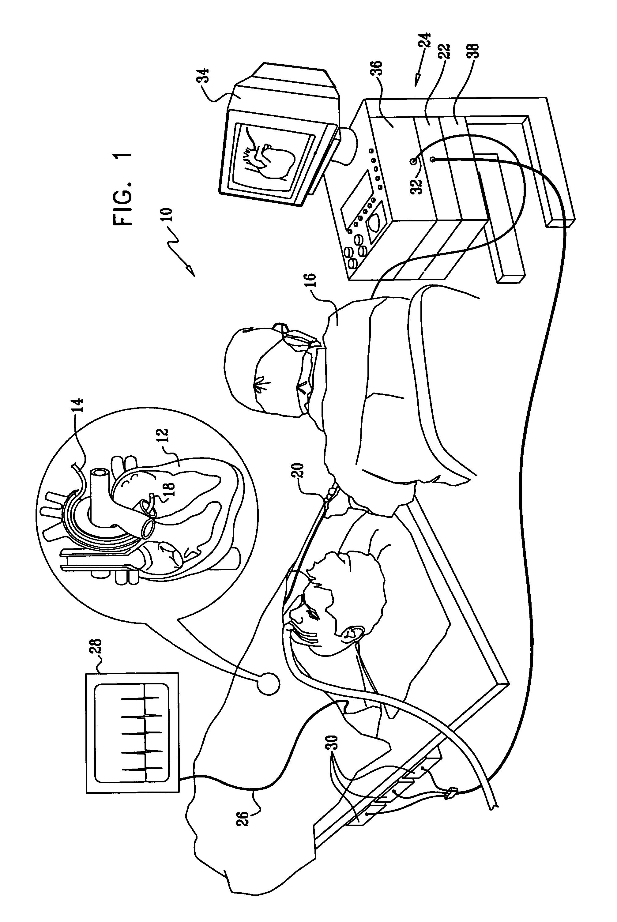
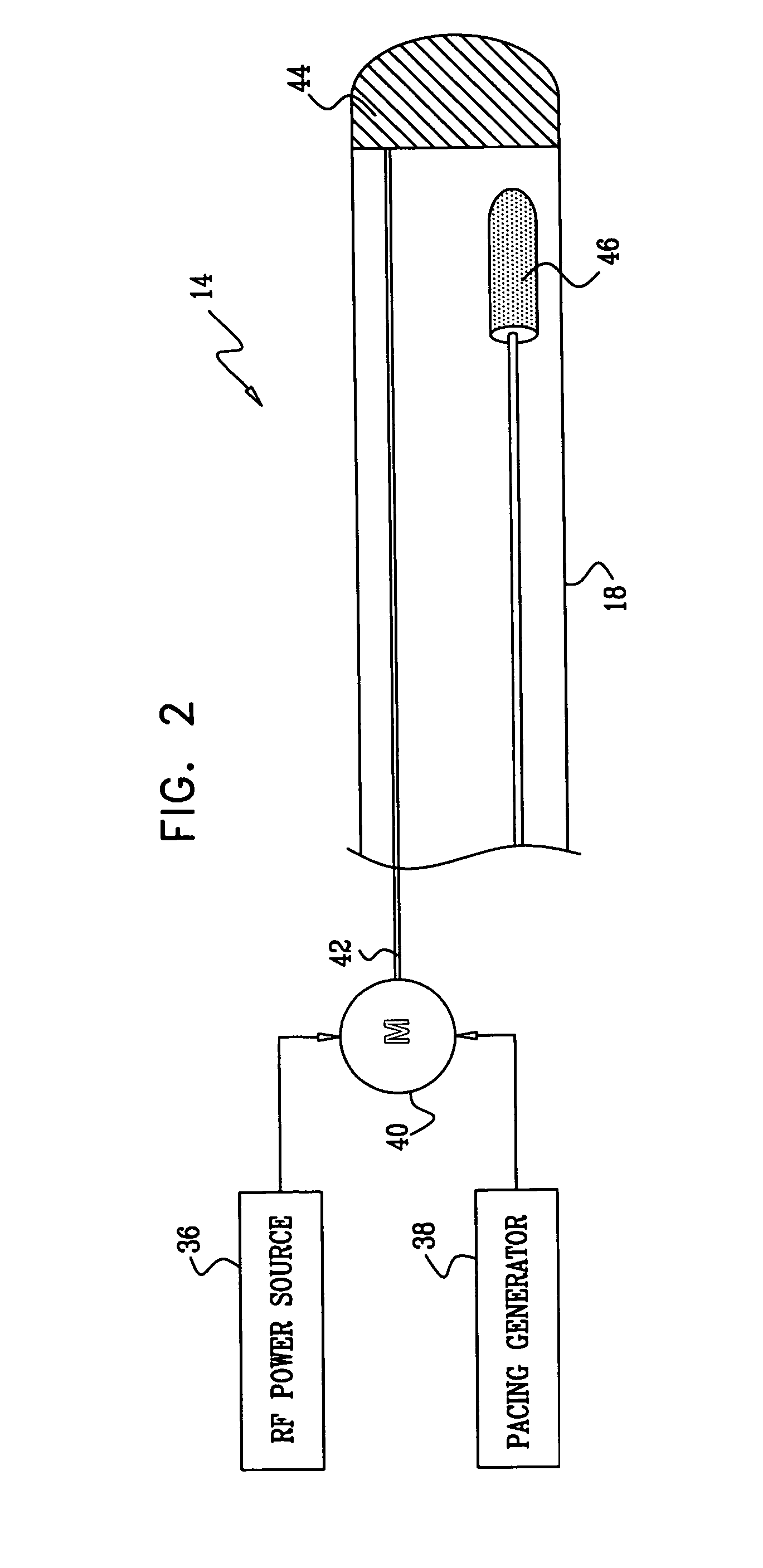
[0035]Turning now to the drawings, reference is initially made to FIG. 1, which is a pictorial illustration of a system 10 for performing ablative procedures on a heart 12 of a living subject in accordance with a disclosed embodiment of the invention. The system comprises a probe, typically a catheter 14, which is percutaneously inserted by an operator 16, who is typically a physician, through the patient's vascular system into a chamber or vascular structure of the heart. The operator 16 brings the catheter's distal tip 18 into contact with the heart wall at a target site that is to be ablated. Radiofrequency electrical current is then conducted through wires in the catheter to one or more electrodes at the distal tip 18, which apply the radiofrequency energy to the myocardium. The energy is absorbed in the tissue, heating it to a point (typically about 50° C.) at which it permanently loses its electrical excitability. When successful, this procedure creates non-conducting lesions ...
embodiment 2
[0061]The method disclosed above with reference to FIG. 2 may be combined with other lesion assessment techniques. Reference is now made to FIG. 4, which is a schematic of the distal tip 18 of the catheter 14 (FIG. 1) in accordance with an alternate embodiment of the invention. The distal tip 18, shown in juxtaposition to target tissue 74, is now provided with an array of ultrasound transducers 76 and a temperature sensor 78, for additional lesion production and assessment as described, for example, in U.S. Pat. Nos., 5,443,489, 6,321,109, 6,083,170, 6,301,496 and U.S. Patent Application Publication Nos. 2004 / 0143258 and 2004 / 0147920, whose disclosures are herein incorporated by reference. The transducers 76 and the temperature sensor 78 are connected to suitable signal processing circuitry in the console 24 (FIG. 1), which can be realized as the above-noted Carto-Biosense Navigation System. Lesion assessment may be conducted simultaneously using feedback from electrical obtained us...
embodiment 3
[0069]Reference is now made to FIG. 5, which is an end view of a distal portion 90 of a catheter suitable for use in the system 10 (FIG. 1), in accordance with an alternate embodiment of the invention. In this embodiment, the tip is provided with an ablation electrode 92 having a central aperture 94 measuring about 1-1.5 mm in diameter.
[0070]Reference is now made to FIG. 6, which is a sectional view taken along line 6-6 of the distal portion 90 of the catheter shown in FIG. 5. The electrode 92 extends a short distance behind the tip of the catheter, being delimited by a broken line 96. An ultrasound transducer 98 is positioned a short distance behind the aperture 94, being separated from the distal end of the catheter by a plug 100 of a sonolucent material such as silicon, and having an interface 102 with the plug 100. The transducer 98 is forward looking, and has a field of view 104, indicated by broken lines, with an operational range of about 8 mm. The field of view 104 extends t...
PUM
 Abstract
Description
Claims
Application Information
 - R&D
- Intellectual Property
- Life Sciences
- Materials
- Tech Scout
- Unparalleled Data Quality
- Higher Quality Content
- 60% Fewer Hallucinations
Browse by: Latest US Patents, China's latest patents, Technical Efficacy Thesaurus, Application Domain, Technology Topic, Popular Technical Reports.
© 2025 PatSnap. All rights reserved.Legal|Privacy policy|Modern Slavery Act Transparency Statement|Sitemap|About US| Contact US: help@patsnap.com


