
Magnetic resonance imaging concepts

a magnetic resonance imaging and concept technology, applied in the field of magnetic resonance imaging concepts, can solve the problems of degrading the effectiveness of the data, affecting the accuracy of the data, so as to achieve accurate motion estimation, effective navigation, and accurate motion estimation.

Active Publication Date: 2011-05-10
NGUYEN THANH +4
View PDF30 Cites 66 Cited by
  • Summary
  • Abstract
  • Description
  • Claims
  • Application Information

AI Technical Summary

Benefits of technology

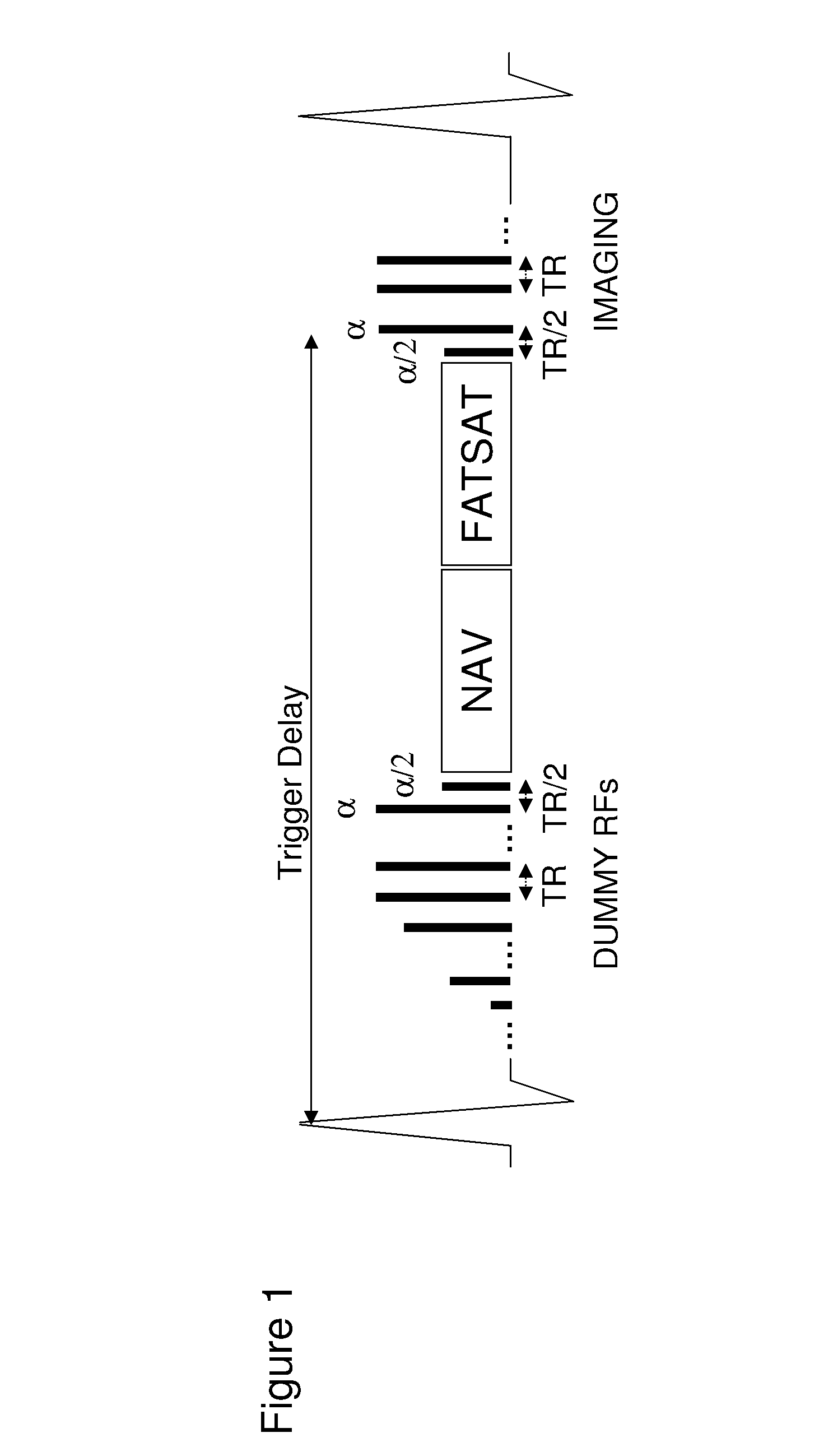
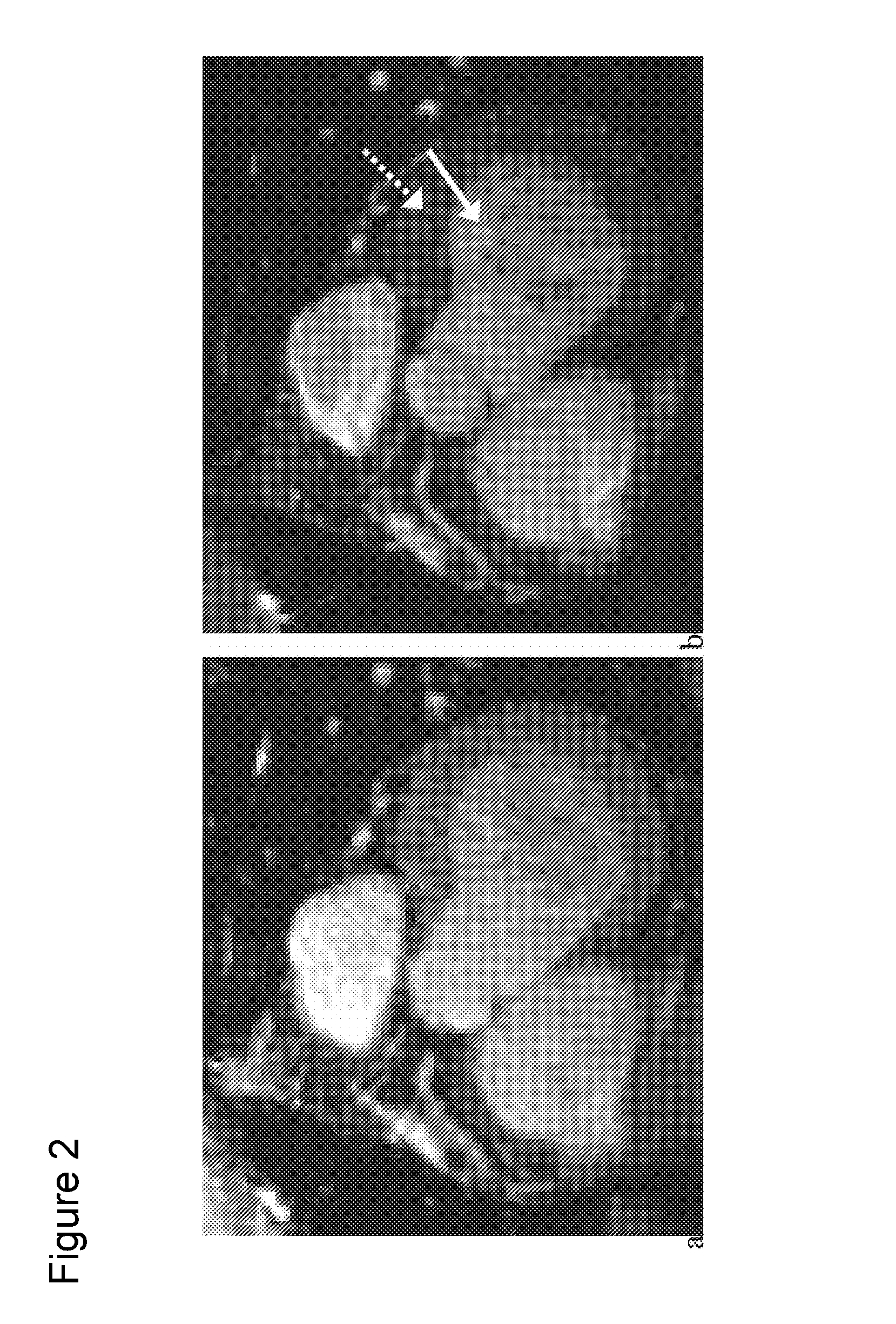
[0021]One aspect of the present invention is an effective navigator method for MRI with magnetization preparation and navigator by minimizing the delay between the navigator echo and the image echo, allowing accurate motion estimation and correspondingly effective gating and correction of motion artifacts in image echoes. One embodiment of navigator steady-state free precession (SSFP) is three-dimensional coronary MRA. In this preferred embodiment, the navigator and fat saturation pulses are preferably executed in steady state after the dummy or preparatory RF pulses so that the delay between the magnetization preparation and image echoes are minimized. Compared with previous preparation schemes, the present invention provides more effective motion suppression, significantly improved blood-to-myocardium contrast-to-noise ratio at slightly but insignificantly decreased blood SNR, significantly reduced fat SNR, and better overall image quality.
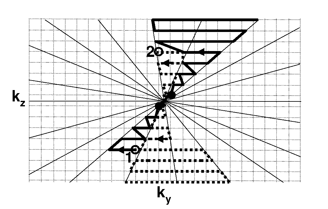
[0023]The present invention further provides for a method of time-resolved MRI data sampling that improves upon prior art methods by increasing temporal resolution and SNR through using non-linear k-space trajectories. This aspect of the present invention includes sliding window reconstruction with complex subtraction of precontrast mask data and improved off-resonance correction for time resolved 3D spiral MRA. The non-linear trajectories may be spiral, and the scan time and sliding window duration are reduced compared to the linear k-space sampling trajectory used in the prior art. That aspect of the present invention also includes cardiac-phased resolved study of the heart using bright or black blood pulse sequences.
[0025]The present invention also provides for methods that allow for the self-calibration of coil sensitivity during data collection runs, thus greatly increasing the SNR and temporal resolution of MRI. The present invention further provides methods for rapid reconstruction of a large number of image volumes from all temporal phases of data collected with multiple RF coils using parallel computing over multiple processors.

Problems solved by technology

That delay may be too long to provide accurate motion information, leading to artifacts in the collected data.
Furthermore, the separation between the fat saturation and the image echoes makes the fat suppression dependent on the field inhomogeneity, thereby degrading its effectiveness.
Finally, the number of preparatory RFs is limited to approximately 20, which may not be a large enough number for adequate SSFP signal contrast and sufficient reduction of signal oscillation in the case of a set of linearly ramped preparatory RF pulses.
These timing procedures complicate the task of performing CEMRA and, though quite reliable, are not without possible error.
However, typical 3D k-space sampling of the required high spatial frequency for depicting vascular details takes too long (10-20 seconds) to provide acceptable temporal resolution.
However, the lack of depth resolution limits the clinical utility of the 2D projection MRDSA.
Furthermore, the radial sampling is limited to sampling one straight spoke per RF excitation, which results in suboptimal SNR efficiency.
However, the repetition-to-repetition changes in phase encoding gradients cause spin phase variations associated with gradient-induced eddy currents and consequently lead to artifacts in SSFP imaging.
However, the temporal variations in blood and background signals after each inversion pulse may cause image artifacts and abate background suppression.
The reference scan for coil sensitivity increases the total scan time and may cause misregistration artifacts if the patient moves between the reference scan and the accelerated data acquisition.

Method used


Image

  • Magnetic resonance imaging concepts
  • Magnetic resonance imaging concepts
  • Magnetic resonance imaging concepts

Examples


Embodiment Construction

[0058]It is to be understood that the figures and descriptions of the present invention have been simplified to illustrate elements that are relevant for a clear understanding of the invention, while eliminating, for purposes of clarity, other elements that may be well known. The detailed description will be provided hereinbelow with reference to the attached drawings.

[0059]One aspect of the present invention relates to an improved navigator protocol for magnetization prepared steady-state free precession 3D coronary MRA. In a presently-preferred embodiment, the magnetization preparation method that executes the navigator echo and the fat saturation just prior to the image echoes. The present scheme allows more effective motion and fat suppression and improved contrast-to-noise ratio for coronary MRA.

[0060]In one implementation of navigator steady-state free precession 3D coronary MRA, the navigator and fat saturation pulses are executed during steady state after the preparatory RFs...


PUM


Abstract

Methods of acquiring magnetic resonance imaging (MRI) data for angiography. The present invention includes novel magnetization preparation schemes where the navigator and fat saturation pulses are executed in steady state after the preparatory pulses in order to minimize the delay between the magnetization preparation and the image echoes. The present invention also provides for improved methods of contrast-enhanced MRI where data are collected along non-linear trajectories through k-space and may also involve novel view ordering. In addition, the present methods employ novel motion corrections that minimize motion artifacts. The present invention further provides novel methods of self-calibrated sensitivity-encoded parallel imaging that allow for accurate and rapid scanning of subjects.

Description

[0001]“This invention was made with government support under Grant Nos. HL064647, HL062994, and HL060879 awarded by the National Institutes of Health. The government has certain rights in this invention.” This statement is included solely to comply with 37 C.F.R. §401.14(f)(4) and should not be taken as an assertion or admission that the application discloses and / or claims only one invention.BACKGROUND OF THE INVENTION[0002]The present application claims the benefit under 35 U.S.C. 119(e) of U.S. Provisional Application No. 60 / 628,614 filed Nov. 16, 2004.[0003]1. Field of the Invention[0004]The field of the present invention generally relates to improvements in methods of magnetization preparation for collection of magnetic resonance imaging data and to improvements in methods of data collection of magnetic resonance imaging data from physiological tissue. In certain preferred embodiments, the present invention is directed to improved methods for performing magnetic resonance angiog...

Claims


Application Information

Patent Timeline
IPC IPC(8): A61B5/05G01V3/00
CPCG01R33/4824A61B5/055G01R33/5614G01R33/561G01R33/5602G01R33/5607G01R33/5635G01R33/5676
Inventor WANG, YISPINCEMAILLE, PASCALNGUYEN, THANH D.
Owner NGUYEN THANH
Learn More