Diagnostic method for brain damage-related disorders

a brain damage and diagnostic method technology, applied in the direction of drugs, instruments, peptide sources, etc., can solve the problems of poor sensitivity and specificity, affecting the diagnostic value of brain damage biomarkers, and unable to accurately predict clinical status and functional outcomes

Inactive Publication Date: 2011-06-07
UNIVERSITY OF GENEVA +1
View PDF2 Cites 2 Cited by
  • Summary
  • Abstract
  • Description
  • Claims
  • Application Information

AI Technical Summary

Problems solved by technology

Most of them have not proved useful indicators of the extent of brain damage and accurate predictors of clinical status and functional outcome.
In fact, the diagnostic value of biomarkers for brain damage-related disorders has been hampered by their late appearance and a delayed peak after the damage event, their poor sensitivity and specificity, and the limited understanding of the mechanisms governing the release of these molecules into the CSF and ultimately in the blood.
As a result of these limitations, the use of brain damage-related disorder biomarkers is currently limited to research settings and none has been recommended for routine assessment [8].

Method used


Image

  • Diagnostic method for brain damage-related disorders
  • Diagnostic method for brain damage-related disorders
  • Diagnostic method for brain damage-related disorders

Examples


example 1

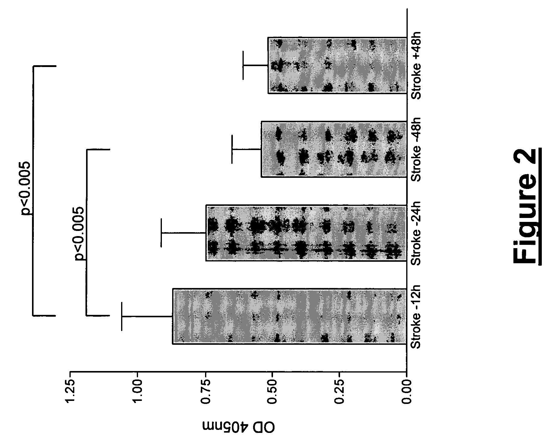
[0166]Using two-dimensional gel electrophoresis (2-DE) separation of cerebrospinal fluid (CSF) proteins and mass spectrometry techniques, 15 polypeptides named in Table 1 were found elevated or decreased in the CSF of deceased patients, used as a model of massive brain damage.

[0167]Study Population and Samples Handling

[0168]Eight CSF samples were used for the proteomics-based approach aiming at discovering brain damage-related disorder markers. Four of these samples were obtained at autopsy from deceased patients 6 hours after death with no pathology of the central nervous system. Four others were collected by lumbar puncture from living patients who had a neurological workup for benign conditions unrelated to brain damage (atypical headache and idiopathic peripheral facial nerve palsy). CSF samples were centrifuged immediately after collection, aliquoted, frozen at −80° C. and stored until analysis.

[0169]CSF 2-DE

[0170]All reagents and apparatus used have been described in detail el...

example 2

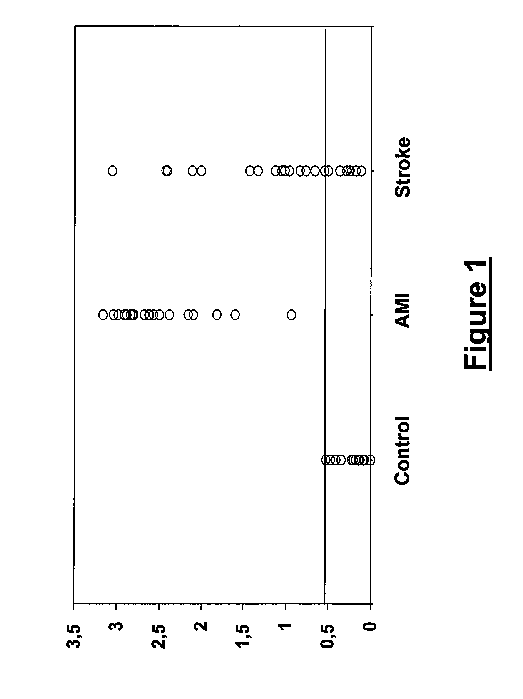
[0174]Using two-dimensional gel electrophoresis (2-DE) separation of cerebrospinal fluid (CSF) proteins and mass spectrometry techniques, FABP was found elevated in the CSF of deceased patients, used as a model of massive brain damage. Since H-FABP, a FABP form present in many organs, is also localised in the brain, an enzyme-linked immunosorbant assay (ELISA) was developed to detect H-FABP in stroke vs. control plasma samples. However, H-FABP being also a marker of acute myocardial infarction (AMI), Troponin-I and creatine kinase-MB (CK-MB) levels were assayed at the same time in order to exclude any concomitant heart damage. NSE and S100B levels were assayed simultaneously.

[0175]Study Population and Samples Handling

[0176]The population used for the assessment in plasma of the various markers detailed below included a total of 64 prospectively studied patients (Table 2) equally distributed into three groups: (1) a Control group including 14 men and 8 women aged 65 years (ranges: 34...

example 3

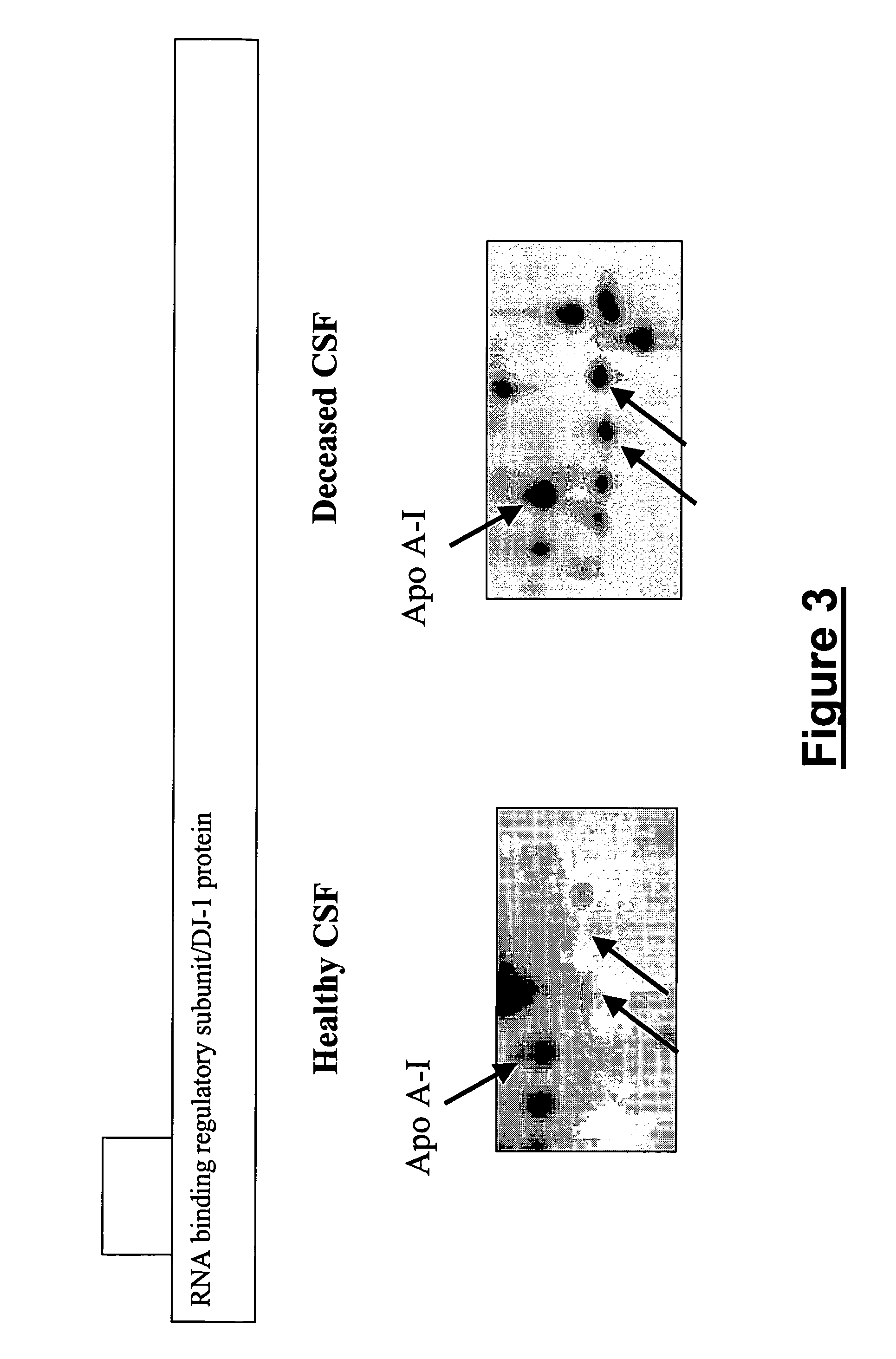
[0195]Three new proteins have been identified on 2-DE gels prepared with CSF samples from deceased patients. These proteins correspond to spots that have been previously shown increased in CSF samples from deceased patients relative to healthy controls. However, previous attempts to identify these proteins using MALDI-TOF mass spectrometry were unsuccessful. The current experiments were performed by μLC-MS-MS using ESI-Ion Trap device (DecaLCQ XP, ThermoFinnigan). Furthermore, the increasing amount of data in databases could lead to the successful identification of previously uncharacterized spots.

[0196](1) RNA-binding protein regulatory subunit (014805) / DJ-1 protein (Q99497): RNA-binding protein regulatory subunit has been previously described in deceased CSF samples (see Example 1 above). Here, we have obtained the same identification with an adjacent spot (FIG. 3). We also confirmed the previous identifications. FIG. 1 shows enlargements of healthy CSF and deceased CSF 2-DE maps....


PUM

PropertyMeasurementUnit
pHaaaaaaaaaa
timeaaaaaaaaaa
pHaaaaaaaaaa

Abstract

A brain damage-related disorder is diagnosed in a subject by detecting at least one polypeptide, or a variant or mutant thereof, selected from A-FABP, E-FABP, PGP 9.5, GFAP, Prostaglandin D synthase, Neuromodulin, Neurofilament L, Calcyphosine, RNA binding regulatory subunit, Ubiquitin fusion degradation protein 1 homolog, Nucleoside diphosphate kinase A, Glutathione S tranferase P, Cathepsin D, DJ-1 protein, Peroxiredoxin 5 and Peptidyl-prolyl cis-trans isomerase A (Cyclophilin A) in a sample of body fluid taken from the subject.

Description

CROSS-REFERENCE TO RELATED APPLICATION[0001]This application is a continuation of Great Britain Patent Application No. PCT / GB2004 / 050012 filed Sep. 20, 2004, the entire specification claims and drawings of which are incorporated herewith by reference.BACKGROUND OF THE INVENTION[0002]1 Field of the Invention[0003]This invention relates to a diagnostic method for brain damage-related disorders. No biological marker is currently available for the routine diagnosis of brain damage-related disorders including cerebrovascular, dementia and neurodegenerative diseases. This invention relates to the use of cerebrospinal fluid from deceased patients as a model for the discovery of brain damage-related disorder markers, and to the use of such markers in diagnosis.[0004]2. Description of the Related Art[0005]Over the last two decades, a number of biological markers (biomarkers) have been studied in the cerebrospinal fluid (CSF) and serum of patients with brain damage-related disorders, includin...

Claims


Application Information

Patent Timeline
Patent Type & Authority Patents(United States)
IPC IPC(8): G01N33/48G01N33/50G01N33/49G01N33/68G01N33/96C07K14/02C07K14/16
CPCC07K14/005G01N33/6896C12N2730/10122C12N2740/16222G01N2800/28G01N2333/705G01N2333/912G01N2333/948A61P9/10A61P19/00A61P21/02A61P25/00A61P25/02A61P25/06A61P25/08A61P25/14A61P25/22A61P25/24A61P25/28A61P27/02A61P29/00
Inventor HOCHSTRASSER, DENIS F.SANCHEZ, JEAN-CHARLESLESCUYER, PIERREALLARD, LAURE
Owner UNIVERSITY OF GENEVA
Why Eureka
Try Eureka