Inhibitors of HIV membrane fusion
a technology of hiv membrane and inhibitors, which is applied in the direction of antibody medical ingredients, peptide sources, instruments, etc., can solve the problems of difficult to emerge drug-escape mutants, and achieve the effects of enhancing stability, bioavailability, and bioavailability
- Summary
- Abstract
- Description
- Claims
- Application Information
AI Technical Summary
Benefits of technology
Problems solved by technology
Method used
Image
Examples
example 1
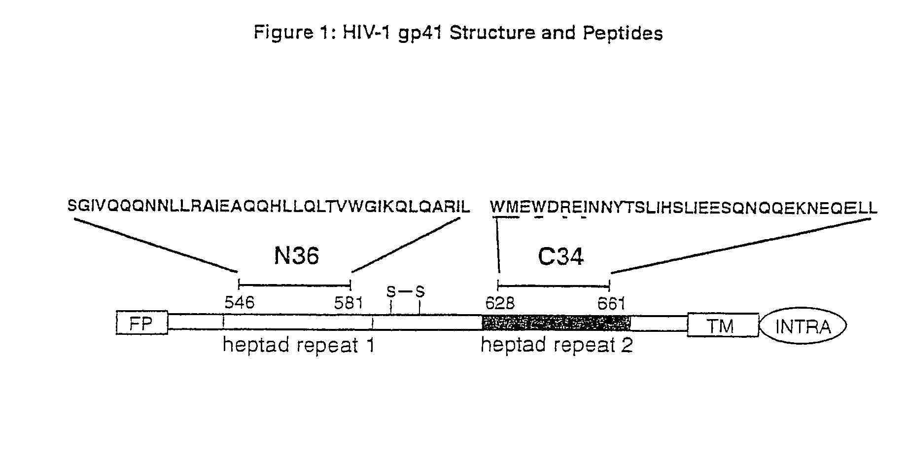
Synthesis of Variants of the C34 Peptide
[0118]Mutant peptides were synthesized by solid-phase FMOC peptide chemistry and have an acetylated amino terminus and an amidated carboxy terminus. After cleavage from the resin, peptides were desalted with a Sephadex G-25 column (Pharmacia), and then purified by reverse-phase high-performance liquid chromatography (Waters, Inc.) on a Vydac C18 preparative column using a linear water-acetonitrile gradient and 0.1% trifluoroacetic acid. Peptide identities were verified by MALDI mass spectrometry (Voyager Elite, PerSeptive Biosystems). Peptide concentrations were measured by tryptophan and tyrosine absorbance in 6 M GuHCl [H. Edelhoch, Biochemistry, 6:1948 (1967)].
example 2
Quantitation of Helical Content and Thermal Stability of Mutant N36 / C34 Complexes
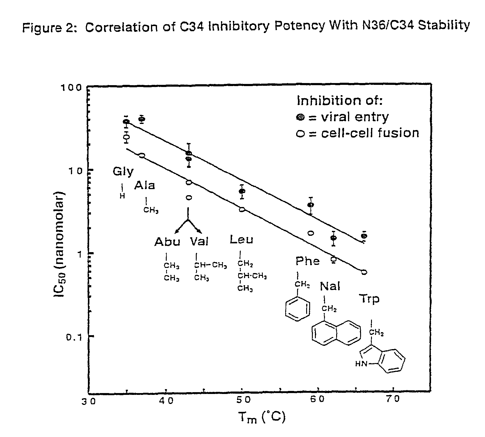
[0119]CD measurements were performed in phosphate-buffered saline (50 mM sodium phosphate, 150 mM NaCl, pH 7.0) with an Aviv Model 62DS spectrometer as previously described (M. Lu, S. C. Blacklow, P. S. Kim, Nat. Struct. Biol., 2:1075 (1995)). The apparent melting temperature of each complex was estimated from the maximum of the first derivative of [θ]222 with respect to temperature.
[0120]The mean residue ellipticities ([θ]222, 103 deg cm2 dmol−1) at 0° C. were as follows: wildtype, −31.7; Met629→Ala; −32.0; Arg633→Ala, −30.7; Ile635→Ala, −25.9; Trp628→Ala, −27.0; Trp631→Ala, −24.9. In the case of the Trp628→Ala and Trp631→Ala mutations, the decrease in [θ]222 is likely to overestimate the actual reduction in helical content. The removal of tryptophan residues from model helices has been reported to significantly reduce the absolute value of [θ]222 even when there is little change in helical content (A....
example 3
Identification of Peptides Which Bind to a Pocket on the Surface of the N-helix Coiled-Coil of HIV-1 gp41
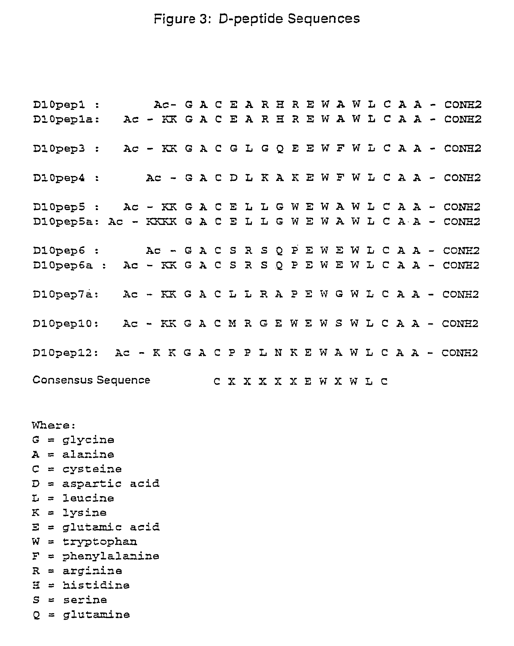
[0121]Methods are available to identify D-peptides which bind to a cavity on the surface of the N-helix coiled-coil of HIV envelope glycoprotein gp41. As described in detail below, D-peptides which bind to a cavity on the surface of the N-helix coiled-coil of HIV-1 envelope glycoprotein gp41 were identified by mirror-image phage display. This method involves the identification of ligands composed of D-amino acids by screening a phage display library. D-amino acid containing ligands have a chiral specificity for substrates and inhibitors that is the opposite of that of the naturally occurring L-amino ligands. The phage display library has been used to identify D-amino acid peptide ligands which bind a target or desired L-amino acid peptide (Schumacher et al. Science, 271:1854-1857 (1996)).
[0122]D-peptides that bind to the hydrophobic pocket of gp41 were identified using a target t...
PUM
| Property | Measurement | Unit |
|---|---|---|
| TOCSY mixing time | aaaaa | aaaaa |
| Tm | aaaaa | aaaaa |
| melting temperature | aaaaa | aaaaa |
Abstract
Description
Claims
Application Information
 - R&D
- Intellectual Property
- Life Sciences
- Materials
- Tech Scout
- Unparalleled Data Quality
- Higher Quality Content
- 60% Fewer Hallucinations
Browse by: Latest US Patents, China's latest patents, Technical Efficacy Thesaurus, Application Domain, Technology Topic, Popular Technical Reports.
© 2025 PatSnap. All rights reserved.Legal|Privacy policy|Modern Slavery Act Transparency Statement|Sitemap|About US| Contact US: help@patsnap.com


