Three-dimensional impedance imaging device

a three-dimensional impedance imaging and imaging device technology, applied in the field of biological imaging devices, can solve the problems of insufficient x-ray mammography for the early detection of breast cancer, inconvenient use, and inability to produce consistent quality,

Inactive Publication Date: 2011-08-30
STEVENS INSTITUTE OF TECHNOLOGY
View PDF11 Cites 18 Cited by
  • Summary
  • Abstract
  • Description
  • Claims
  • Application Information

AI Technical Summary

Problems solved by technology

X-ray mammography is often insufficient for the early detection of breast cancer.
Consistent quality is technically difficult to produce and interpretations are variable and subjective.
Therefore, although conventional screening methods have been proven to reduce mortality in women above age fifty, the efficacy of mammography as a life-saving measure in young women is uncertain.
Especially problematic among young women is the higher rate of false-positive and false-negative results.
False positives result in over-diagnosis and over-treatment (e.g., 75 percent of biopsied lesions resulting from suspicious mammogram findings turn out to be benign).
Furthermore, the false-negative rate has been determined to be as high as 34 percent, increasing the potential mortality for one third of the screened population.
Each film requires uncomfortable compression of the mammary tissue.
However, conventional mammography cannot distinguish between tissue types or distinguish between in situ lesions or invasive cancer.
When the biopsy proves that the anomalous tissue detected by x-ray mammography is indeed healthy, the mammography is said to have produced a false positive.
Studies show that testing costs for false positives may be near one-third of the entire mammography cost per year.
While the monetary cost is one consideration, the emotional burden ensuing from false-positive results provided by the mammogram cannot be neglected.
Women who experience false positives suffer from impaired emotional states for up to three months, with symptoms including impaired moods and limited daily functions.
In addition, fear of breast cancer is instilled in most patients receiving false-positive results.
In addition, radiologists' interpretations are not 100 percent accurate and can miss lesions that appear on the film.
Poor film quality caused by inadequate x-ray mammographic techniques can also lead to false negatives.
Delay in treatment and uncontrolled progression of the disease are possible outcomes of false negatives.
In fact, the leading cause of action in medical malpractice lawsuits arises from late or missed breast cancer diagnoses.
Furthermore, EIT mammography can create three-dimensional images, which is beyond the capabilities of x-ray mammography.
There are several challenges to creating an effective EIT system.
However, two-dimensional imaging is insufficient to yield clinically accurate results.
One obstacle to be overcome is the integration of a three-dimensional electrode array with imaging software.
Errors in placing the electrodes, which cannot be avoided even when the electrodes are applied by a skilled technician, can lead to errors in determining the size and location of anomalous tissues.
Further, the electrical signals detected at the electrodes can be distorted by reflective and refractive noise at the skin surface that electrode placement gels neglect.
For example, if an inflated rubber glove were used as the image of a hand, no person's hand could be accurately imaged.
Furthermore, the degree of inaccuracy would vary by patient, making variations difficult to correct by means of software.

Method used


Image

  • Three-dimensional impedance imaging device
  • Three-dimensional impedance imaging device
  • Three-dimensional impedance imaging device

Examples


Embodiment Construction

[0032]The technology disclosed herein uses an electrically-conductive mediating fluid to provide an electrical contact between the electrodes of an EIT device and the skin of a body part to be examined. In the EIT method discussed herein, the height of the fluid is raised or lowered between impedance measurements, enabling tomographic images of the tissue under examination to be resolved mathematically for subsequent viewing.

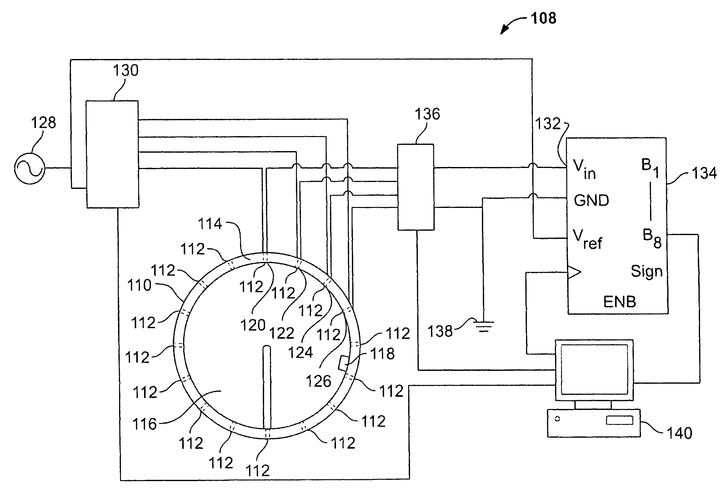
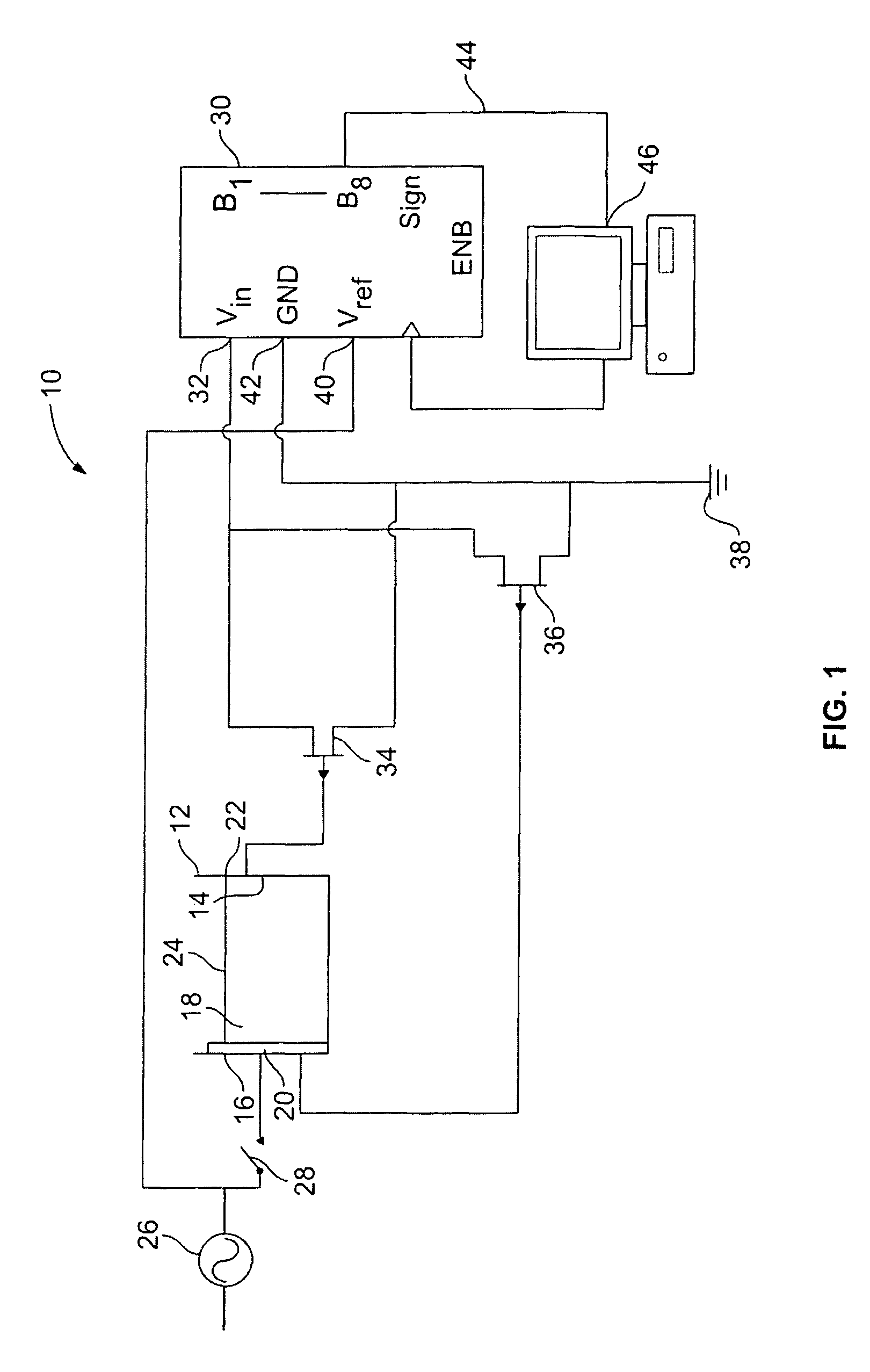
[0033]FIG. 1 illustrates a conceptual example of an apparatus 10 for implementing an EIT method according to the present invention. For the purpose of illustration, the apparatus of FIG. 1 is presented with elements related to three electrodes (not shown). In practice, more than three electrodes should be used, as discussed elsewhere herein. The nature of such electrodes is discussed elsewhere herein.

[0034]Referring to FIG. 1, the electrodes are provided as part of testing tank 12 along the inner surface 14 of the tank wall 16. The tank 12 contains an electrical...


PUM


Abstract

In a method of electrical impedance tomography (EIT), a mediating fluid provides electrical contact between the electrodes of an EIT device and the skin of a body part to be examined. The height of the fluid is raised or lowered between impedance measurements, enabling tomographic images of the tissue under examination to be resolved mathematically for subsequent viewing. Tomographic planes are isolated by calculating differences between Cartesian models generated from impedance values measured at the plane of interest and at an adjacent plane.

Description

CROSS-REFERENCE TO RELATED APPLICATIONS[0001]This application claims the benefit of U.S. Provisional Patent Application No. 61 / 023,561, filed on Jan. 25, 2008, which is incorporated herein by reference in its entirety.FIELD OF THE INVENTION[0002]The present invention relates to devices and methods for imaging, and, more particularly, to a device and method for biological imaging.BACKGROUND OF THE INVENTION[0003]Breast cancer is the most common cancer among women in America, accounting for 15 percent of all cancer deaths. Survival depends strongly on early diagnosis. The foremost breast cancer screening tool is the x-ray mammography. It is estimated that, each year, over 34 million mammograms are conducted in the United States. X-ray mammography can be used to detect variations in tissue density, and when an abnormality is detected, further tests are employed to detect the exact cause of the anomaly.[0004]X-ray mammography is often insufficient for the early detection of breast cance...

Claims


Application Information

Patent Timeline
Patent Type & Authority Patents(United States)
IPC IPC(8): A61B5/05
CPCA61B5/0536A61B5/4312
Inventor FREED, KATHERINECALDERIA, MEGANOSTROFF, RACHELEUBANKS, ESTHERMARTINI, RAINERHAZELWOOD, VIKKI
Owner STEVENS INSTITUTE OF TECHNOLOGY
Why Eureka
Try Eureka