Differential interference phase contrast X-ray imaging system

a phase contrast and imaging system technology, applied in imaging devices, instruments, applications, etc., can solve the problems of insufficient contrast of x-ray, insufficient x-ray absorbing contrast, etc., to achieve high radiation flux, wide emission angle, and high photon energy

Inactive Publication Date: 2011-12-06
SHENZHEN UNIV
View PDF24 Cites 94 Cited by
  • Summary
  • Abstract
  • Description
  • Claims
  • Application Information

AI Technical Summary

Benefits of technology

[0015]Two key innovations are proposed in present invention. The first innovation is the novel x-ray tube. The focal spot of the x-ray tube is a parallel line array, which dispenses with the absorption grating. This tube can work in the voltage range of 40-120 kV. By inserting an x-ray filter in the x-ray passage, the x-ray photon energy of the central wavelength of the x-ray beam emitted from the tube can be adjusted in the range of 20-100 keV, with a bandwidth of ±20%. Most importantly, this novel X-ray tube has several advantages like one dimension spatial coherence, high radiant flux, high photon energy and wide emission angle. Thus, invention of the tube provides a practicable x-ray source for the x-ray differential interference phase-contrast imaging system.
[0017]The present invention provides a novel x-ray detector with a function of an analyzer grating. This novel X-ray detector has a one-dimensional periodic structure. The direction of the periodic structure corresponds to the interference fringes direction of the x-ray phase grating; and the periodic pitch of the structure corresponds to the interference fringes pitch. In every loop cycle, half of the cycle is insensitive to X-ray; this is equivalent to adding an absorption grating with 100% absorbing ratio. Another half cycle of the periodic structure is the effective part for detection. The structure of the detector and the grating interference fringes generates Moiré fringes. Combining the detector and the analyzer grating to a single optoelectronic device not only reduces the number of optical compartments in the whole system, but diminishes the system complexity also. Moreover, the difficult task in making the absorption grating is avoided. The higher the X-ray photon energy, the harder to make the absorption grating. When an analyzer grating is used in medical imaging, the effective absorption thickness of the grating is usually over 300 μm, and the width of the interference pattern is only 1 μm, the aspect ratio is over 300; it is almost impossible to produce such an analyzer grating under present technology.

Problems solved by technology

But a weakly absorbing object consisting of light elements which does not absorb X-ray so much is too transparent for X-ray to create a sufficient contrast detected by X-ray detector.
However, with respect to those soft tissues, the X-ray absorbing contrast is relatively poor.
Nevertheless, synchrotron radiation source is too large and too expensive in most practical applications.
Moreover, the conventional synchrotron radiation source cannot provide 40-70 keV photon energy that is necessary to generate satisfying contrast for medical applications.
It is usually difficult or even impossible to apply the micro-focus X-ray tube as source in hospitals or any real time examination facility because it cannot provide sufficient radiant flux due to some critical limitations, like difficulty in heat dissipation.
All of these limitations make the differential interference phase-contrast imaging unfeasible in hospitals.
How to realize the function of analyzer grating without such absorption grating in differential phase-contrast imaging system is another challenge.
Although the phase-contrast image of sample can be obtain by the X-ray detector with high spatial resolution, the applications of two absorption grating, which work as generator of structure light and analyzer grating respectively, are significant limiting factors.
With the increasing of X-ray photon energy, an insuperable difficulty in fabricating absorption grating occurs.
Moreover, additional critical problems like view field, radiant flux, image distortion also occur.
Therefore, although the principle of differential interference phase-contrast imaging based on low luminance X-ray source has been introduced several years ago, and the imaging system is able to generate excellent result on small experimental animals, the applications in medical diagnosis and other related fields are still technically challenged, or practically infeasible.

Method used


Image

  • Differential interference phase contrast X-ray imaging system
  • Differential interference phase contrast X-ray imaging system
  • Differential interference phase contrast X-ray imaging system

Examples


Embodiment Construction

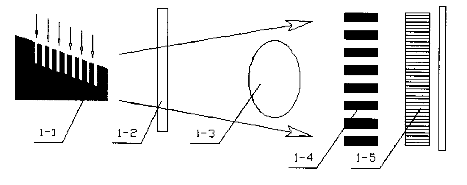
[0039]As shown in FIG. 1, in accordance with the direction of X-ray propagation, the basic components of present system include 1-1 X-ray tube, 1-2 filter, 1-3 object platform, 1-4 X-ray phase grating, 1-5 detector. System administration software and computer control the system. The key factors are: 1) The coherent X-ray beam from source array whose focal spots are linearly parallel arranged, has high energy and wide angle of de angle of divergence with 30-50 degree 2) The novel X-ray detector adopted in present invention plays dual roles of conventional analyzer grating and conventional detector. The basic structure of the detector includes a set of parallel array of X-ray scintillator screen, optical coupling system, area array detector or photoconductive X-ray detector. The structure and size of the X-ray scintillator screen and the photoconductive X-ray detector are consistent with the parallel-arranged linear X-ray beam with good coherence and high energy.

[0040]The novel X-ray ...


PUM

PropertyMeasurementUnit
emission angleaaaaaaaaaa
density distributionaaaaaaaaaa
photon energyaaaaaaaaaa

Abstract

A differential phase-contrast X-ray imaging system is provided. Along the direction of X-ray propagation, the basic components are X-ray tube, filter, object platform, X-ray phase grating, and X-ray detector. The system provides: 1) X-ray beam from parallel-arranged source array with good coherence, high energy, and wider angles of divergence with 30-50 degree. 2) The novel X-ray detector adopted in present invention plays dual roles of conventional analyzer grating and conventional detector. The basic structure of the detector includes a set of parallel-arranged linear array X-ray scintillator screens, optical coupling system, an area array detector or parallel-arranged linear array X-ray photoconductive detector. In this case, relative parameters for scintillator screens or photoconductive detector correspond to phase grating and parallel-arranged line source array, which can provide the coherent X-rays with high energy.

Description

RELATED APPLICATION[0001]This application claims the benefit of Peoples' Republic of China application Serial No. 200810216469.3 filed on Oct. 10, 2008, hereby specifically incorporated by reference in its entirety.FIELD OF THE INVENTION[0002]This invention provides a differential interference phase-contrast X-ray imaging system.DESCRIPTION OF RELATED ART[0003]Noninvasive imaging plays an important role and has been widely applied in many fields like medicine, life science, material science, industrial application, and security inspection etc. X-ray imaging is one of the most important methods for noninvasive imaging. The contrast of conventional X-ray radiography based upon the attenuation of X-ray is obtained through difference in the absorption cross-section of the constituents of the object. But a weakly absorbing object consisting of light elements which does not absorb X-ray so much is too transparent for X-ray to create a sufficient contrast detected by X-ray detector. Especi...

Claims


Application Information

Patent Timeline
Patent Type & Authority Patents(United States)
IPC IPC(8): G01N23/083
CPCA61B6/00A61B6/4035A61B6/4233A61B6/484H01J35/08A61B6/4291G21K2207/005H01J2235/086
Inventor NIU, HAN-BENGUO, JIN-CHUANLIU, XIN
Owner SHENZHEN UNIV
Why Eureka
Try Eureka