Treatment of ocular disease

a technology for ocular disease and ocular surgery, applied in the field of treatment of glaucoma, can solve the problems of high training skills and the risks of ocular surgery, and achieve the effect of suitable contact surfa

Inactive Publication Date: 2012-02-28
NOVA EYE INC
View PDF2 Cites 91 Cited by
  • Summary
  • Abstract
  • Description
  • Claims
  • Application Information

AI Technical Summary

Benefits of technology

[0086]The handpiece incorporating the locating device and the surgical device presents a suitable contacting surface to the eye. A slight curvature of the tissue contacting surfaces of the apparatus approximating the radius of the eye aids mechanical stability for the precise surgical placement of the surgical device. The contacting surfaces may also be modified to hold or deliver a coupling fluid or gel to aid energy transfer for the locating device.

Problems solved by technology

Such surgical intervention requires highly trained skills and presents the risks associated with ocular surgery.

Method used


Image

  • Treatment of ocular disease
  • Treatment of ocular disease
  • Treatment of ocular disease

Examples


example 1

[0102]An experiment was performed to determine the target resolution of high frequency ultrasound. An ultrasound phantom was prepared to emulate micro channels of various diameters. The phantom was prepared by placing stainless steel tubing (Small Parts, Inc. Miami Lakes, Fla.) of various diameters across a standard 80 mm styrene petri dish, spaced 10 mm apart. A 10% solution of gelatin, 250 bloom (Woburn, Edible Pork Skin Gelatin) was prepared by heating gelatin powder in distilled water until fully in solution. The gelatin solution was poured into the petri dish until the tubes were covered to a depth of approximately 1 mm. The gelatin was allowed to solidify by cooling and subsequently the tubes were withdrawn from the petri dish leaving open channels of various diameters. Channel diameters of 110, 150, 205, 230 and 255 microns were created in this manner. Luer hub tubing connectors were bonded to the ends of the channels to allow for injection of fluids in the channels.

[0103]The...

example 2

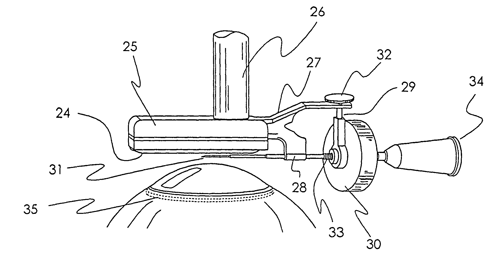
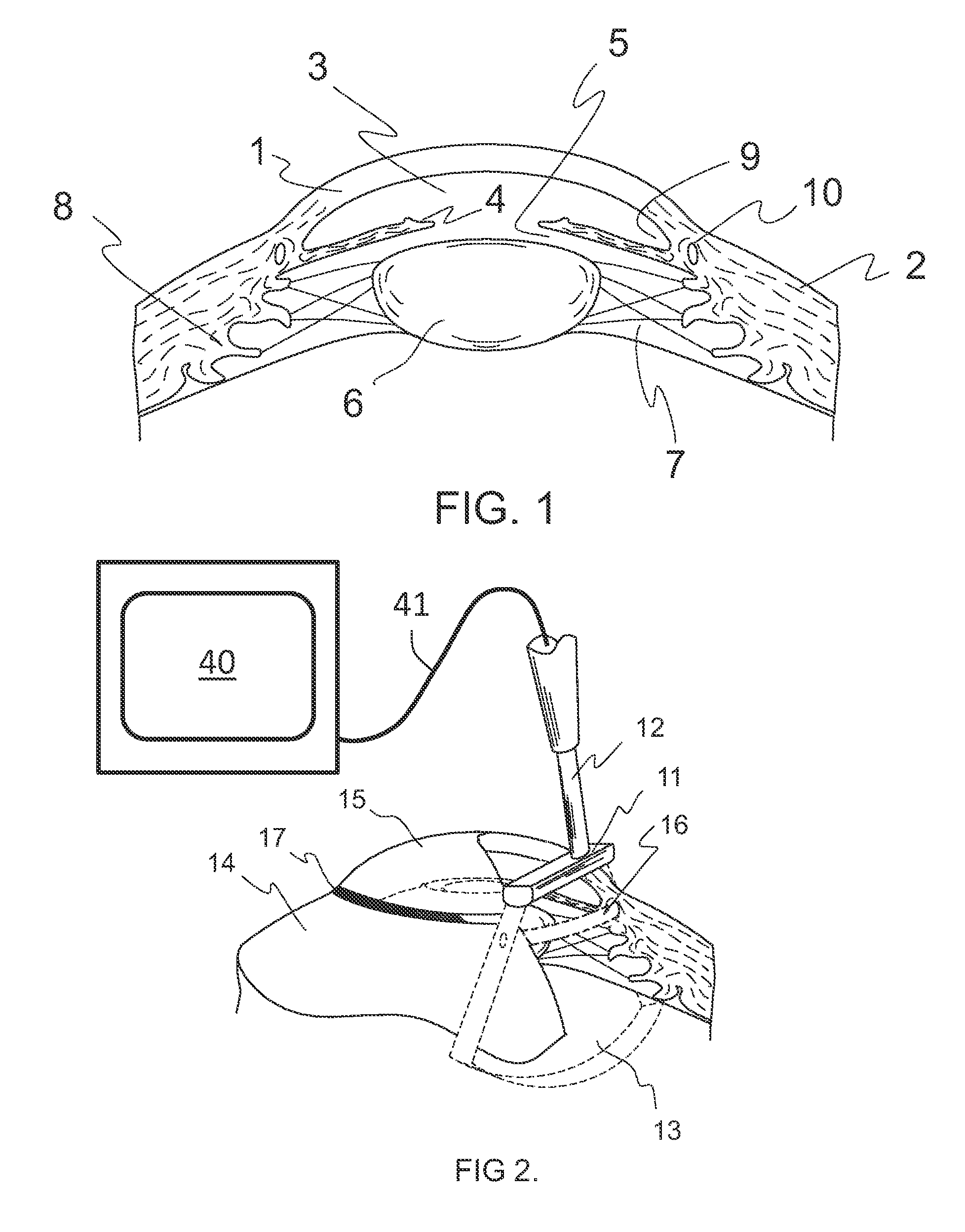
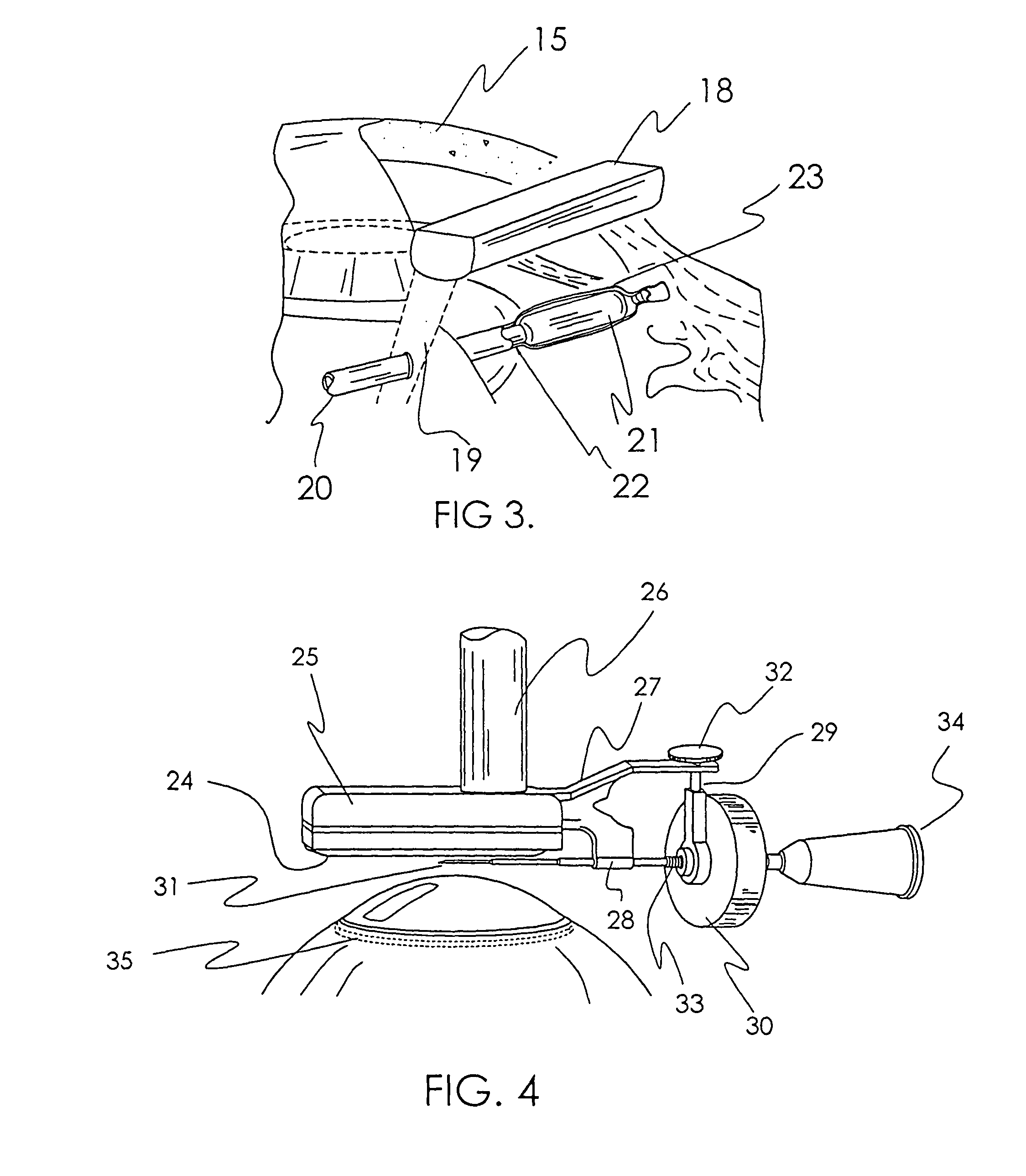
[0104]A unitary system is constructed comprised of a focused ultrasound transducer mounted at a right angle to the handpiece and an injection system coupled to the handpiece and whose axis is disposed in the same plane as the scan wedge of the transducer. The transducer is connected to a hardware system comprised of a signal generator, signal receiver, an image processing system and a display. The ultrasound imaging system is used to determine the location of Schlemm's Canal. The ultrasound transducer operating between 40 and 150 MHz transmit frequency is used to image the episcleral tissues near Schlemm's Canal. Preferably, the ultrasound system has an axial and lateral resolution of at least 60 microns for imaging of fine structures and is capable of discriminating Schlemm's Canal, whose central axis is disposed between 450-600 microns beneath the scleral surface. The tissue contacting surface of the transducer is curved to accommodate the curvature of the eye and a slight raised ...

example 3

[0107]A microsurgical system as described in Example 2 is adapted for introduction of the microcannula by mechanized means under guidance from the processed ultrasound signal. The control system is designed to provide introduction and redirection of the microcannula as determined by the known location of the microcannula tip relative to the estimated location of Schlemm's Canal from the ultrasound imaging and analysis system.


PUM


Abstract

The invention relates to a novel apparatus for the treatment of ocular disease, particularly glaucoma. The apparatus consists of a locating device to locate Schlemm's Canal within the anterior portion of the eye and a surgical tool to access the canal for treatment. The apparatus allows for guided, minimally invasive surgical access to Schlemm's Canal to enable surgical procedures to be performed on the canal and trabecular meshwork to reduce intraocular pressure. The apparatus may also deliver devices or substances to Schlemm's Canal in the treatment of glaucoma.

Description

CROSS REFERENCE TO OTHER APPLICATIONS[0001]This application is a divisional of U.S. Utility application Ser. No. 09 / 735,408, filed Dec. 11, 2000, now abandoned which claims benefit of U.S. Provisional Patent Application 60 / 172,693, filed Dec. 10, 1999, the specifications of which are hereby incorporated by reference in their entirety.FIELD OF THE INVENTION[0002]The present invention is directed to methods and apparatus for minimally invasive ocular surgery procedures and, more particularly, to the treatment of glaucoma.BACKGROUND OF THE INVENTION[0003]The anterior chamber of the eye is filled with a fluid known as aqueous humor. This fluid is produced by the ciliary body located adjacent to the lens. The fluid flows around the lens, through the trabecular meshwork and into an anatomical conduit, the sinus venosus sclerae, also known as Schlemm's Canal. The fluid is then expelled from the canal to the episcleral veins. In the disease state known as narrow or open angle glaucoma, the ...

Claims


Application Information

Patent Timeline
Patent Type & Authority Patents(United States)
IPC IPC(8): A61M35/00A61B8/10A61F9/007
CPCA61B8/0841A61B8/10A61F9/00781
Inventor YAMAMOTO, RONALD K.CONSTON, STANLEY R.NASH, MICHAEL F.KOCH, PAUL S.
Owner NOVA EYE INC
Why Eureka
Try Eureka