Medical image management system

a medical image and management system technology, applied in the field of medical imaging, to achieve the effect of reducing the size of image data, reducing image file size, and reducing file siz

Active Publication Date: 2012-04-24
HEART IMAGING TECH
View PDF130 Cites 22 Cited by
  • Summary
  • Abstract
  • Description
  • Claims
  • Application Information

AI Technical Summary

Benefits of technology

[0041]In a preferred embodiment, the converter engine comprises a decoding engine for extracting raw image data; and a physiologic knowledge engine adapted to receive data from the decoding engine. The physiologic knowledge engine adjusts the image quality and reduces the size of the image data, which is then transferred to a post engine. The physiologic knowledge engine is primarily responsible for reducing the image file size without loss of diagnostic data though other aspects of the Present Invention are used to reduce file size while maintaining viability of the data. The encoding engine converts the image data to browser compatible image data.

Problems solved by technology

In certain situations, however, the particular manner in which the images are made available to physicians and their patients introduces obstacles to timely and accurate diagnoses of disease.
These obstacles generally relate to the fact that each manufacturer of a medical imaging system uses different and proprietary formats to store the images in digital form.
Although it is typically possible to “export” the images from a proprietary workstation to an industry-standard format such as “Digital Imaging Communications in Medicine” (DICOM), Version 3.0, several limitations remain as discussed subsequently.
The specialist physician, however, typically has not performed a clinical history and physical examination of the patient and often is not aware of the patient's other test results.
Although this approach does allow for expert interpretation of the images by the specialist physician, several limitations are introduced for the primary physician and for the patient, such as, for example:(1) The primary physician does not see the images unless he travels to another department and makes a request;(2) It is often difficult to find the images for viewing because there typically is no formal procedure to accommodate requests to show the images to the primary physician;(3) Until the written report is forwarded to the primary physician's office, it is often difficult to determine if the images have been interpreted and the report generated;(4) Each proprietary workstation requires training in how to use the software to view the images;(5) It is often difficult for the primary physician to find a technician who has been trained to view the images on the proprietary workstation;(6) The workstation software is often “upgraded” requiring additional training;(7) The primary physician has to walk to different departments to view images from the same patient but different modalities;(8) Images from the same patient but different modalities cannot be viewed side-by-side, even using proprietary workstations;(9) The primary physician cannot show the patient his images in the physician's office while explaining the diagnosis; and(10) The patient cannot transport his images to another physician's office for a second opinion.
A similar barrier to accessing medical information exists for physicians interested in providing real-time training to other physicians, and for physicians seeking real-time advice from other physicians based on the results of medical imaging.
Generally, such training and / or advice can only be obtained by having the second physician physically present in the same room or in an immediately adjacent room while the first physician performs the imaging procedure.
Teleradiology does not, however, allow for the examination of the images from any site other than the central location, precluding examination of the images by the primary physician and the patient.
Several technical limitations of current Internet standards, however, create a situation where straightforward processing of the image data results in images which transfer across the Internet too slowly, lose diagnostic information or both.
One such limitation is the bandwidth of current Internet connections which, because of the large size of medical images, result in transfer times which are unacceptably long.
The problem of bandwidth can be addressed by compressing the image data before transfer, but compression typically involves loss of diagnostic information.
In addition, due to the size of the images the time required to process image data from an original format to a format which can be viewed by Internet browsers is considerable, meaning that systems designed to create Web Pages “on the fly” introduce a delay of seconds to minutes while the person requesting to view the images waits for the data to be processed.
Workstations allow images to be reordered or placed “side-by-side” for viewing, but again, an Internet system would have to create new Web Pages “on the fly” which would introduce further delays.
There are several disadvantages to using “Java” to manipulate the image data.
First, the user must wait additional time while the “Java” code is downloaded.
Third, the user must wait while the “Java” code processes the image data, which is slow because the image files are large.
Fourth, “Java” code is relatively new and often causes browsers to “crash.” Finally, due to the “crashing” problem “Java” programmers typically only test their code on certain browsers and computers, such as Microsoft Explorer on a PC, precluding widespread use by owners of other browsers and other computer platforms.
The approach of Wood, however, creates Web Pages “on the fly,” meaning that the user must wait for the image processing to complete.
In addition, even after processing of the image data into a Web Page the approach of Wood does not provide for processing the images in such as way that excessive image transfer times due to limited bandwidth are addressed or provide for “brightness / contrast” to be addressed without loss of diagnostic information.
In addition, the approach of Wood is limited to ultrasound images generated by scanners manufactured by a single company, and does not enable viewing of images from modalities other than ultrasound.
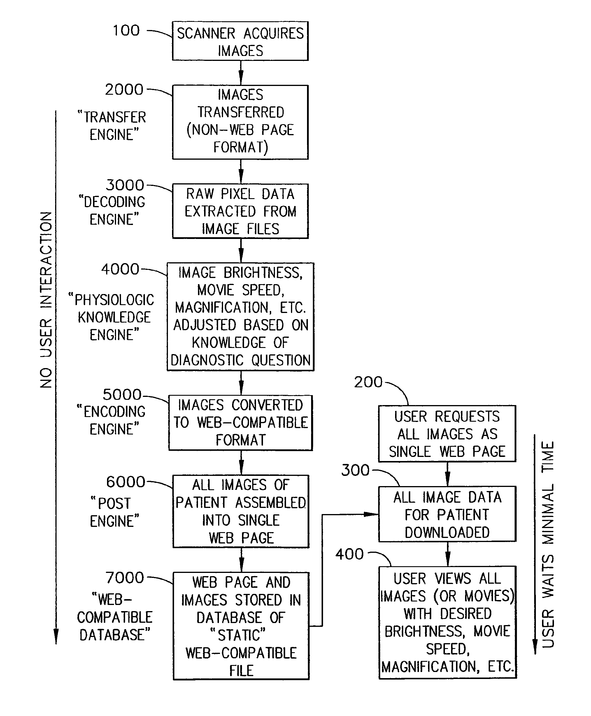
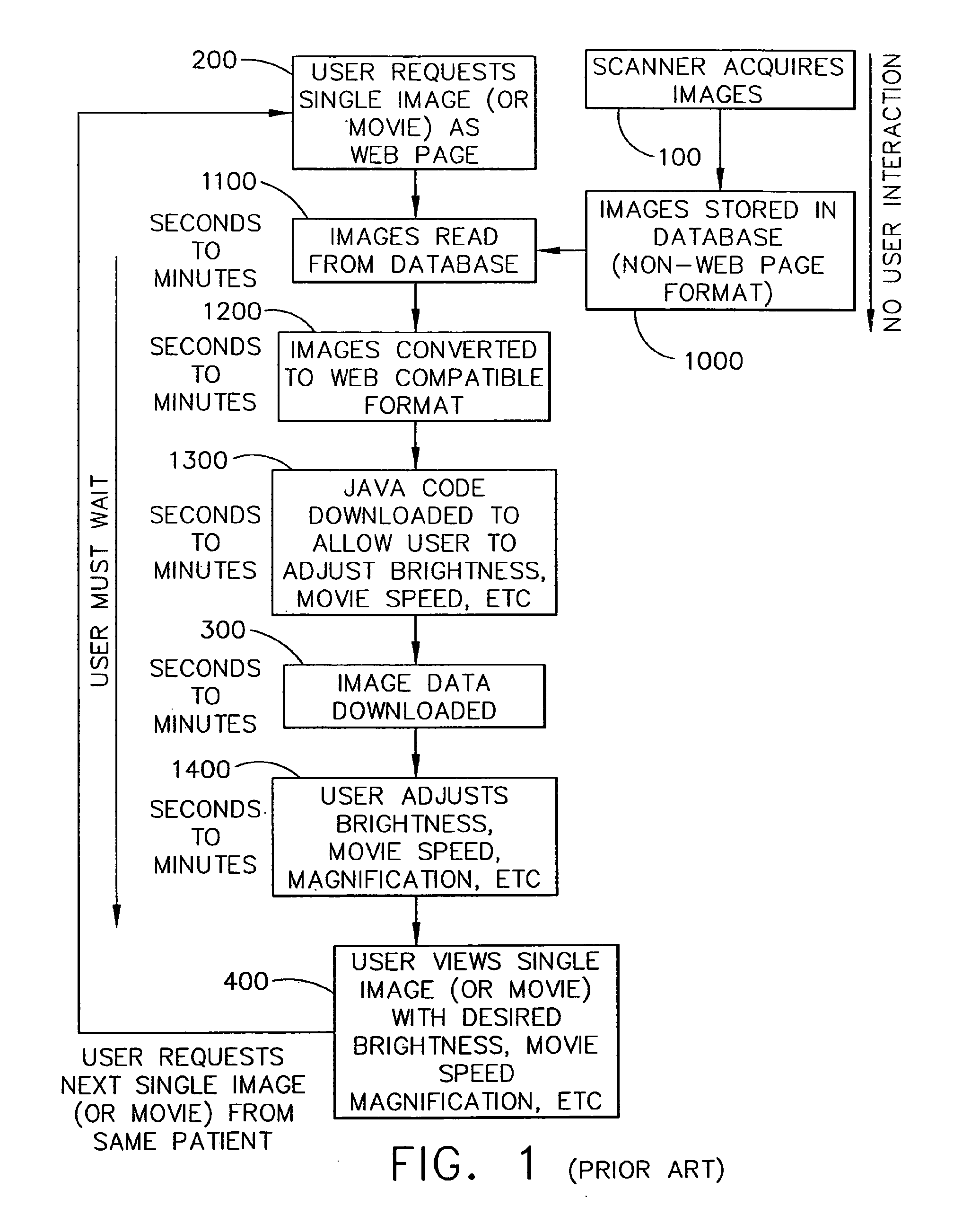
As can be seen in FIG. 1, serial processing of image data “on the fly” combined with extensive user interaction results in a slow, expensive, and unstable system.
Steps 1000-1400 result in extensive user interaction which results in the system being slow, expensive and unstable.

Method used


Image

  • Medical image management system
  • Medical image management system
  • Medical image management system

Examples


Embodiment Construction

[0066]The Present Invention is discussed in relation to imaging with specific applications discussed in relation to cardiac images; however, other uses will be apparent from the teachings disclosed herein. The Present Invention will be better understood from the following detailed description of exemplary embodiments, with reference to the attached figures, wherein like reference numerals and characters refer to like parts, and by reference to the following Claims.

[0067]It will be apparent to one possessing ordinary skill in the art that the structure, methods and systems described herein regarding a medical image management system are additionally and inherently applicable to the management of multiple types of medical information, such as, for example, medical imaging reports, electrocardiograms, medical test results, patient demographics, clinic reports, procedure reports, in-patient summary reports and the like.

[0068]The herein-described Present Invention has been constructed an...


PUM


Abstract

A method of managing medical information is disclosed. Medical image data is received, at a real-time transfer engine, at the same time that the patient is being scanned by a medical imaging device. The medical image data is then converted to a browser-compatible image format at a converter engine connected to receive the medical image data from the real-time transfer engine. The converter engine comprises a decoder engine for extracting image pixel data from the medical image data and an encoding engine for converting the image pixel data to a browser-compatible format connected to receive the image pixel data. The image pixel data may be converted to a browser compatible format without loss of diagnostic data.

Description

CROSS-REFERENCE TO RELATED APPLICATIONS[0001]This application is a continuation-in-part application of U.S. application Ser. No. 10 / 999,544 filed on Nov. 30, 2004 entitled “Medical Image Management System,” which is a continuation application of U.S. application Ser. No. 09 / 742,575, which issued as U.S. Pat. No. 6,934,698 on Aug. 23, 2005 entitled “Medical Image Management System.”FIELD OF THE PRESENT INVENTION[0002]The Present Invention relates to medical imaging. Specific exemplary embodiments discussed relate to cardiac medical imaging.BACKGROUND OF THE PRESENT INVENTION[0003]The description of the references in this Section is not intended to constitute an admission that any reference referred to herein is “Prior Art” with respect to the Present Invention, unless specifically designated as such.[0004]Medical imaging is important and widespread in the diagnosis of disease. In certain situations, however, the particular manner in which the images are made available to physicians a...

Claims


Application Information

Patent Timeline
Patent Type & Authority Patents(United States)
IPC IPC(8): G06F17/00
CPCG06F17/30244G06F17/30569G06F16/258G06F16/50
Inventor JUDD, ROBERT M.CHEN, ENN-LINGKIM, RAYMOND J.
Owner HEART IMAGING TECH
Why Eureka
Try Eureka