
Micro-machined medical devices, methods of fabricating microdevices, and methods of medical diagnosis, imaging, stimulation, and treatment

a micro-machined, medical device technology, applied in the field of micro-machined medical devices, can solve the problems of limiting the use of conventional ultrasound systems to larger vessels, affecting the image resolution of conventional ultrasound systems, and affecting the diagnosis of patients, so as to achieve the effect of reducing the charge buildup

Active Publication Date: 2013-06-04
STC UNM
View PDF15 Cites 0 Cited by
  • Summary
  • Abstract
  • Description
  • Claims
  • Application Information

AI Technical Summary

Benefits of technology

[0012]In some aspects, the present disclosure is directed to methods of fabricating one or more of the medical devices disclosed herein, including, for example, adding a self-aligned silicon dioxide layer under a first polysilicon layer to minimize the charge buildup associated with a silicon nitride-polysilicon interface.

Problems solved by technology

Unfortunately, traditional medical ultrasonic systems are bulky and normally operate outside the body.
However, its size (typically 3-4 millimeters in diameter) limits its use to larger vessels.
Image resolution from conventional ultrasound systems is relatively poor, especially for anatomical structures deep inside body tissue.
For imaging or diagnosis applications, long-wavelength acoustic waves limit the image resolution or the accuracy of the measurement / diagnosis.
For therapeutic stimulation, long-wavelength ultrasounds generally spread over an area larger than needed and could negatively affect the neighboring healthy tissue.
However, limited by their short penetration depth, they cannot penetrate through the body to reach a target deep inside the tissue.
These conventional probes and devices are typically of millimeter to centimeter diameter cross-section and cause significant disruption to the tissue.
They are not suitable for operation in more delicate organs, for example, the brain and liver, because the disruption (and as a result the damage) is too much.
The drawback of the boron-defined dissolving wafer process comes from the diffusion process required for doping the silicon and the high tensile stress in the boron-doped silicon.
The high-temperature boron diffusion process may not be very compatible with transistor and / or MEMS processes.
Additionally, the boron-caused high-stress may warp the device structure.
While this approach may provide good control on the final device dimension and prevents the stress problem, it requires use of a high-cost SOI wafer.

Method used


Image

  • Micro-machined medical devices, methods of fabricating microdevices, and methods of medical diagnosis, imaging, stimulation, and treatment
  • Micro-machined medical devices, methods of fabricating microdevices, and methods of medical diagnosis, imaging, stimulation, and treatment
  • Micro-machined medical devices, methods of fabricating microdevices, and methods of medical diagnosis, imaging, stimulation, and treatment

Examples


Embodiment Construction

[0036]Reference will now be made in detail to embodiments of the invention, examples of which are illustrated in the accompanying drawings. Wherever possible, the same reference numbers will be used throughout the drawings to refer to the same or like parts.

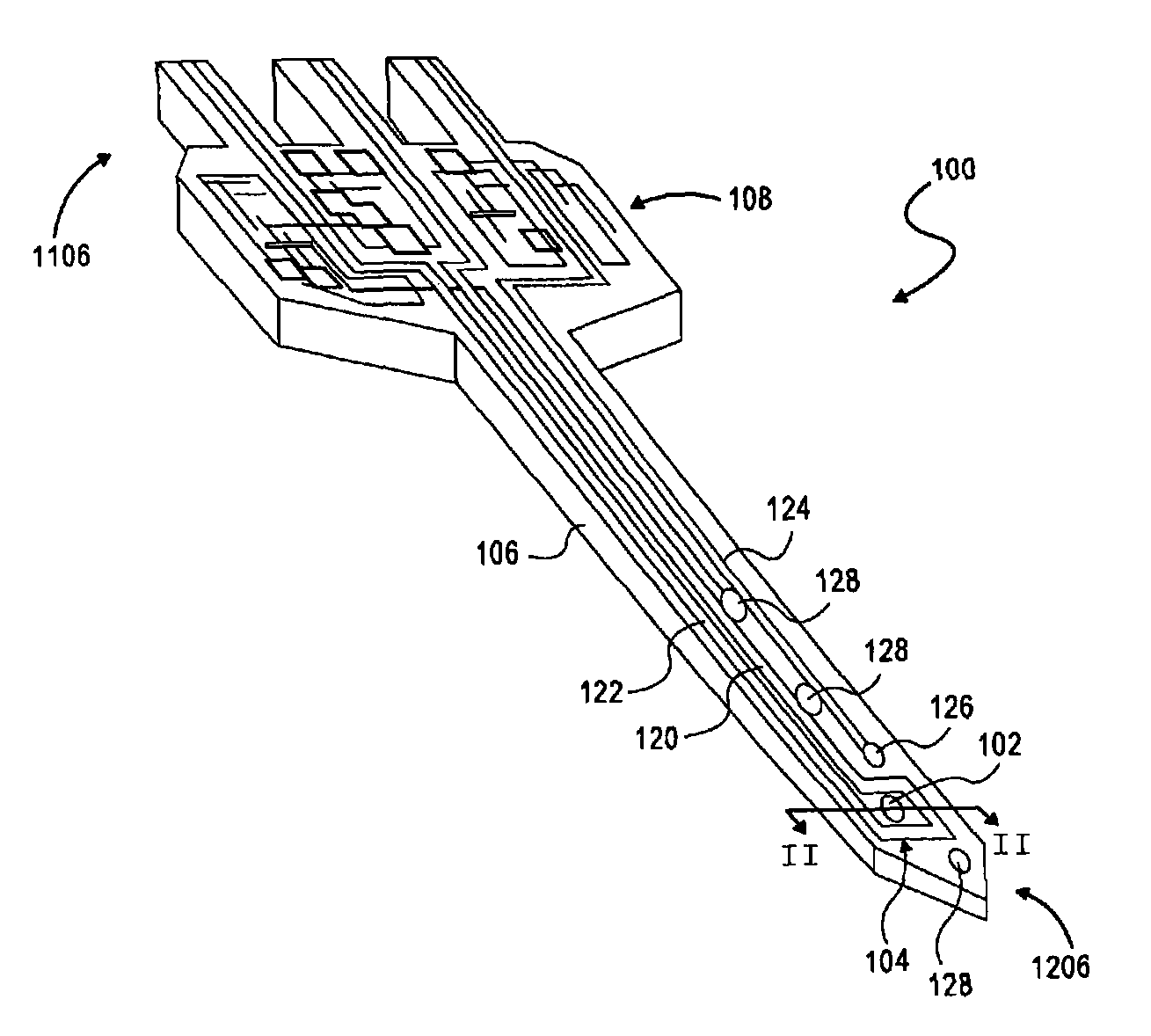
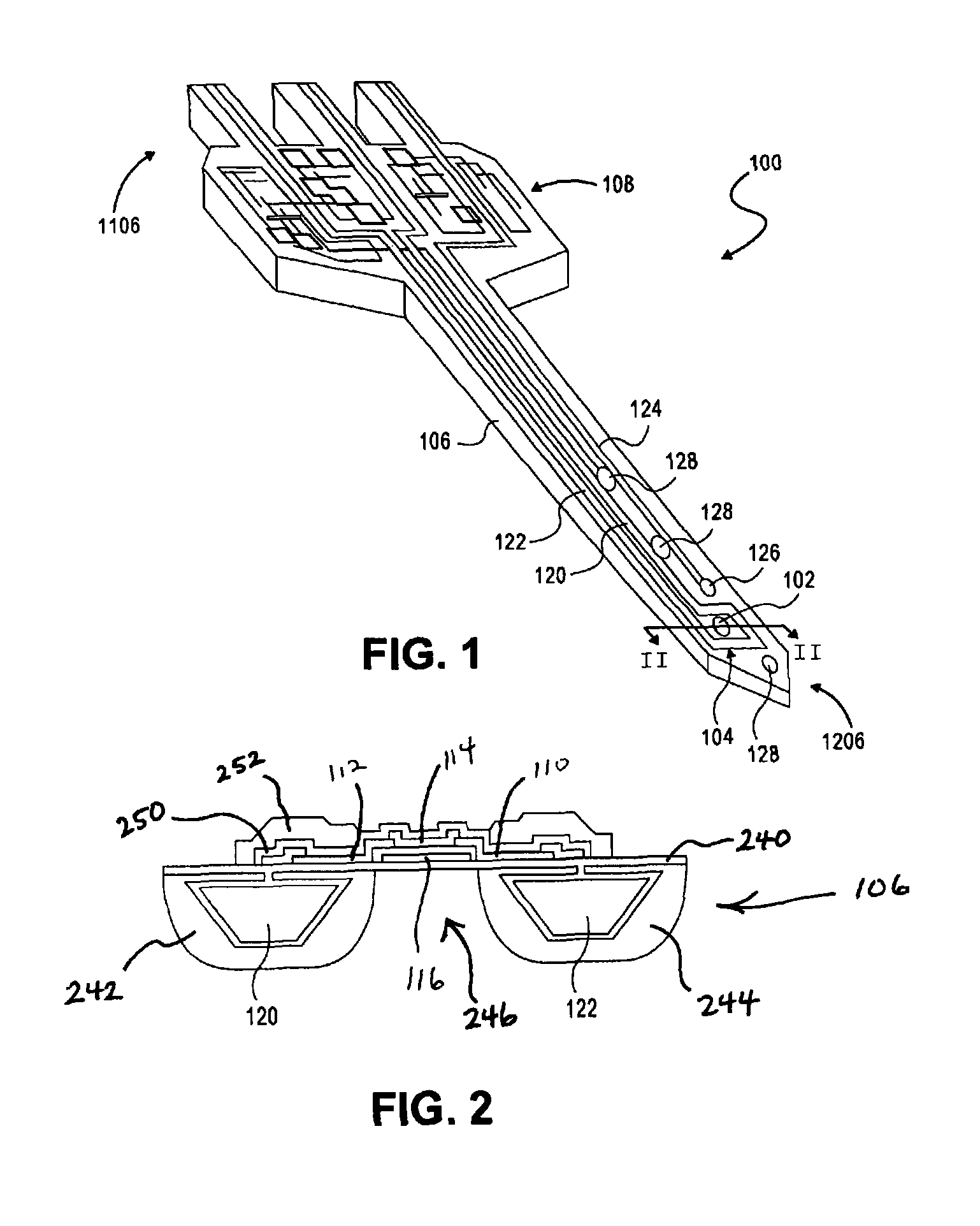
[0037]Referring to FIGS. 1 and 2, an exemplary embodiment of the present invention will be described. FIG. 1 illustrates an exemplary medical device 100 in accordance with various aspects of the invention. The medical device 100 may comprise a thermoelectric assembly 102 and a cooling system 104 associated with a substrate 106, for example, a micro-machined substrate. The substrate 106 may comprise, for example, silicon, silicon germanium, or any other know substrate. The substrate may have a first end 1106 sized and configured for connection to at least one fluid conduit, for example, a pipette, and a second end 1206 sized and configured for insertion into a body tissue, for example, a human body tissue such as, for example, art...


PUM


Abstract

A medical device may include a micro-machined substrate, at least one thermo-electric assembly associated with the substrate, and a cooling system configured to configured to remove heat from the a region of the substrate proximal the substrate. According to various aspects, a method of clearing plaque from a blood vessel may include implanting a micro-device in the blood vessel, wherein the micro-device may include at least one ultrasonic transducer, and operably controlling the micro-device to emit high frequency ultrasonic waves for breaking up said plaque.

Description

CROSS-REFERENCE TO RELATED APPLICATIONS[0001]This application claims the benefit of priority of U.S. provisional application No. 60 / 640,601, filed on Dec. 30, 2004, U.S. provisional patent application No. 60 / 734,385 entitled “Silicon Implantable Transducer Systems for Medical Applications,” filed on Nov. 8, 2005, and U.S. provisional application No. 60 / 739,942, filed on Nov, 28, 2005, the contents of all of which are incorporated herein by reference in their entirety.TECHNICAL FIELD[0002]The present disclosure is directed generally to medical devices and methods. More particularly, the present disclosure is directed to micro-machined medical devices, methods of fabricating micro devices, and methods of medical diagnosis, imaging, stimulation, and treatment.BACKGROUND[0003]Conventional ultrasound devices may be used for medical diagnosis, imaging, and stimulation. For example, ultrasound imaging generally involves transmission of high frequency sound into the body followed by the rec...

Claims


Application Information

Patent Timeline
Patent Type & Authority Patents(United States)
IPC IPC(8): A61B1/12
CPCA61M37/00
Inventor CHEN, JINGKUANGCHENG, XIAOYANG
Owner STC UNM
Learn More