Cytotoxic T-cell epitope peptides that specifically attack epstein-barr virus-infected cells and uses thereof

a cytotoxic, t-cell technology, applied in the field of cytotoxic t-cell epitope peptides, can solve the problem that such nk/t cells cannot process lmp1 and have not been demonstrated

Inactive Publication Date: 2013-07-09
AICHI PREFECTURE +1
View PDF3 Cites 13 Cited by
  • Summary
  • Abstract
  • Description
  • Claims
  • Application Information

AI Technical Summary

Benefits of technology

[0076][14] a method for producing a passive immunotherapeutic agent against Epstein-Barr virus, which comprises the step of obtaining a cytotoxic T cell by reacting a peripheral blood lymphocyte with a major histocompatibility antigen complex and / or major histocompatibility antigen complex-tetramer prepared from the peptide of any one of [1] to [4], allowing the formation of a complex in which said major histocompatibility antigen complex and / or major histocompatibility antigen complex-tetramer are bound with the cytotoxic T cell, and isolating the cytotoxic T cell from said complex;

Problems solved by technology

However, it has not been demonstrated that such NK / T cells can process LMP1 and generate HLA-restricted epitopes.

Method used


Image

  • Cytotoxic T-cell epitope peptides that specifically attack epstein-barr virus-infected cells and uses thereof
  • Cytotoxic T-cell epitope peptides that specifically attack epstein-barr virus-infected cells and uses thereof
  • Cytotoxic T-cell epitope peptides that specifically attack epstein-barr virus-infected cells and uses thereof

Examples


example 1

Preparation of Cell Strains and Dendritic Cells

[0234]The present inventors first induced CTL clones specific to LMP1, an EBV protein, and identified LMP1-derived epitopes.

[0235]The cell strains and dendritic cells used in the experiments for identifying LMP1-derived epitopes (Examples 1 to 8) were prepared by the following methods.

[0236]The study design and purpose, which had been approved by the institutional review board of Aichi Cancer Center (Japan), were fully explained and informed consents were obtained from all the donors.

[0237]EBV-infected LCLs were prepared by known methods (Kuzushima K., et al., Blood, 94:3094-3100 (1999)) and cultured in RPMI1640 medium (Sigma Chemical Co., St. Louis, Mo., USA) supplemented with 10% fetal calf serum (PAA laboratories, Pasching, Austria), 2 mM L-glutamine, 50 U / mL penicillin, 50 μg / mL streptomycin, and 50 μg / mL kanamycin.

[0238]EBV-carrying NK cell lines, SNK-6 (Nagata H., et al., Blood, 97:708-713 (2001)), and SNK-10 (Zhang Y., et al., Br...

example 2

Preparation of Deletion Mutant ΔLMP1 mRNA

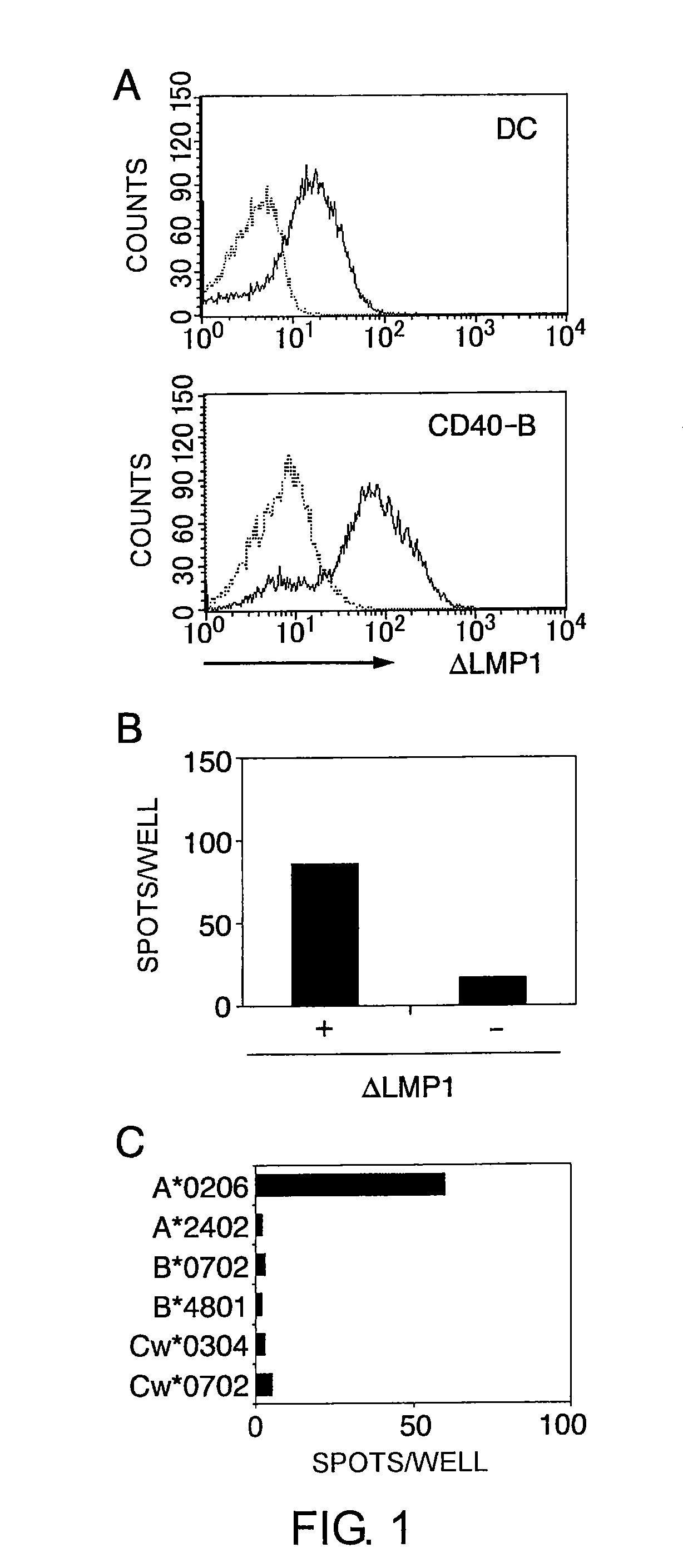
[0244]To generate deletion mutant ΔLMP1-expressing antigen-presenting cells, the present inventors first produced deletion mutant ΔLMP1 mRNA by using an in vitro transcription system from the pcDNA / ΔLMP1 plasmid.

[0245]To construct a 43-amino acid N-terminal deletion mutant, ΔLMP1 (Gottschalk S., Blood, 101: 1905-1912 (2003)), PCR was performed using a sense primer 5′-AAGCTTGCCACCATGAGTGACTGGACTGGA-3′ (SEQ ID NO: 23), an antisense primer 5′-TTGAATTCTTAGTCATAGTAGCTTAGCTGA-3′ (SEQ ID NO: 24), and EBV strain B95-8 (NCBI accession no. V01555) cDNA as a template. The amino acid sequence and the oligonucleotide sequence of the deletion mutant ΔLMP1 are shown as SEQ ID NOs: 34 and 35, respectively. The resultant DNA fragment was cloned into pcDNA 3.1(+) (Invitrogen, Carlsbad, Calif., USA) using its HindIII and EcoRI restriction enzyme recognition sites (pcDNA / ΔLMP1). For constructing further C-terminal and N-terminal deletion mutants of the ΔLMP1, tr...

example 3

Transduction of ΔLMP1 mRNA into Cells and Verification of ΔLMP1 Expression in Cells

[0248]Fragments comprising the T7 promoter region and the ΔLMP1 coding region were prepared by PCR using pcDNA / ΔLMP1 as a template. The amplified DNA was used as a template for in vitro transcription of 5′-capped mRNA. The in vitro transcription of 5′-capped mRNA was performed using a mMESSAGE mMACHINE kit (Ambion, Austin, Tex., USA).

[0249]The 3′ polyA tail was added using polyA polymerase (Ambion) followed by purification with an RNeasy kit (Qiagen, Tokyo Japan).

[0250]Prior to electroporation, the dendritic cells and CD40-B cells were washed twice with serum-free RPMI1640 medium and adjusted to a final concentration of 2.5×10⁷ cells / mL. The cells in 40 μL RPMI 1640 medium were mixed with 20 μg of mRNA, and electroporated in a 2 mm cuvette using an Electro Square Porator ECM 830 (Harvard Apparatus, Holliston, Mass.). The conditions were 450 V and 500 μS for dendritic cells and 350 V and 350 μS for CD4...


PUM

PropertyMeasurementUnit
concentrationaaaaaaaaaa
frequencyaaaaaaaaaa

Abstract

The present inventors introduced mRNAs for the Epstein-Barr virus proteins LMP1 and EBNA1 into antigen-presenting cells, and as a result, demonstrated that the cells induced Epstein-Barr virus-specific cytotoxic T cells. The present inventors also demonstrated that the cytotoxic T cells recognized epitope peptides presented via HLA-A*0206, HLA-Cw*0303, or HLA-Cw*0304, inhibited the outgrowth of Epstein-Barr virus-infected B cells, and lysed Epstein-Barr virus-infected NK lymphomas and NK cells.

Description

CROSS-REFERENCES TO RELATED APPLICATIONS[0001]This application is a U.S. National Phase of PCT / JP2006 / 321479, filed Oct. 27, 2006, which claims priority to Japanese Patent Application No. 2005-315306, filed Oct. 28, 2005, the contents of which are herein incorporated by reference.TECHNICAL FIELD[0002]The present invention relates to Epstein-Barr virus-specific cytotoxic T cell epitope peptides, vaccines using such peptides for treating or preventing Epstein-Barr virus infection or Epstein-Barr virus-positive cancers, and passive immunotherapeutic agents against Epstein-Barr virusBACKGROUND ART[0003]The Epstein-Barr virus (a γ herpes virus that stays dormant (latent) for a long period in memory B cells; hereinafter may be abbreviated as EBV) is involved in many malignancies, for example, Burkitt's lymphoma, Hodgkin's disease (HD) and nasopharyngeal carcinoma (NPC), as well as post-transplant lymphoproliferative disorder (Non-patent Document 1). In latent infections, viral protein exp...

Claims


Application Information

Patent Timeline
Patent Type & Authority Patents(United States)
IPC IPC(8): A61K39/00C07K14/00A61K38/00A61K35/14A61K35/17A61K38/08A61K39/245A61K48/00A61P35/00A61P37/04C07K7/06C07K14/05C12N15/09C12Q1/06
CPCA61K38/00A61K39/00A61P35/00A61P37/04C07K7/06C07K14/005C12N2710/16222G01N33/505
Inventor KUZUSHIMA, KIYOTAKAITO, YOSHINORIOKAMURA, AYAKOAKATSUKA, YOSHIKIMORISHIMA, YASUO
Owner AICHI PREFECTURE
Why Eureka
Try Eureka