System and method for remote melanoma screening

a technology of computer image analysis and remote melanoma, applied in the field of system and method for computer image analysis for screening for skin cancer, can solve the problems of increasing devastating and deadly, aggressive invasion of surrounding tissues, abnormal growth of cells,

Inactive Publication Date: 2013-09-24
HEALTH DISCOVERY CORP
View PDF43 Cites 34 Cited by
  • Summary
  • Abstract
  • Description
  • Claims
  • Application Information

AI Technical Summary

Benefits of technology

[0021]In a preferred embodiment, the support vector machine is trained using the pre-processed training data set. In this manner, the additional representations of the training data provided by the preprocessing may enhance the learning machine's ability to discover knowledge therefrom. In the particular context of support vector machines, the greater the dimensionality of the training set, the higher the quality of the generalizations that may be derived therefrom. When the knowledge to be discovered from the data relates to a regression or density estimation or where the training output comprises a continuous variable, the training output may be post-processed by optimally categorizing the training output to derive categorizations from the continuous variable.
[0022]A test data set is pre-processed in the same manner as was the training data set. Then, the trained learning machine is tested using the pre-processed test data set. A test output of the trained learning machine may be post-processed to determine if the test output is an optimal solution. Post-processing the test output may comprise interpreting the test output into a format that may be compared with the test data set. Alternative postprocessing steps may enhance the human interpretability or suitability for additional processing of the output data.

Problems solved by technology

As melanoma progresses, it becomes increasingly more devastating and deadly.
If not detected at treated at an early stage, these cells become abnormal, grow uncontrollably, and aggressively invade surrounding tissues.
The MelaFind® scanner is designed for use by medical professionals and is not intended for general consumer use, which means that the patient must have already suspected a problem and consulted a physician before such a scanner would be available for use on the patient.
However, the quality of such images tends to be fairly low.
Optimized extraction and reconstruction of data within an image can be problematic where sources of noise and other factors can negatively impact the ability to efficiently extract data from the image, thus impairing the effectiveness of the imaging method for its intended use.
Examples of areas in which image analysis can be problematic include astronomical observation and planetary exploration, where sources can be faint and atmospheric interference introduce noise and distortion, military and security surveillance, where light can be low and rapid movement of targets result in low contrast and blur, and medical imaging, which often suffers from low contrast, blur and distortion due to source and instrument limitations.
Adding to the difficulty of image analysis is the large volume of data contained within a digitized image, since the value of any given data point often cannot be established until the entire image is processed.
A significant drawback of back-propagation neural networks is that the empirical risk function may have many local minimums, a case that can easily obscure the optimal solution from discovery.
Standard optimization procedures employed by back-propagation neural networks may converge to a minimum, but the neural network method cannot guarantee that even a localized minimum is attained, much less the desired global minimum.
In particular, the skill of the practitioner implementing the neural network determines the ultimate benefit, but even factors as seemingly benign as the random selection of initial weights can lead to poor results.
Furthermore, the convergence of the gradient-based method used in neural network learning is inherently slow.
A further drawback is that the sigmoid function has a scaling factor, which affects the quality of approximation.
Possibly the largest limiting factor of neural networks as related to knowledge discovery is the “curse of dimensionality” associated with the disproportionate growth in required computational time and power for each additional feature or dimension in the training data.

Method used


Image

  • System and method for remote melanoma screening
  • System and method for remote melanoma screening
  • System and method for remote melanoma screening

Examples


example 1

Classifier Variation

[0180]Using a set of cropped images obtained from the DERMNET™ skin disease image atlas (available on the World Wide Web at dermnet.com), the recognizer that was used in the ABCD analysis described above with reference to FIG. 14, was re-trained and tested. Malignant samples (Malignant Melanoma and Lentigo Maligna) were separated from all other samples, including some very ambiguous ones that mimic melanoma.

[0181]Five-fold cross-validation experiments were performed using the same four classifiers that were used in the garbage image classification processing described above. The four classifiers were Naïve Bayes, Linear kernel, Polynomial—2nd degree (Poly 2) kernel, and RBF kernel, were used to process the previously-described data representation of ABC features. The best result was obtained using a linear kernel, with AUC=0.78. An ensemble classifier voting was tested on classifiers trained independently on A, B, and C features yielding AUC=0.75. An AUC of 0.80 ...

example 2

Modified Pre-Processing

[0212]The DERMATLAS image dataset from Johns Hopkins University (available on the World Wide Web at dermatlas.org) was used to extract 1000 images of skin diseases with various diagnoses, including herpes, acnea, drug reactions, insect bites, squamous cell carcinoma, basal cell carcinoma, congenital nevi, other benign moles, and melanoma. Associate with the images are annotations both in the form of MS access database and MS Excel spreadsheets. The annotations include age and gender of the patient, data source information, and diagnosis.

[0213]Software was written to crop the images into 1717 cropped pictures (some coming from the same image). The software also allows a framing box to be drawn around the lesion, such as described above with reference to FIG. 17a. The framing box facilitates segmentation of the mole from the background and is in a similar form to the data that would be collected using the smart phone application.

[0214]The original annotations do...


PUM


Abstract

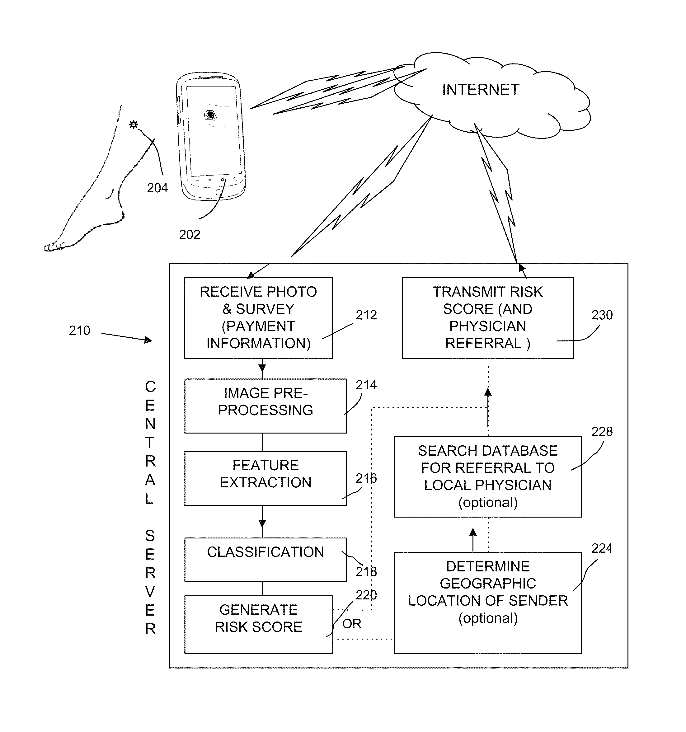
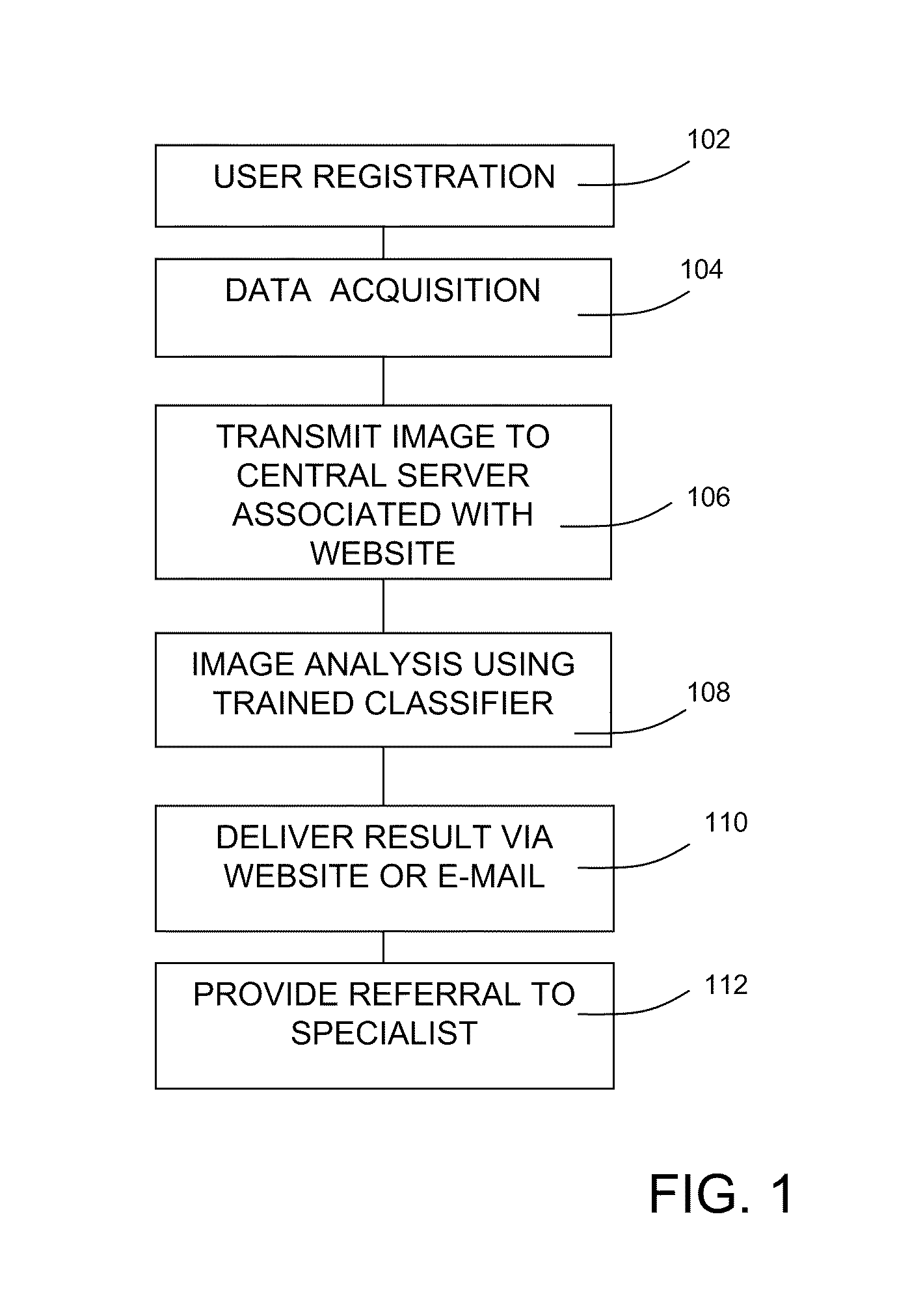
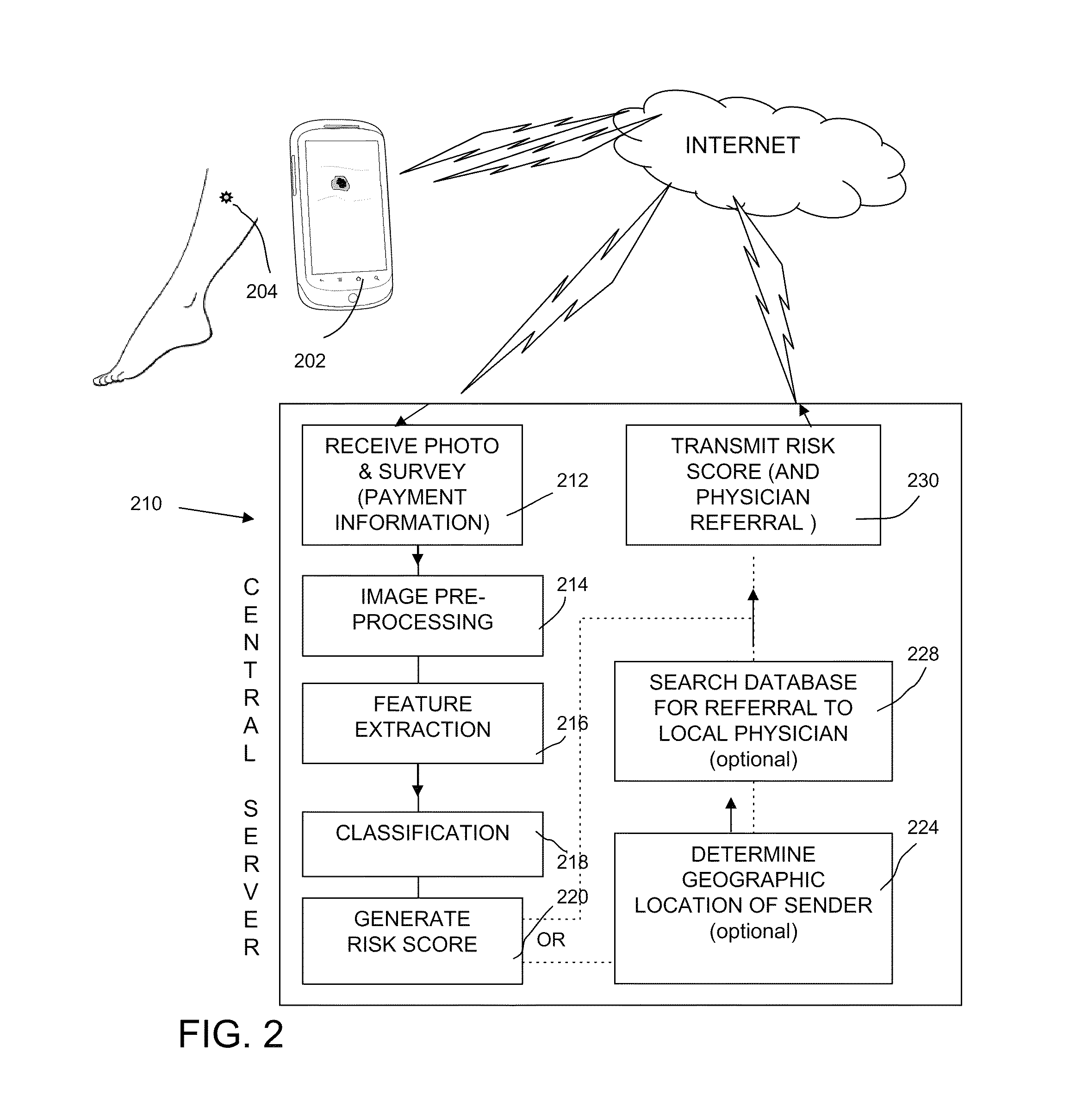
A system and method are provided for diagnosing diseases or conditions from digital images taken by a remote user with a smart phone or a digital camera and transmitted to an image analysis server in communication with a distributed network. The image analysis server includes a trained learning machine for classification of the images. The user-provided image is pre-processed to extract dimensional, shape and color features then is processed using the trained learning machine to classify the image. The classification result is postprocessed to generate a risk score that is transmitted to the remote user. A database associated with the server may include referral information for geographically matching the remote user with a local physician. An optional operation includes collection of financial information to secure payment for analysis services.

Description

RELATED APPLICATIONS[0001]This application claims the priority of each of U.S. Provisional Application No. 61 / 289,372, filed Dec. 22, 2009, U.S. Provisional Application No. 61 / 293,987, filed Jan. 11, 2010, U.S. Provisional Application No. 61 / 304,796, filed Feb. 15, 2010, and U.S. Provisional Application No. 61 / 308,792, filed Feb. 26, 2010, and is a continuation-in-part of U.S. application Ser. No. 12 / 814,431, filed Jun. 11, 2010, which is a continuation of application Ser. No. 11 / 926,129, filed Oct. 29, 2007, now issued as U.S. Pat. No. 7,797,257, which is a continuation of U.S. application Ser. No. 11 / 033,570, filed Jan. 11, 2005, which is a continuation of U.S. application Ser. No. 09 / 633,410, filed Aug. 7, 2000, now issued as U.S. Pat. No. 6,882,990. The disclosure of each of the related applications is incorporated herein by reference.FIELD OF THE INVENTION[0002]The present invention relates to a system and method for computer image analysis for screening for skin cancer using p...

Claims


Application Information

Patent Timeline
Patent Type & Authority Patents(United States)
IPC IPC(8): G06F15/18G06N20/10G06N20/20
CPCG06F19/345G06N99/005G06T7/0012G06T7/602G06T7/606G06T7/608G06F19/321G06F19/3431G06T2207/10024G06T2207/20081G06T2207/30088G06T7/62G06T7/66G06T7/68G16H50/30G16H50/20G06N20/00G16H30/40G16H40/67G06N20/20G06N20/10
Inventor GUYON, ISABELLEBARNHILL, STEPHEN D.
Owner HEALTH DISCOVERY CORP
Why Eureka
Try Eureka