Radioactive emission detector equipped with a position tracking system

a technology of position tracking and radioactive emission, which is applied in the direction of sensors, radiotherapy, diagnostics, etc., can solve the problems of affecting the methods and outcomes of minimally invasive surgical procedures, affecting the safety of patients, and causing tremendous blunt trauma and blood loss

Active Publication Date: 2013-10-22
SPECTRUM DYNAMICS MEDICAL LTD
View PDF784 Cites 78 Cited by
  • Summary
  • Abstract
  • Description
  • Claims
  • Application Information

AI Technical Summary

Benefits of technology

[0033]The present invention successfully addresses the shortcomings of the presently known configurations by providing a radioactive emission probe in communication with a position tracking system and the use thereof in a variety of systems and methods of medical imaging and procedures. Specifically, wide aperture collimation—deconvolution algorithms are provided, for obtaining a high-efficiency, high resolution image of a radioactivity emitting source, by scanning the radioactivity emitt

Problems solved by technology

The use of minimally invasive surgical techniques has dramatically affected the methods and outcomes of surgical procedures.
Physically cutting through tissue and organs to visually expose surgical sites in conventional “open surgical” procedures causes tremendous blunt trauma and blood loss.
Exposure of internal tissues and organs in this manner also dramatically increases the risk of infection.
The result of such open surgical procedures is more pain and suffering, higher procedural costs, and greater risk of adverse outcomes.
However, there still exist a host of technical hurdles that limit the efficacy and increase the difficulty of minimally invasive procedures, some of which were overcome by the development of sophisticated imaging techniques.
The main limitation of the system is that once the body is penetrated, scanning capabilities are limited to a translational movement along the line of penetration.
An effective collimator for gamm

Method used


Image

  • Radioactive emission detector equipped with a position tracking system
  • Radioactive emission detector equipped with a position tracking system
  • Radioactive emission detector equipped with a position tracking system

Examples


Embodiment Construction

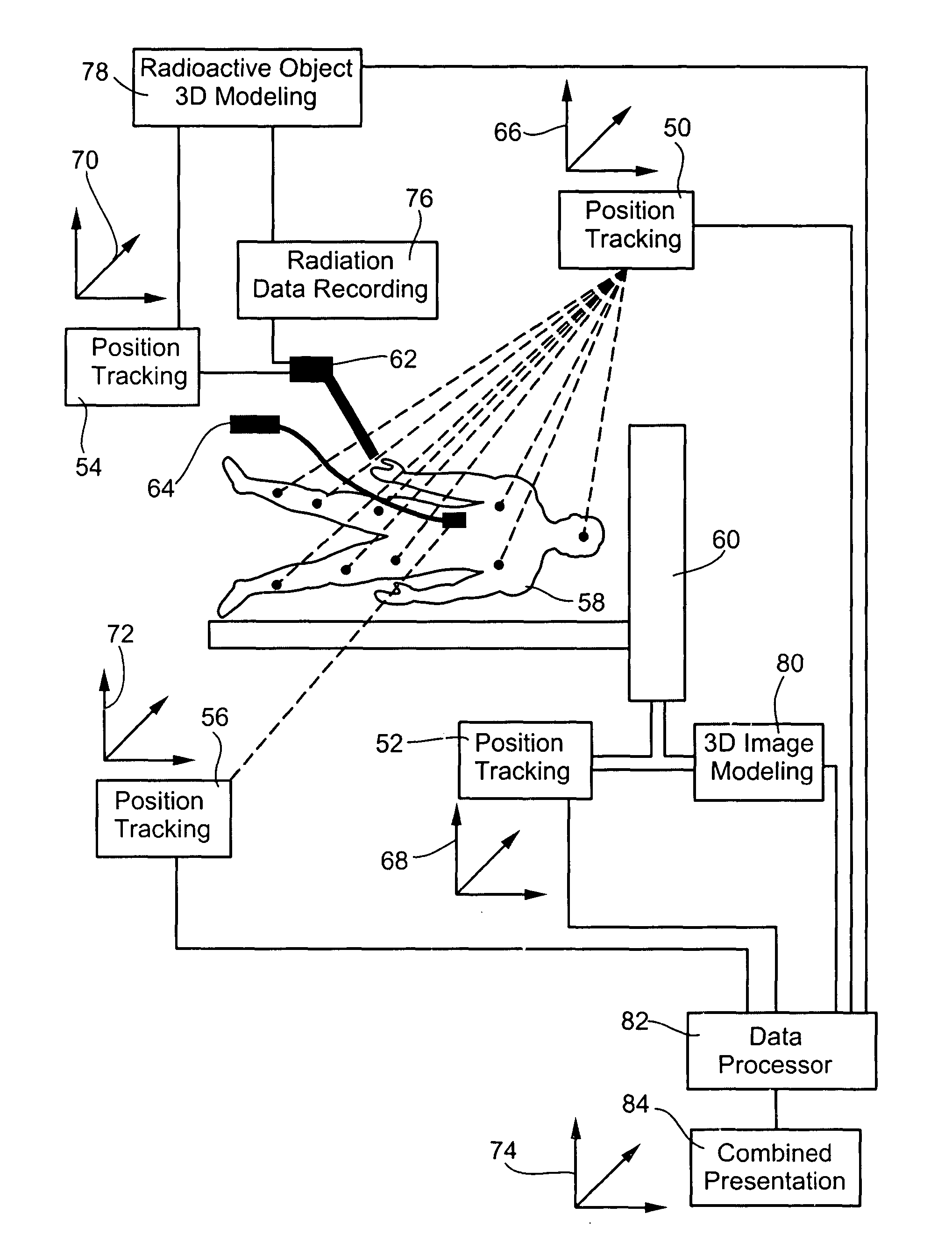
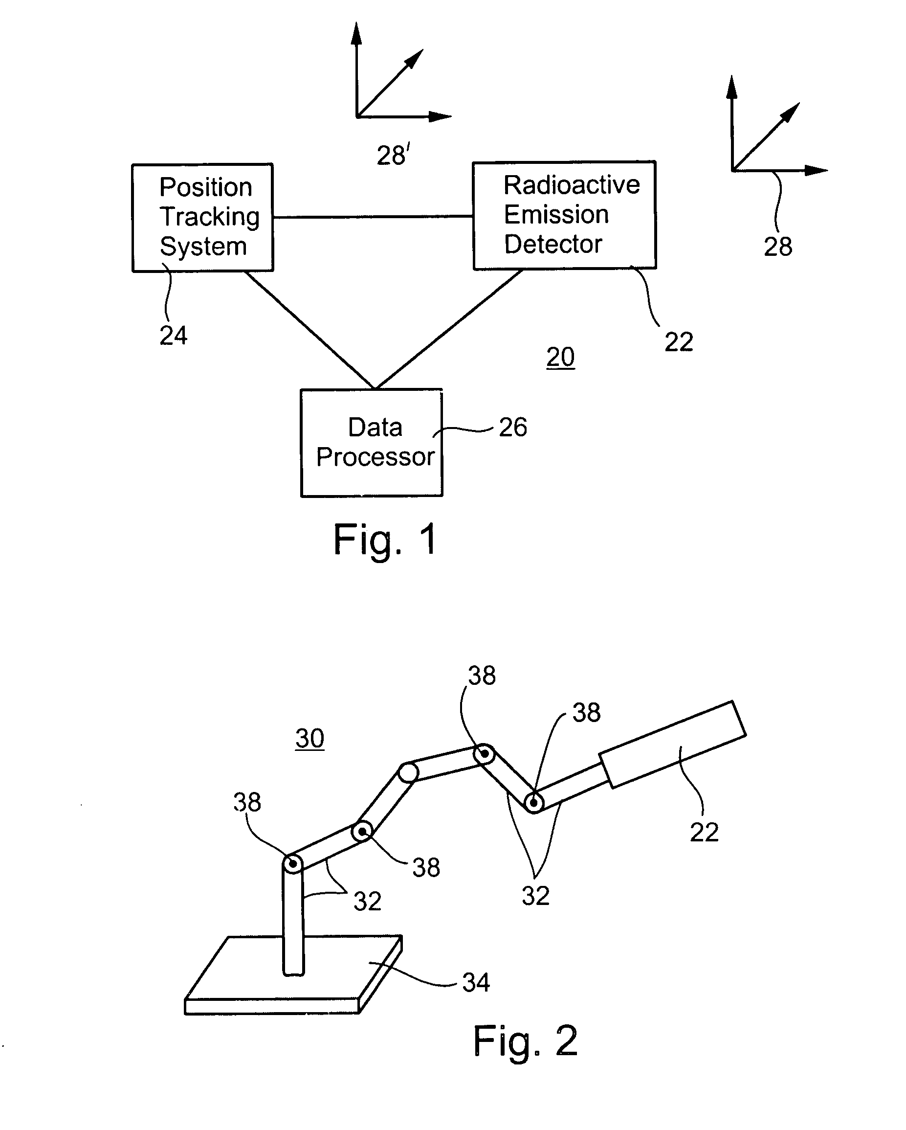
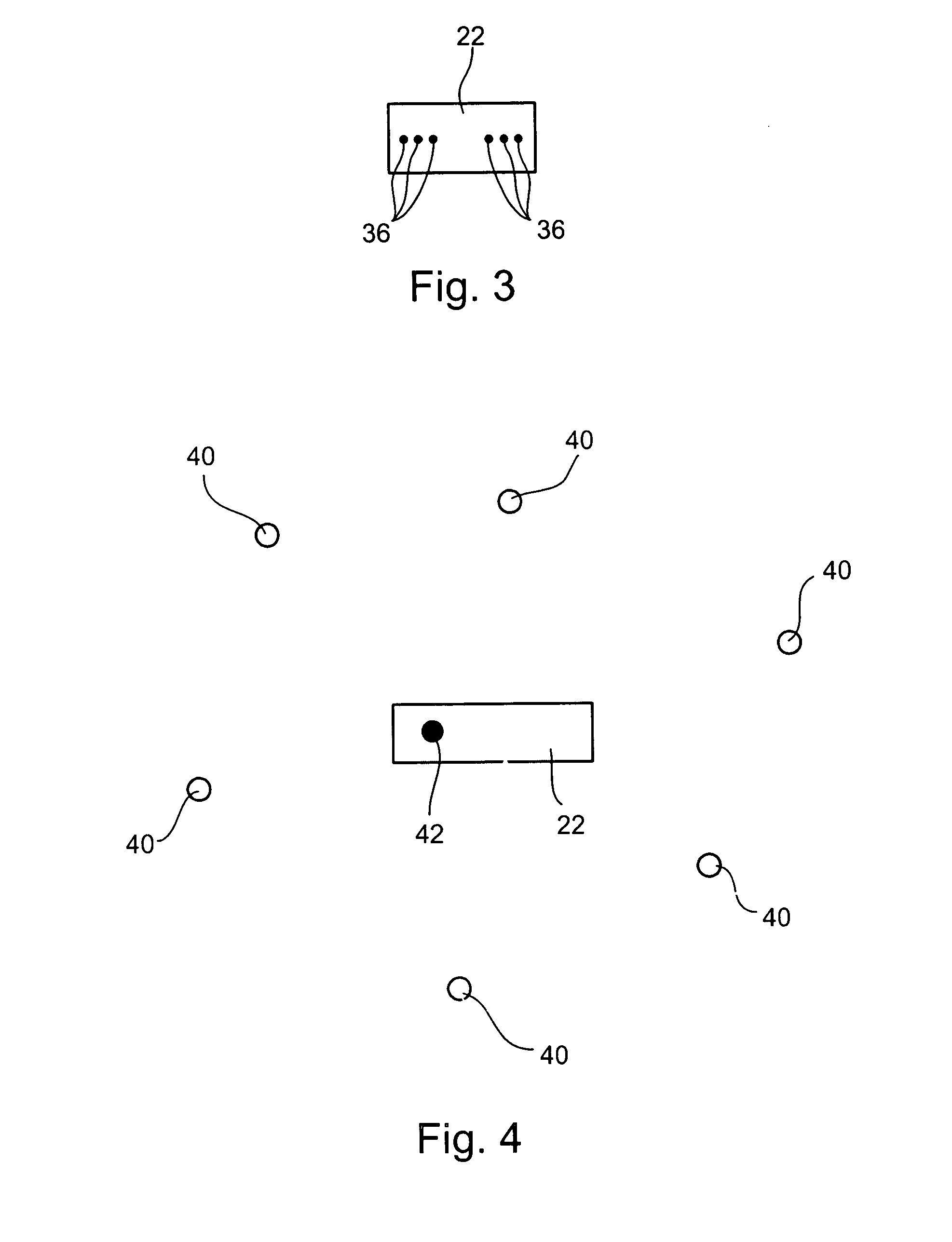
[0073]The present invention relates to a radioactive emission probe in communication with a position tracking system and the use thereof in a variety of systems and methods of medical imaging and procedures. Specifically, wide-aperture collimation—deconvolution algorithms are provided, for obtaining a high-efficiency, high resolution image of a radioactivity emitting source, by scanning the radioactivity emitting source with a probe of a wide-aperture collimator, and at the same time, monitoring the position of the radioactive emission probe, at very fine time intervals, to obtain the equivalence of fine-aperture collimation. The blurring effect of the wide aperture is then corrected mathematically. Furthermore, an imaging method by depth calculations is provided, based on the attenuation of photons of different energies, which are emitted from the same source, coupled with position monitoring.

[0074]The principles and operation of the present invention may be better understood with ...


PUM


Abstract

A radioactive emission probe in communication with a position tracking system and the use thereof in a variety of systems and methods of medical imaging and procedures, are provided. Specifically, wide aperture collimation—deconvolution algorithms are provided, for obtaining a high-efficiency, high resolution image of a radioactivity emitting source, by scanning the radioactivity emitting source with a probe of a wide-aperture collimator, and at the same time, monitoring the position of the radioactive emission probe, at very fine time intervals, to obtain the equivalence of fine-aperture collimation. The blurring effect of the wide aperture is then corrected mathematically. Furthermore, an imaging method by depth calculations is provided, based on the attenuation of photons of different energies, which are emitted from the same source, coupled with position monitoring.

Description

[0001]This is a continuation-in-part of U.S. patent application Ser. No. 10 / 343,792, filed on Jul. 11, 2001, which is a National Phase application of PCT / IL01 / 00638, filed on Jul. 11, 2001, which is a continuation-in-part of U.S. patent application Ser. No. 09 / 727,464 filed Dec. 4, 2000, which is a continuation-in-part of U.S. patent application Ser. No. 09 / 714,164, filed Nov. 17, 2000, which is a continuation-in-part of U.S. patent application Ser. No. 09 / 641,973, filed Aug. 21, 2000. Additionally, this application is filed concurrently with co-pending application “INGESTIBLE PILL,” whose disclosure is incorporated herein by reference, and both applications derive priority from U.S. Provisional application No. 60 / 394,936, filed on Jul. 11, 2002.FIELD AND BACKGROUND OF THE INVENTION[0002]The present invention relates to a radioactive emission probe equipped with a position tracking system. More particularly, the present invention relates to the functional integration of a radioactiv...

Claims


Application Information

Patent Timeline
IPC IPC(8): A61B6/00A61N5/00
CPCA61B5/055A61B6/4057A61B5/064A61B5/07A61B5/415A61B5/418A61B5/6835A61B6/482G01T1/161A61B6/4258A61B6/037A61B5/06A61B6/12A61B8/0833A61B2019/542A61B6/4417A61B6/4423A61B6/583A61B8/4245A61B8/4254A61B2090/392
Inventor KIMCHY, YOAVAMRAMI, RONIBOUSKILA, YONAANTEBI, UDISIDORENKO, NICKBEN-DAVID, GALZILBERSTEIN, YOEL
Owner SPECTRUM DYNAMICS MEDICAL LTD
Why Eureka
Try Eureka