Methods of isolating genes encoding proteins of specific function and of screening for pharmaceutically active agents

Inactive Publication Date: 2006-10-26
YEDA RES & DEV CO LTD
View PDF0 Cites 2 Cited by
  • Summary
  • Abstract
  • Description
  • Claims
  • Application Information

AI Technical Summary

Benefits of technology

[0062] The present invention successfully addresses the shortcomings of the presently known configurations by providing novel screening methods, which enable on the one hand, to elucidate new cellular targets, based on their subcellular localization, and on the other hand to discover new therapeutic leads. The methods of the present invention are readily applicable for high-throughput screening assays based on robotic preparations of samples in multi-wells and their automated imaging and real-time analysis by computerized microscopy. Combination of these methods and adaptation of present capacities in microscope technologies to fast screening of microsamples are estimated to test in exceess of 100,000 samples per day. This allows to obtain fast feedback from screens of gene and drug libraries.

Problems solved by technology

Although the two hybrid method has evolved considerably since first presented (4), it is still mostly limited to proteins that can be localized to the nucleus, thus preventing efficient use with certain extracellular proteins.
In addition, the two hybrid system suffers from several other inherent limitations; first, proteins must be able to fold and exist stably in yeast cells and to retain activity as fusion proteins; second, interactions dependent on post-translational modification that do not occur in yeast, or occur inefficiently, will not be detected; third, many proteins, including those not normally involved in transcription, will activate transcription when fused to a DNA-binding domain and fourth, interactions involving a third non-peptidic factor might not be detected.
Although a promising approach, the mass spectroscopy method is limited by high costs of operation and equipment and a need for highly skilled technicians.
More significantly, this method is also limited by the need for isolated protein complexes, which are oftentimes difficult or nearly impossible to obtain.
The identification of proteins that are localized in a given compartment typically requires a lengthy procedure of cell-disruption and ultracentrifugation.
These procedures disrupt the membranes of the cell but if carefully applied, leave organelles such as nuclei, mitochondria, lysosomes, and peroxisomes intact.
Fractionation approaches suffer from several inherent limitations.
First, such approaches depend on the yield and enrichment achievable.
Second, purified fractions can be devoid of functionally relevant components lost during purification, while being contaminated with various cell components not normally associated with the fraction.
Third, such methodology can only be applied to compartments, which are amenable to cell fractionation, making components exhibiting transient or restricted localization difficult to isolate.
Finally, the relative amount and the biochemical characteristics of protein components can impose an additional burden.
However radioactive-based screening methods are limited not only by the cost of reagents and as such the cost per assay, but also by the inherent limitations associated with miniaturization of radioactive assays.
However, although fluorescence labeling is inherently sensitive, it does not provide adequate performance for large-scale screens since it is susceptible to background effects, both from the biological milieu and from photophysical effects such as light scattering.
However LnTRF labeling suffers from several major drawbacks.
Thus, the sensitivity of FRET to distances on the molecular scale sets a major limitation on its feasibility.

Method used


Image

  • Methods of isolating genes encoding proteins of specific function and of screening for pharmaceutically active agents
  • Methods of isolating genes encoding proteins of specific function and of screening for pharmaceutically active agents
  • Methods of isolating genes encoding proteins of specific function and of screening for pharmaceutically active agents

Examples


example 1

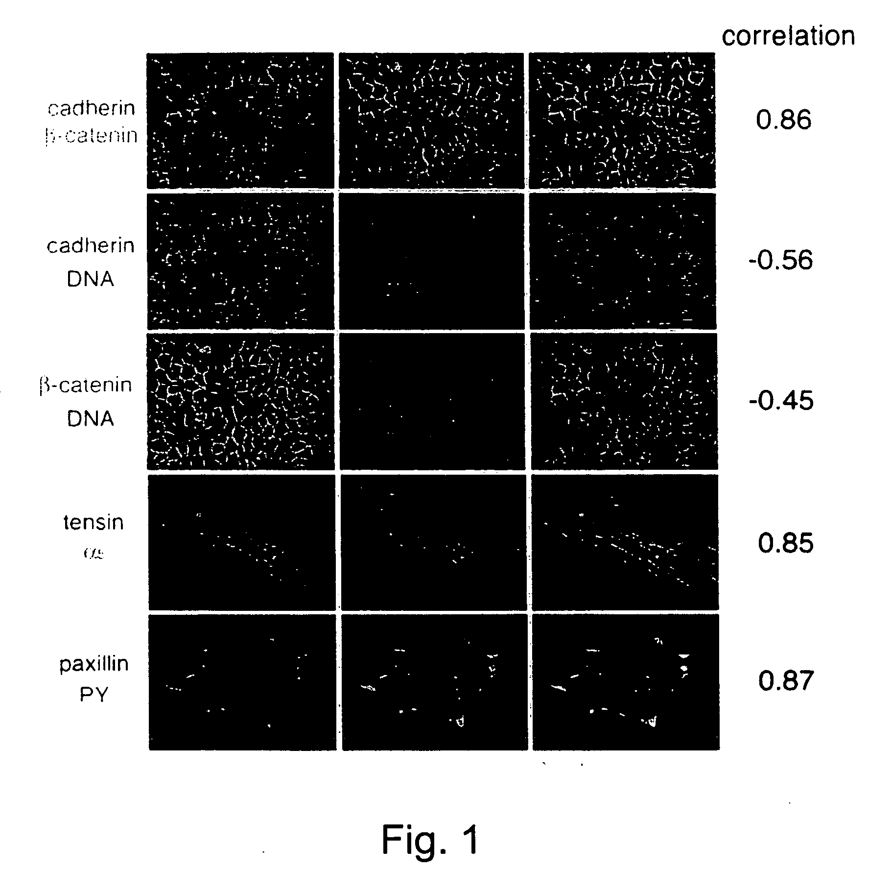
Co-Localization Analysis of Pairs of Proteins in Cells

[0157] To determine the spatial relationships between different proteins in cells, images of the two labels were acquired and subjected to correlation analysis (according to Eq. 1, where N and M are the images of the two labels).

[0158]FIG. 1 illustrates 10 examples for such comparison. It is shown that for largely overlapping structures (i.e., cadherin / catenin; tensin / a5; paxillin / PY) high values (>˜0.7-0.8) were obtained. No correlation (i.e., cadherin or catenin / DAPI) were close to zero. Partial overlaps gave intermediate values.

example 2

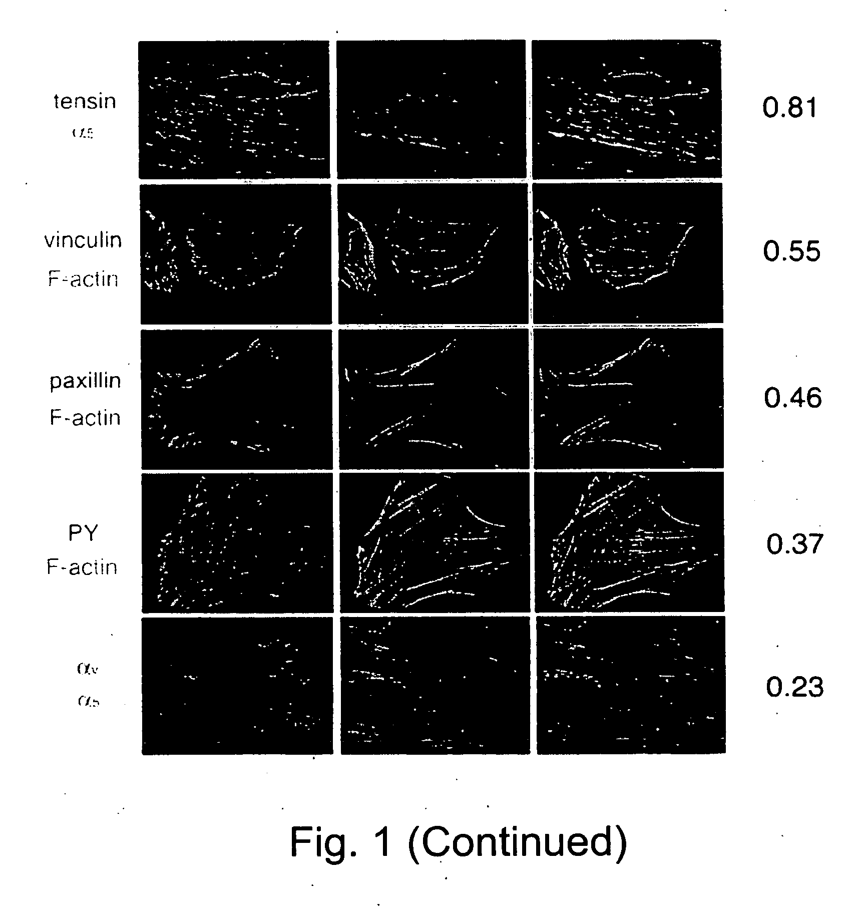
Effect of Actomyosin Inhibitors on Tensin Motility in Live Cells

[0159] Inhibitors of acto-myosin contractility, like latrunculin-A (Lat-A), H-7 and ML-7, block the translocation of fibrilar adhesion (Zamir et al., Nature Cell Biology, Vol. 2, 191-196, 2000). In order to follow the kinetics of this effect a correlation analysis according to Eq. 1 was applied on digital movies of GFP-tensin transfected cells before, during and after treatments with inhibitors. Before treatment, fibrilar adhesions were highly dynamic and thus the correlation between two sequential frames was low (80 minutes).

example 3

Identifying Distortions Between Images of the Same Field

[0160] To examine the ability of correlation analysis to characterize distortions between images a rotated, translated or shrunken variants from one original image were created, and two composites were made: one from 4 original images and a second from the original, translated, contracted and rotated variants (FIG. 3). The composite images were divides to 784 square frames and for each frame the translation vector that maximizes the correlation between the two images (Eq. 4, [amax, bmax]) was found. The results found for each frame are displayed by a line with size and direction equal to the translation vector and a color proportional to the maximal correlation found (Eq. 5, Cmax). The results indicate that the translation vectors indeed tracked successfully the local translation of structures, and, as a whole, indicated the type of global distortion performed (contraction, rotation, translation and not-distorted) in the four ...


PUM

PropertyMeasurementUnit
Fluorescenceaaaaaaaaaa

Abstract

A method of isolating polynucleotides encoding polypeptides affecting an organization of a subcellular organelle or structure of interest is provided. The method comprises: (a) expressing within a plurality of cells an expression library including a plurality of expression constructs each encoding a polypeptide of interest; (b) highlighting the subcellular organelle or structure of interest of the plurality of cells; and (c) isolating a cell or cells of the plurality of cells in which a cellular distribution and / or level of the subcellular organelle or structure of interest is altered to thereby isolate polypeptides capable of affecting the organization of the subcellular organelle or structure of interest.

Description

RELATED APPLICATIONS [0001] This Application is a Divisional of U.S. patent application Ser. No. 10 / 333,680, filed on Jan. 23, 2003, which is a National Phase of PCT Patent Application No. PCT / IL01 / 00813, filed on Aug. 29, 2001, which claims the benefit of U.S. Provisional Patent Application No. 60 / 306,457, filed on Jul. 20, 2001, and U.S. Provisional Patent Application No. 60 / 228,420, filed on Aug. 29, 2000. The contents of the above applications are all incorporated by reference.FIELD AND BACKGROUND OF THE INVENTION [0002] The present invention relates to methods of isolating genes encoding proteins of specific function, such as, proteins which are localized to specific subcellular organelles or structures, proteins involved in the formation, organization and / or maintenance of specific subcellular organelles or structures and proteins which bind other proteins. The present invention further relates to a method of screening for pharmaceutically active agents, capable of at least pa...

Claims


Application Information

Patent Timeline
IPC IPC(8): C12Q1/00C12N5/10C12N15/09C12N15/10C12Q1/02G01N33/15G01N33/50
CPCC12N15/1051G01N33/5076C12N15/1086
Inventor GEIGER, BENJAMINKAM, ZVIZAMIR, ELIBERSHADSKY, ALEXANDERSHTUTMAN, MICHAELBEN-ZE'EV, AVRI
Owner YEDA RES & DEV CO LTD
Why Eureka
Try Eureka