Selection of snapshots of a medical image sequence

a medical image and sequence technology, applied in image enhancement, instruments, ultrasonic/sonic/infrasonic diagnostics, etc., can solve the problem of high operator dependence on ultrasound image acquisition

Active Publication Date: 2013-12-03
KONINKLIJKE PHILIPS ELECTRONICS NV
View PDF15 Cites 55 Cited by
  • Summary
  • Abstract
  • Description
  • Claims
  • Application Information

AI Technical Summary

Benefits of technology

[0009]The image selector, which selects an image in which the object has been detected, helps to achieve a more operator independent selection of acquired images. In existing hand-held medical image acquisition systems, the operator manually stores snapshots of images in which the object of clinical relevance is displayed. However, this task may be automated using the object detector and image selector. Since this may decrease the operator dependence, it may increase the reproducibility of image acquisition. Also, because the operator does not have to spend time and attention on the task of selection and storage of snapshots, the image acquisition may be more efficient and / or easier for the operator. Also, in an embodiment the images that were not selected as snapshots are discarded.
[0011]In an embodiment, the image selector is arranged for selecting the image while the temporal sequence is being acquired with the hand-held medical imaging device. The image selection may be performed ‘on-line’, during the acquisition of the images. This makes the acquisition especially efficient, since only the selected images need to be stored. Also, the operator does not need to manually select image snapshots.
[0015]An embodiment comprises means for establishing a size of the object in an image, and wherein the image selector comprises means for selecting an image having a size above a predetermined threshold and / or having a maximum size among a plurality of sizes. The size may be measured as the longest axis, the shortest axis, the total area, or in another way. By selecting the image showing the object in a large size, it may be enforced that the cross sectional view of the object intersects the object in the middle. Alternatively, it helps to find an image showing the object in a clinically relevant way.
[0017]In an embodiment, the hand-held medical imaging device comprises an ultrasound probe, the sequence of medical images comprising a sequence of 2D ultrasound images obtained from the ultrasound probe. Ultrasound is a modality typically applied in a hand-held device, and the image selection system decreases operator dependence of ultrasound snapshots. The object may, for example, comprise an axillary lymph node.
[0018]An embodiment comprises an annotator for providing annotation of the selected image, the annotation indicating or describing the object detected in the selected image by the object detector, the snapshot means comprising means for storing the annotation of the selected image. This is a useful feature for enabling a radiologist to quickly see the object which provided the reason why the image was stored. It avoids having to do the detection again at a later stage.
[0019]The annotator may comprise means for including in the annotation at least one measurement value relating to the object. This improves the usefulness of the annotation and avoids having to perform the calculations again. Examples of measurements include size and aspect ratio of the lymph node or tumor.

Problems solved by technology

Both for the purpose of computer-aided detection and manual evaluation, the high level of operator dependence on the image acquisition of ultrasound images poses a problem.

Method used


Image

  • Selection of snapshots of a medical image sequence
  • Selection of snapshots of a medical image sequence
  • Selection of snapshots of a medical image sequence

Examples


Embodiment Construction

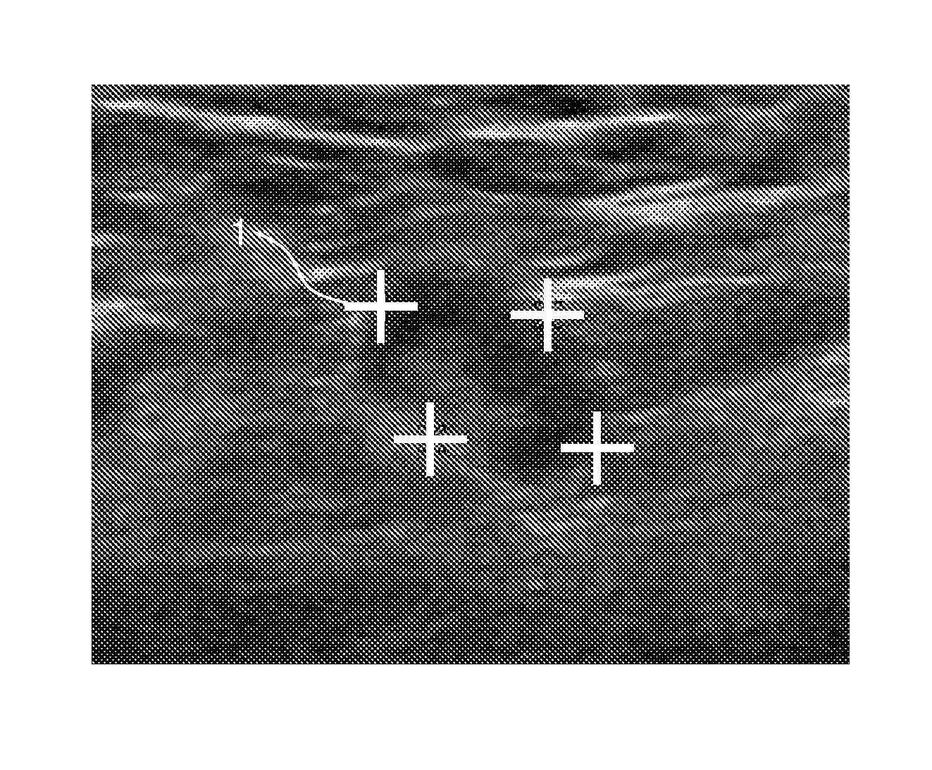
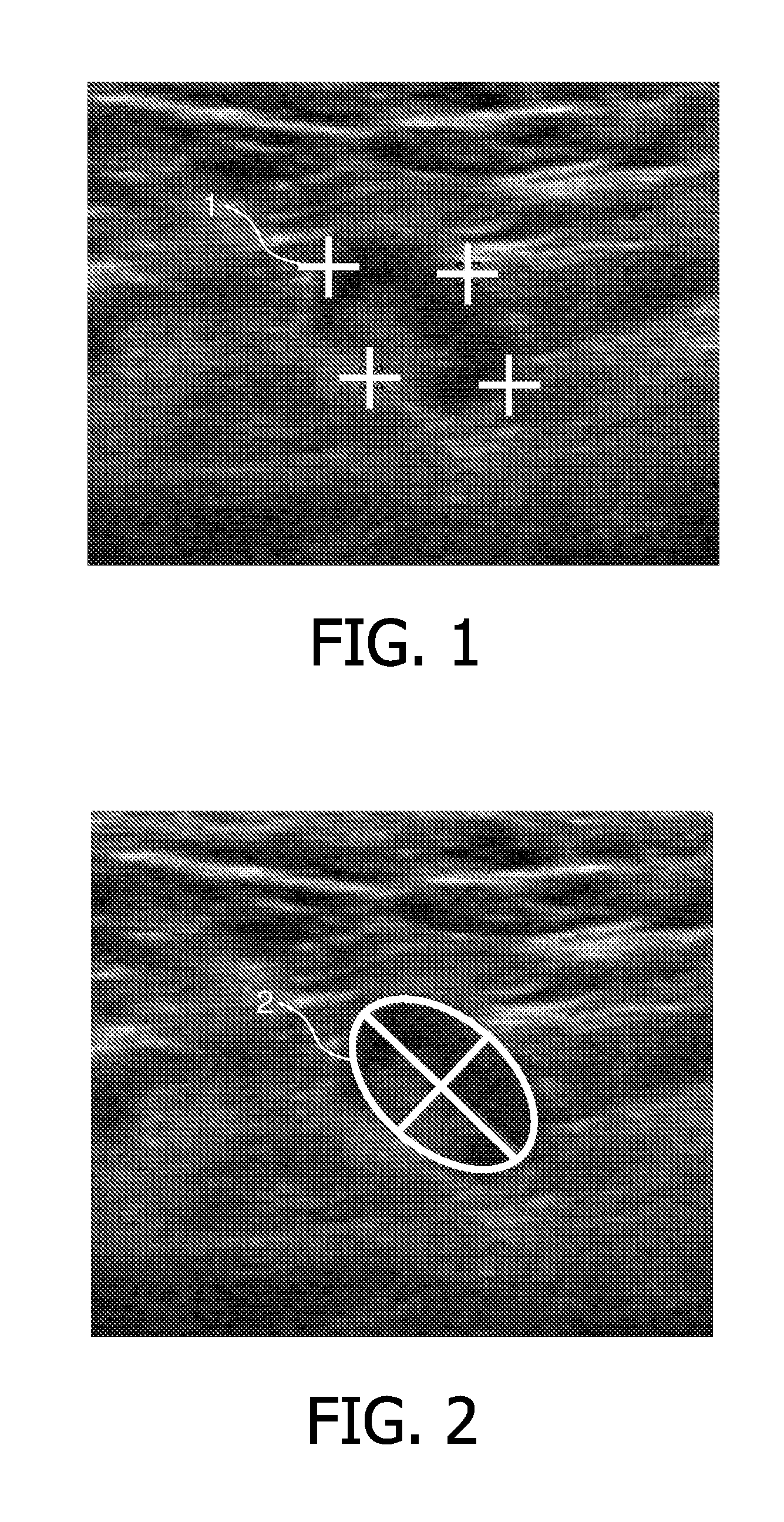
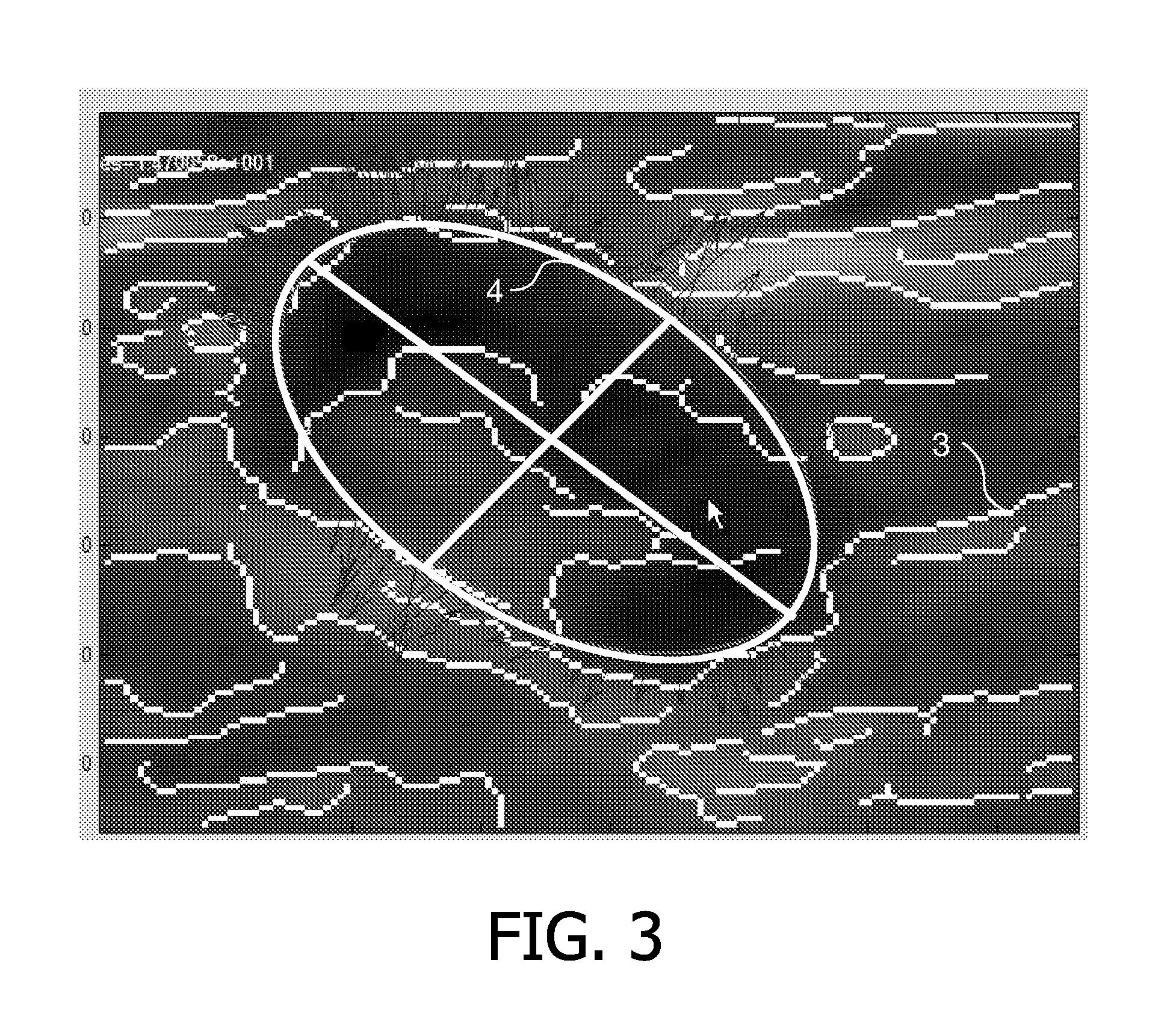
[0036]An automatic segmentation and tracking algorithm may be employed in order to segment and track lymph nodes and / or breast cancer lesions in medical images. Based on the quality of the segmentation and additional quality criteria, acquired images may be automatically selected for storing. Moreover, the selected images may automatically be labeled using the available segmentation information. An automatic segmentation and tracking algorithm may be used for segmenting the lymph node initially in a 2D ultrasound image and for tracking the lymph node in the image, while the operator moves the hand held ultrasound probe. Using image quality measures, which are based on the current segmentation result, 2D snap-shots may be selected automatically for storage. The selected snap-shots may be annotated to represent the segmentation result and stored automatically by the system. This does not only reduce the workload of the operator, as there is no need for manual annotations, but at the s...


PUM


Abstract

A system is provided for automatic selection of medical images. The system comprises an input (302) for receiving a temporal sequence of medical images acquired with a hand-held medical imaging device. An object detector (303) is provided for detecting an object in at least one of the images. An image selector (304) is provided for selecting an image in which the object has been detected by the object detector. A snapshot means (305) is provided for storing the selected image as a snapshot. Moreover, a hand-held medical imaging device is provided for generating the sequence of medical images.

Description

FIELD OF THE INVENTION[0001]The invention relates to selection of a medical image. More particularly, the invention relates to a system, method, and computer program product for automatically selecting an image for storing the image as a snapshot.BACKGROUND OF THE INVENTION[0002]In ultrasound scans of lymph nodes or breast cancer lesions, it is common practice that the operator moves a handheld ultrasound probe and manually selects images from the resulting video stream for storing. Furthermore, the operator can optionally place graphical annotations or markers on the image in order to identify the position and extent of the structure under investigation.[0003]Staging of axillary lymph nodes is an important step in the assessment of the spread of the disease in newly diagnosed breast cancer patients. An image-based way of pre-surgical lymph node staging consists of using ultrasound images, potentially in combination with other imaging modalities, such as MR.[0004]The ultrasound imag...

Claims


Application Information

Patent Timeline
Patent Type & Authority Patents(United States)
IPC IPC(8): G06K9/00
CPCG06T7/0012G06F19/321G06T2207/10016G06T2207/10132G06T2207/30004A61B8/5223G16H30/20
Inventor BUELOW, THOMASBERGTHOLDT, MARTINMEINEL, LINA ARBASH
Owner KONINKLIJKE PHILIPS ELECTRONICS NV
Why Eureka
Try Eureka