Nanoparticles for imaging and treating chlamydial infection
a technology of chlamydia and nanoparticles, applied in the field of nanoparticles and targeting moieties for imaging and treating chlamydia, can solve the problem of ineffective antimicrobial drugs that exis
- Summary
- Abstract
- Description
- Claims
- Application Information
AI Technical Summary
Benefits of technology
Problems solved by technology
Method used
Image

Examples
example 1
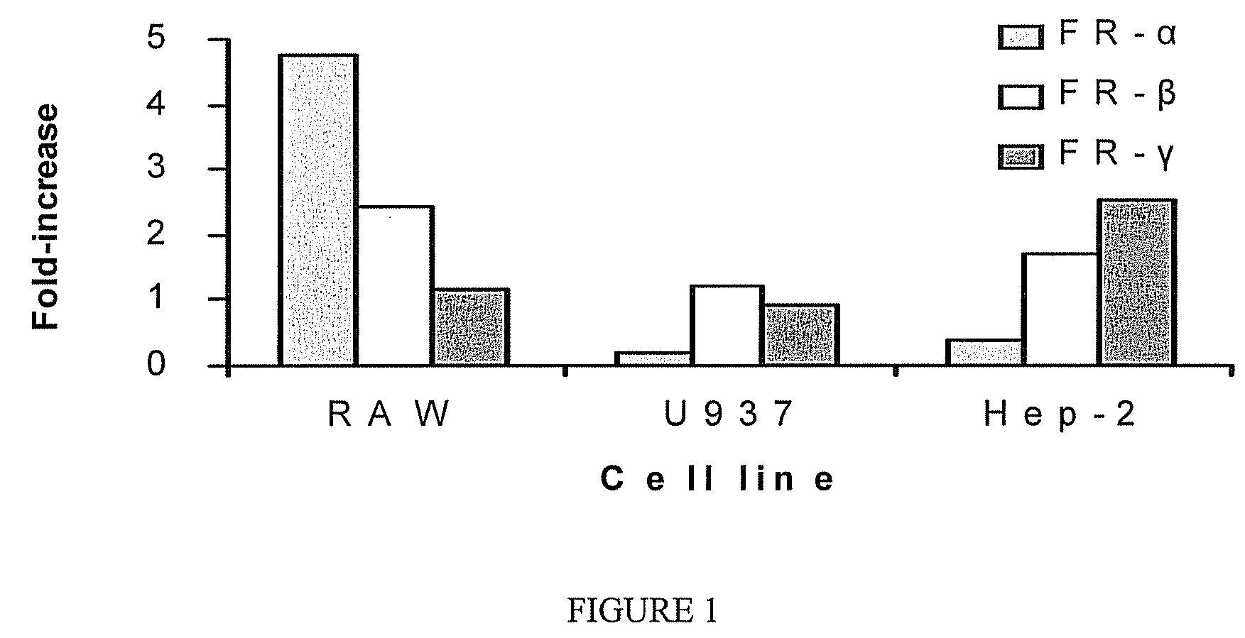
Chlamydia-Infected Cells Overexpress Folic Acid Receptors
[0089]Mammalian cells encode multiple folic acid receptors, designated α, β, and γ (Ross, J. F. et al. (1992) Cancer 73:2432-2443). The present Example was performed to examine the expression of those receptors in Chlamydia infected cells. Nearly confluent monolayers of cycloheximide-treated cells were infected in vitro at MOI 5:1 with K serovar C. trachomatis. The cell lines employed in these experiments were RAW 264.7 and U937 (murine and human macrophage lines, respectively), and HEp-2 (human epithelial cell line).
[0090]At 24 hr post-infection, infected and uninfected control cultures were harvested, and RNA / cDNA was prepared for real time RT-PCR analysis to determine relative levels of mRNA encoding each of the three receptor subtypes in infected versus uninfected cells. As shown in FIG. 1, C. trachomatis infection resulted in increased expression of the β, and γ receptors in human cells. The over-expressed isoform was dep...
example 2
Preparation of Nanoparticles with Folic Acid on the Surface
[0091]This example uses techniques developed by applicants to anchor PEG and PEG-folate conjugate on the surface of nanoparticles, and described in Provisional Application Ser. No. 60 / 871,404, filed Dec. 12, 2006, incorporated herein by reference. The technique relies on the interfacial activity of PEG-X block copolymer conjugate, where X is any hydrophobic polymer (example, polylactide, polypropylene oxide, etc). Most nanoparticle formulations involve an emulsion step in the preparation. Following the formation of the emulsion, a solution of PEG-containing block copolymer (for example PLA-PEG (1000 / 5000 Da), with or without conjugated ligand (folic acid, for example) in an organic solvent (methanol, chloroform, etc), is added to the emulsion. PLA-PEG is a surface active block copolymer, composed of hydrophobic PLA chains and hydrophilic PEG chains.
[0092]Addition of the block copolymer to the emulsion results in the hydropho...
example 3
Folic Acid-Derivatized Nanoparticles Accumulate More in Infected Cells
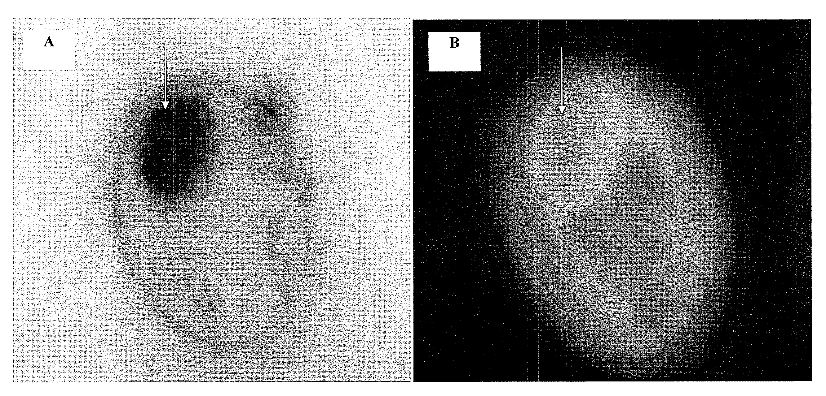
[0096]This example was performed to study the targeting of fluorescently-labeled nanoparticles to chlamydial inclusions within HEp-2 host cells infected with C. trachomatis serovar K. Nearly confluent monolayers of cycloheximide-treated cells were infected at MOI 5:1, and at 24 hr post-infection, infected cells were pulsed with folic acid-conjugated poly(D,L-lactide-co-glycolide) (PLGA) nanoparticles labeled with 6-coumarin. Quantitative studies indicated that infected cells accumulate significantly more folic acid-conjugated nanoparticles than uninfected cells (FIG. 2), and correlates well with increased expression of folic acid receptors in infected cells (FIG. 1).
[0097]This discovery was then used to deliver plasmid DNA into Chlamydial inclusions (FIG. 4). HEp-2 cells were infected with Chlamydia (CT) and then pulsed with nanoparticles containing plasmid DNA (FIG. 4). Delivery to inclusions was imaged using sep...
PUM
| Property | Measurement | Unit |
|---|---|---|
| concentration | aaaaa | aaaaa |
| concentration | aaaaa | aaaaa |
| concentration | aaaaa | aaaaa |
Abstract
Description
Claims
Application Information