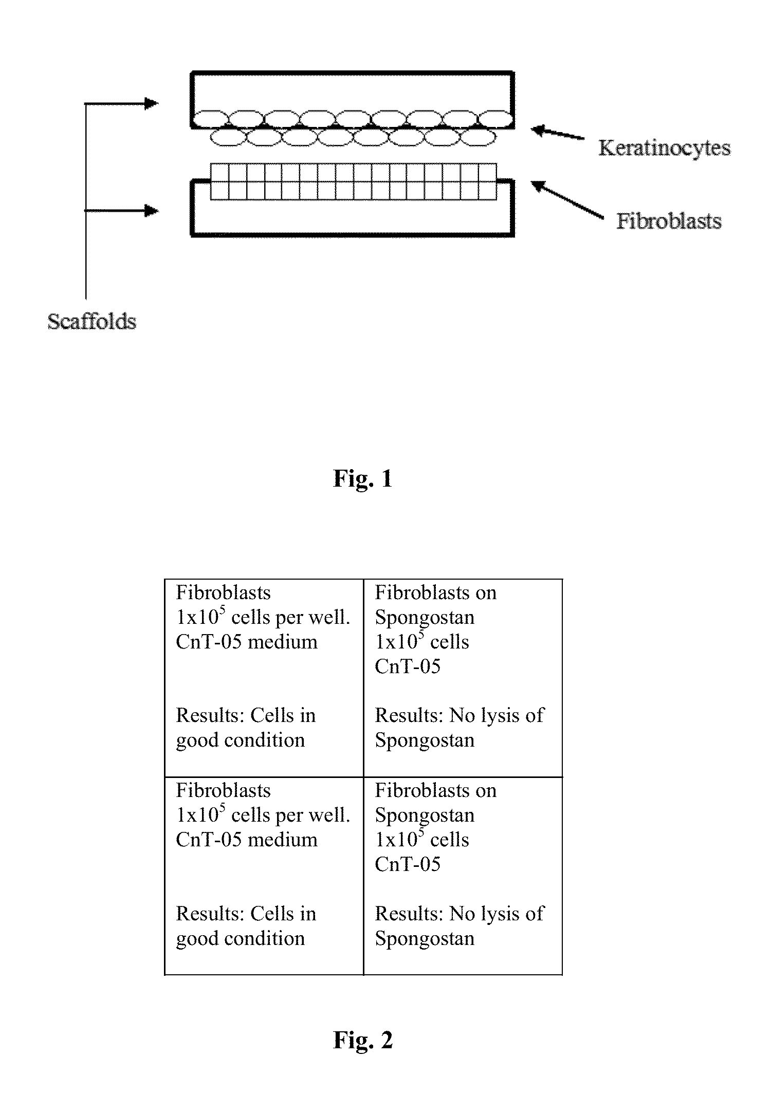
Cellular scaffold

a cell and tissue technology, applied in the field of tissue engineering, can solve the problems of fibroblast culture, clear undesirable effects, and a challenge to the production of therapeutic tissue models

Active Publication Date: 2014-04-29
STEMEDICA CELL TECH
View PDF32 Cites 1 Cited by
  • Summary
  • Abstract
  • Description
  • Claims
  • Application Information

AI Technical Summary

Problems solved by technology

The use of fibroblasts also presents a challenge to the production of therapeutic tissue models.
Although fibroblasts provide growth factors and other cell-to-cell contacts that facilitate cell division, their proliferation may outpace epidermal cell division resulting in a culture that is overgrown with fibroblasts.
This is clearly undesirable as therapies aimed at the regeneration of epidermal tissues must be carried out using carriers rich in epidermal cells.
However this technique requires the presence of dermal components which is undesirable in therapeutic applications.
Thus, seeding collagen scaffolds with precursor keratinocytes results in scaffold degredation unless fibroblasts are present.

Method used


Image

  • Cellular scaffold
  • Cellular scaffold
  • Cellular scaffold

Examples


example 1

Equipment and Material

[0174]Laminar flow hood[0175]CO2 incubator[0176]Inverted microscope[0177]PCT medium (Progenitor Cell Targeting Medium) (Cat#CnT-07, Chemicon)[0178]Epidermal Keratinocyte Medium (Cat#CnT-02,Chemicon)[0179]1M Calcium Chloride (CaCl2) (Cat#SH30289.01, Lot #NAQS24 656, HyClone)[0180]Dermal Fibroblasts Medium (Cat#CnT-05, Chemicon)[0181]PBS (Ca-free) (Lot#1332722, Cat #20012,Gibco)[0182]Hank's solution Ca-free (Lot#1326012, Cat#14170,Gibco)[0183]0.25% Trypsin-0.5 mM EDTA solution (Lot#1329679,Cat#252000-106 Invitrogen.Inc)[0184]Trypan blue solution (Lot #36K2353, Cat. 18154, Sigma)[0185]12, 24-well plates[0186]Culture flask, 25 cm2 [0187]15, 50 ml sterile plastic tubes[0188]Spongostan (Johnson@Johnson)[0189]Live / Dead Viability / Cytotoxicity Kit:[0190]Caleein AM (component A), two vials, 40 μl each, 4 mM in anhydrous DMSO, Ethidium Homodimer-1 (component B), two vials, 200 μl each, 2 mM in DMSO / H2O 1:4 (v / v) (Molecular Probes Invitrogen detection technologies).

example 2

Primary Cell Culture

[0191]An abdominal skin biopsy from a 44-45 year old Hispanic female was washed 10 times in 100 ml of Gentamicin solution in Ca-free PBS. The washed skin was trimmed to remove the fat and to expose the dermal layer. The trimmed skin was cut with scissors on pieces 3 mm thick and 3 cm long. The pieces were washed 3 times in 30 ml of Gentamicin solution in Ca-free PBS. The washed pieces were incubated in 0.125% Dispase solution in Ca-free PBS for 18 hr at +4 C. Following the incubation the epidermis was peeled off by forceps and placed in Ca-free PBS. The isolated epidermis was digested in 0.125% Trypsin-0.53 mM EDTA solution for 3 min in the water bath at 37 C. The digestion was stopped with ×2 amount of 1% human albumin solution RPMI-1640 culture medium. The cell suspension was filtered through 200μ a nylon mesh, centrifuged, resuspended in Ca-free Hank's solution and the cell count was determined.

[0192]The cells were centrifuged again, resuspended in Keratinocyt...

example 3

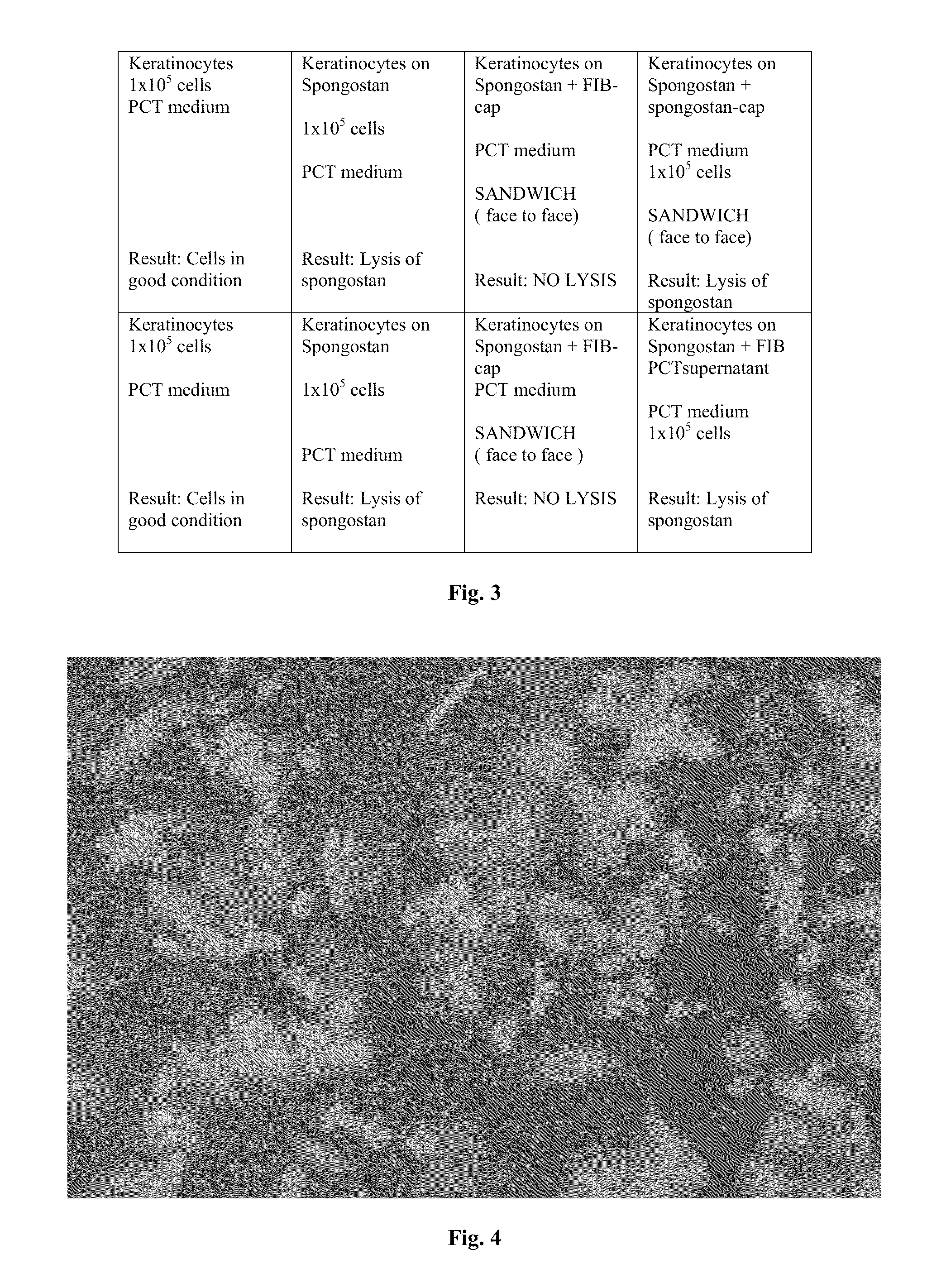
Preparation of Spongostan for Fibroblast Seeding

[0193]1. Cut a circle from Spongostan film 2.5 cm in diameter.[0194]2. Put circle of Spongostan into a well of the 24-well plate.[0195]3. Add 500 μl of Dermal Fibroblasts Medium (Cat#CnT-05, Chemicon) to well with Spongostan to prepare for cell culturing in CnT-05 medium[0196]4. Place 24-well plate in a 37 C, CO2 incubator for 30 minutes.[0197]5. Remove 24-well plate from an incubator.[0198]6. Remove the medium from the well.[0199]7. Add 500 μl of CnT-05 medium to a well with Spongostan.[0200]8. Place 24-well plate in a 37 C, CO2 incubator for 30 minutes[0201]9. Repeat 3-8 steps (washing) 2 times.


PUM


Abstract

A cellular scaffold that is suitable for tissue regeneration, cell culture and in vitro assays. The invention relates to a layered cell scaffold that is seeded with mesenchymal and ectodermal cells. The layered cellular scaffold comprises an inoculum of mesenchymal cells and ectodermal cells positioned between two opposing scaffolds in a sandwich configuration. The layered cell scaffold provides a functional skin equivalent that is suitable for transplantation and in vitro cell-based assays.

Description

CROSS-REFERENCE TO RELATED APPLICATIONS[0001]This application is a continuation in part of application Ser. No. 11 / 616,787, filed Dec. 27, 2006 now U.S. Pat. No. 8,105,380, which claims priority to provisional application Ser. No. 60 / 854,059 filed Oct. 23, 2006. The entire contents of these applications are incorporated herein by reference in their entirety.FIELD OF THE INVENTION[0002]The present invention generally relates to tissue engineering and specifically relates to scaffolding for cell and tissue culture. Particularly, the invention relates to a cellular scaffold that is seeded with regenerative cells. More particularly, the invention relates to a layered cellular scaffold that has ectodermal cells and mesenchymal cells seeded between at least two biocompatible substrates. The invention also relates to methods of using a layered scaffold for cell transplantation, cell culture and cell-based in vitro assays.BACKGROUND[0003]The tissue engineering field is well developed and a ...

Claims


Application Information

Patent Timeline
Patent Type & Authority Patents(United States)
IPC IPC(8): A61F2/10
CPCC12N5/0698A61L27/3804A61K2035/124A61L27/60C12N2502/1323C12N2502/094A61L27/3891
Inventor KHARAZI, ALEXANDERKHARAZI, LUDMILLATANKOVICH, NIKOLAI
Owner STEMEDICA CELL TECH
Why Eureka
Try Eureka