Method for identifying cells based on DNA replication domain timing profiles

a technology of dna replication domain and dna replication, applied in the field of identifying cells based on dna replication domain timing profiles, can solve the problems of insufficient information to distinguish various types of cells, difficult to distinguish most cells based on appearance alone, and inability to apply in a high-throughput manner

Active Publication Date: 2014-05-20
FLORIDA STATE UNIV RES FOUND INC
View PDF0 Cites 2 Cited by
  • Summary
  • Abstract
  • Description
  • Claims
  • Application Information

AI Technical Summary

Problems solved by technology

This approach is useful in cases in which cells display a distinctive shape (e.g., long axons in neurons) and / or an easily recognizable feature (e.g., a lipid vesicle stained for fats), but most cells are difficult to distinguish based on their appearance alone.
Histology-based procedures to cell identification also require a highly trained person making them impossible to apply in a high-throughput manner.
However, because each antibody only recognizes one particular protein antigen, such approaches generally do not provide sufficient information to distinguish various types of cells.
In other words, a single protein marker is rarely a guarantee of a particular cell type.
On the other hand, larger scale protein detection methods (e.g., proteomics) suffer from insufficient sensitivity and a lack of capability for automation.
However, the decisive drawback of this system is the instability of RNA.
This is especially problematic when working with archived samples (e.g., preserved biopsies) or with limited amounts of cellular material.
A further problem with RNA-based approaches is that mRNA fluctuates in response to temporary changes in environmental conditions.
Therefore, RNA-based approaches for cell identification are limited by perturbations in gene expression caused by transient cell culture conditions, cell-to-cell heterogeneity in gene expression, and random degradation of mRNA in cell-derived extracts or samples that adversely affect the robustness, reproducibility, and interpretation of such techniques.
In general, RNA-based arrays are useful discovery tools, but they are not yet widely applicable as a clinical or large scale assay method for the identification of cells.
However, these techniques based on epigenetic chemical modifications to identify cell states are limited by the fact that (1) they require very high resolution (200 bp nucleosomal units), (2) they reflect dynamic chromatin states that can change or become heterogeneous within a homogeneous cell type, (3) there is a large diversity of histone modifications that would need to be individually investigated to gain a comprehensive profile, and (4) these rely on the use of different and expensive antibodies and other reagents that would create challenges for high-throughput analysis.

Method used


Image

  • Method for identifying cells based on DNA replication domain timing profiles
  • Method for identifying cells based on DNA replication domain timing profiles
  • Method for identifying cells based on DNA replication domain timing profiles

Examples


example 1

Materials and Methods

ESC Culture, Neural Differentiation, and BrdU-Labeling

[0174]D3 cells (see, e.g., Doetschman et al., “The in vitro development of blastocyst-derived embryonic stem cell lines: formation of visceral yolk sac, blood islands and myocardium,”J. Embryol. Exp. Morphol. 87:27-45 (1985), the entire contents and disclosure of which are hereby incorporated by reference), 46C cells (see, e.g., Ying et al., “Conversion of embryonic stem cells into neuroectodermal precursors in adherent monoculture,”Nat. Biotechnol. 21:183-186 (2003), the entire contents and disclosure of which are hereby incorporated by reference), and TT2 cells (see, e.g., Yagi et al., “A novel ES cell line, TT2, with high germline-differentiating potency,”Anal Biochem. 214:70-76 (1993), the entire contents and disclosure of which are hereby incorporated by reference) are male ESC lines with a normal karyotype that are cultured in the presence of LIF (leukemia inhibitory factor) as described in, for example...

example 2

Replication Domain Structure in Embryonic Stem Cells

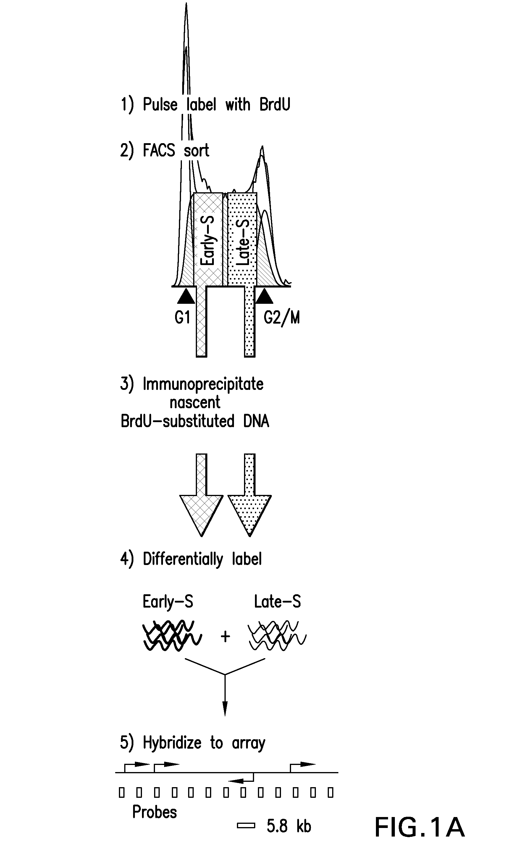
[0185]Replication timing is mapped in mESCs using high-density oligonucleotide arrays, adapting a previously developed retroactive synchronization method. See, e.g., Schubeler et al., “Genome-wide DNA replication profile for Drosophila melanogaster: a link between transcription and replication timing,”Nat. Genet. 32:438-442 (2002); and Gilbert D M, “Temporal order of replication of Xenopus laevis 5S ribosomal RNA genes in somatic cells,”PNAS USA 83:2924-2928 (1986), the entire contents and disclosures of which are hereby incorporated by reference. ESCs are chosen because they provide the opportunity to directly evaluate dynamic changes in the replication program in response to changes in growth conditions (see, e.g., Hiratani et al., “Differentiation-induced replication-timing changes are restricted to AT-rich / long interspersed nuclear element (LINE)-rich isochores,”PNAS USA 101: 16861-16866 (2004); and Perry et al., “A dynamic swi...

example 3

Domain Structure is Conserved Between Independent mESC Lines

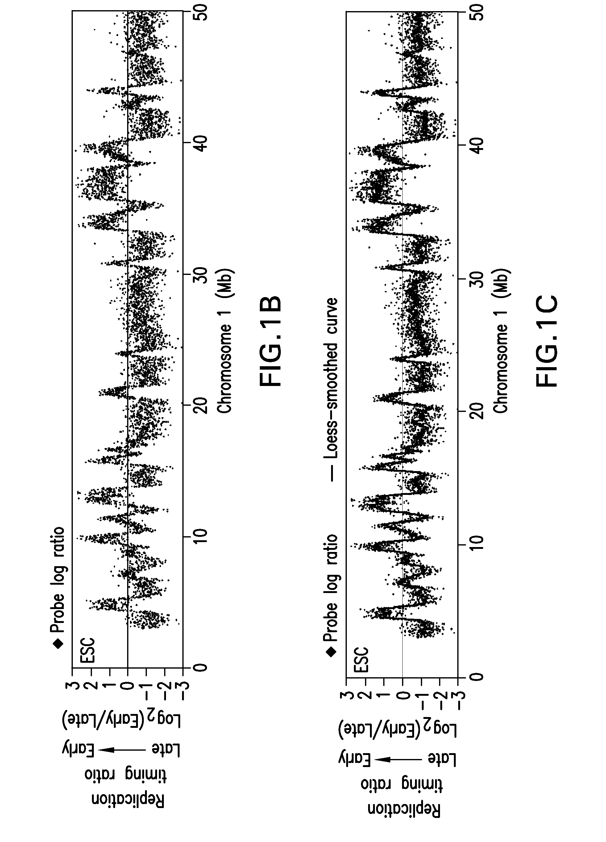
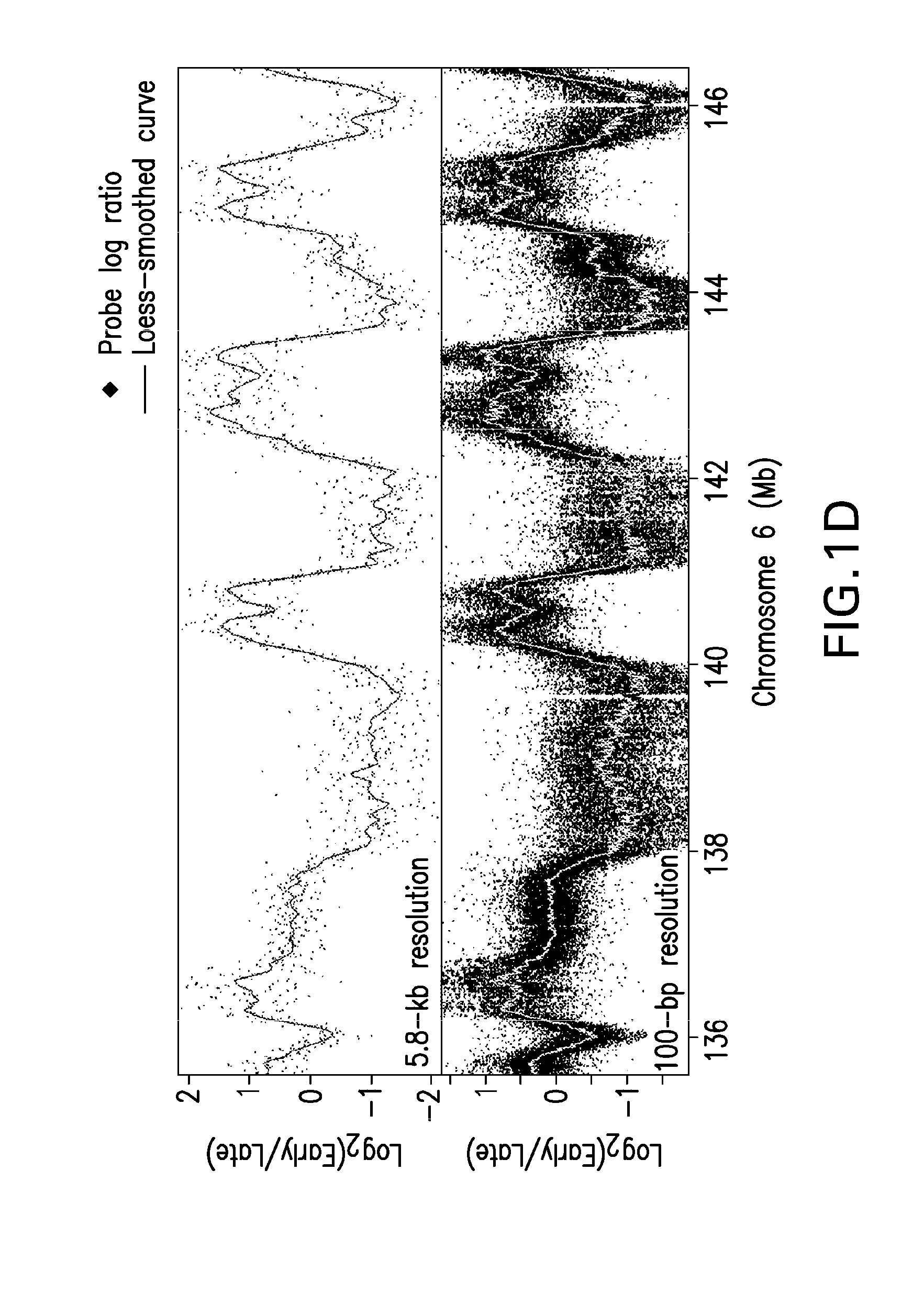
[0189]The results described above demonstrate that coordinately replicated regions (replication domains) constitute functional units of chromosomes whose boundaries may be molecularly defined. The fact that replication domain boundaries may be so precisely mapped in populations of cells demonstrates that their positions are highly stable from cell cycle to cell cycle. To evaluate whether these boundaries are a conserved property of chromosomes in multiple mESCs, three mESC lines from two independently established mouse inbred strains are compared. Lines D3 and 46C are both derived from the 129 mouse strain and so are nearly identical genetically, but are separated by more than 20 years in cell culture, while TT2 is derived 15 years ago from a C57BL / 6xCBA hybrid mouse and is therefore genetically polymorphic (see, e.g., Doetschman et al., (1985), supra; Yagi, et al., (1993), supra; and Ying et al., (2003), supra). Despite th...


PUM

PropertyMeasurementUnit
replication domain sizesaaaaaaaaaa

Abstract

Methods for identifying and / or distinguishing a homogeneous population of cells based on their replication domain timing profile using high resolution genomic arrays or sequencing procedures are provided. These methods may be used to compare the replication timing profile for a population of cells to another replication timing profile(s), a replication timing fingerprint, and / or one or more informative segments of a replication timing fingerprint, which may be simultaneously or previously determined and / or contained in a database, to determine whether there is a match between them. Based on such information, the identity of the population of cells may be determined, or the identity of the population of cells may be distinguished from other populations of cells or cell types. Methods for determining a replication timing fingerprint for particular cell types are also provided.

Description

CROSS-REFERENCE TO RELATED APPLICATIONS[0001]This application claims the priority date of Prov. App. No. 60 / 969,399, entitled “METHOD FOR IDENTIFYING CELLS BASED ON DNA REPLICATION DOMAIN TIMING PROFILES,” filed Aug. 31, 2007, and the entire contents and disclosure of this provisional application are hereby incorporated by reference.GOVERNMENT INTEREST STATEMENT[0002]This invention was made with government support under contract numbers GM083337 and GM085354-015319 awarded by the National Institutes of Health. The government has certain rights in the invention.BACKGROUND[0003]1. Field of the Invention[0004]The present invention broadly relates to a method for identifying cells based on their replication domain timing profiles using a high resolution genomic array. The present invention also broadly relates to a method for distinguishing cells from other cells based on replication timing profiles using a high resolution genomic array. The present invention further broadly relates to ...

Claims


Application Information

Patent Timeline
Patent Type & Authority Patents(United States)
IPC IPC(8): C12Q1/68C40B30/04
CPCC12Q1/6804C12Q1/6837C12Q1/6806C12Q1/6809C12Q2565/626C12Q2565/501C12Q2525/101G01N33/5005
Inventor GILBERT, DAVID M.RYBA, TYRONEHIRATANI, ICHIRO
Owner FLORIDA STATE UNIV RES FOUND INC
Why Eureka
Try Eureka