
Methods and apparatus for transesophageal microaccess surgery

a technology for transesophageal microaccess and surgery, applied in the field of methods and apparatus for transesophageal microaccess surgery, can solve the problems of difficult details that need improvement, require new approaches, etc., and achieve the effect of preventing contamination of the perforation si

Inactive Publication Date: 2014-07-08
MICROACCESS
View PDF61 Cites 128 Cited by
  • Summary
  • Abstract
  • Description
  • Claims
  • Application Information

AI Technical Summary

Benefits of technology

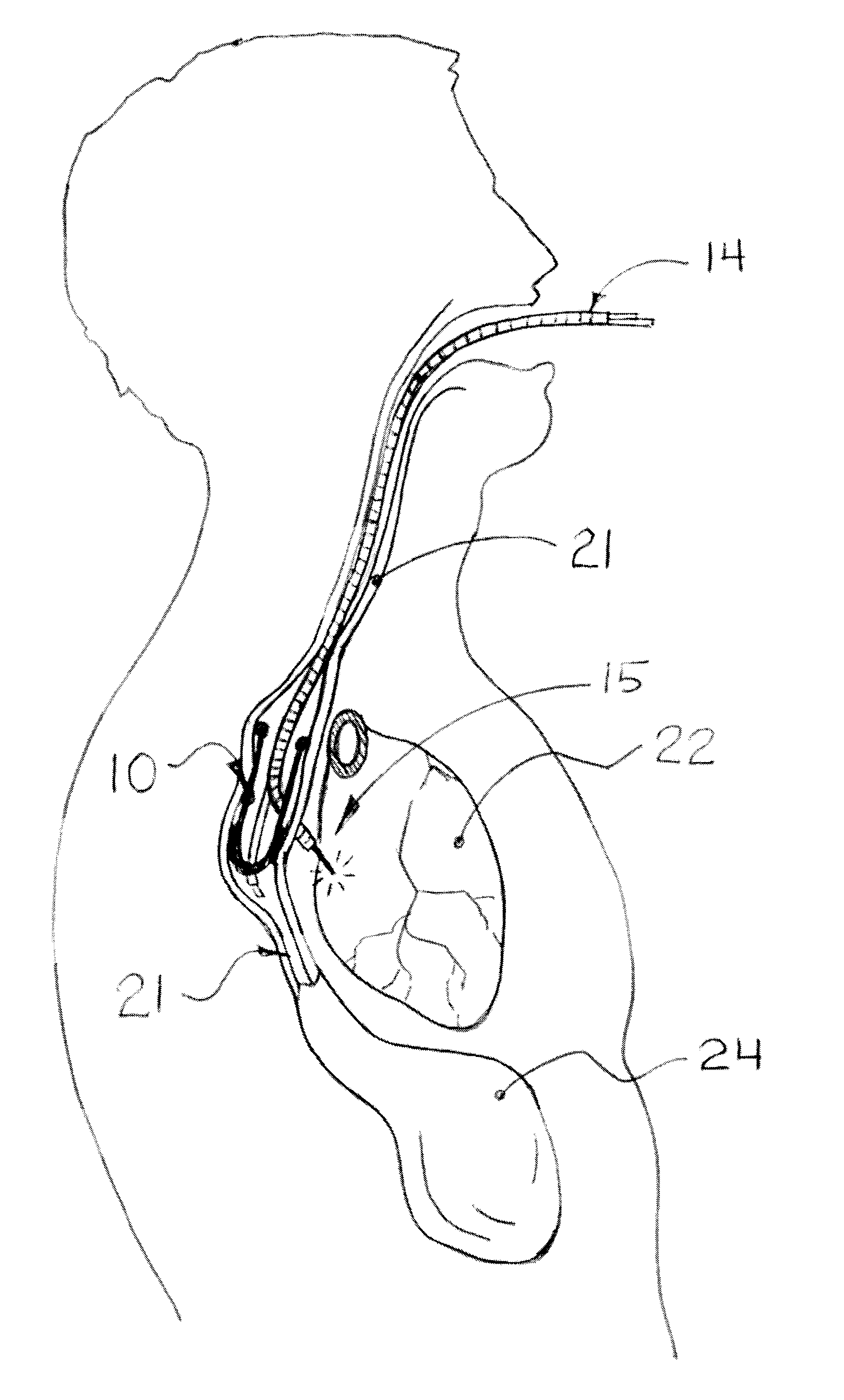
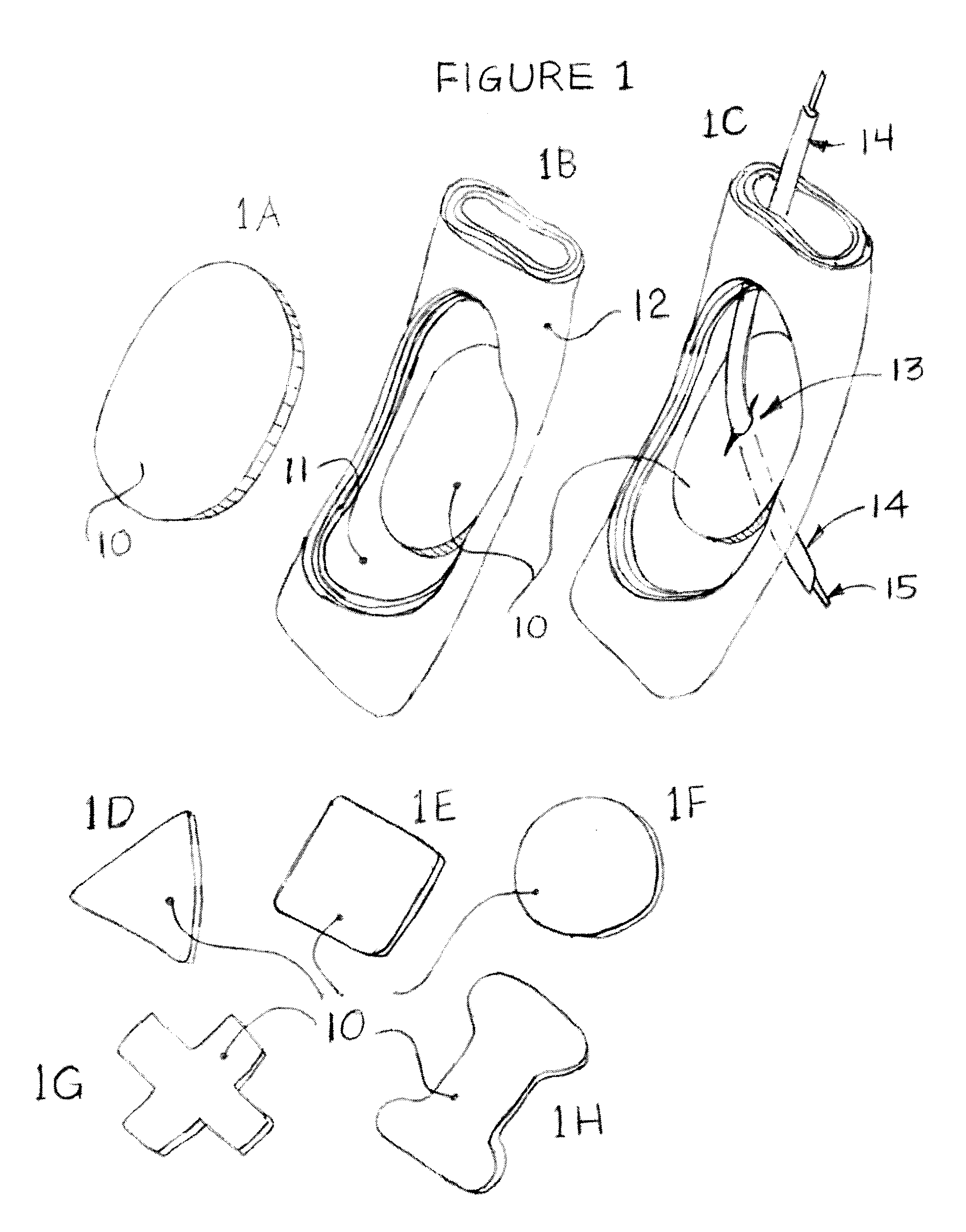
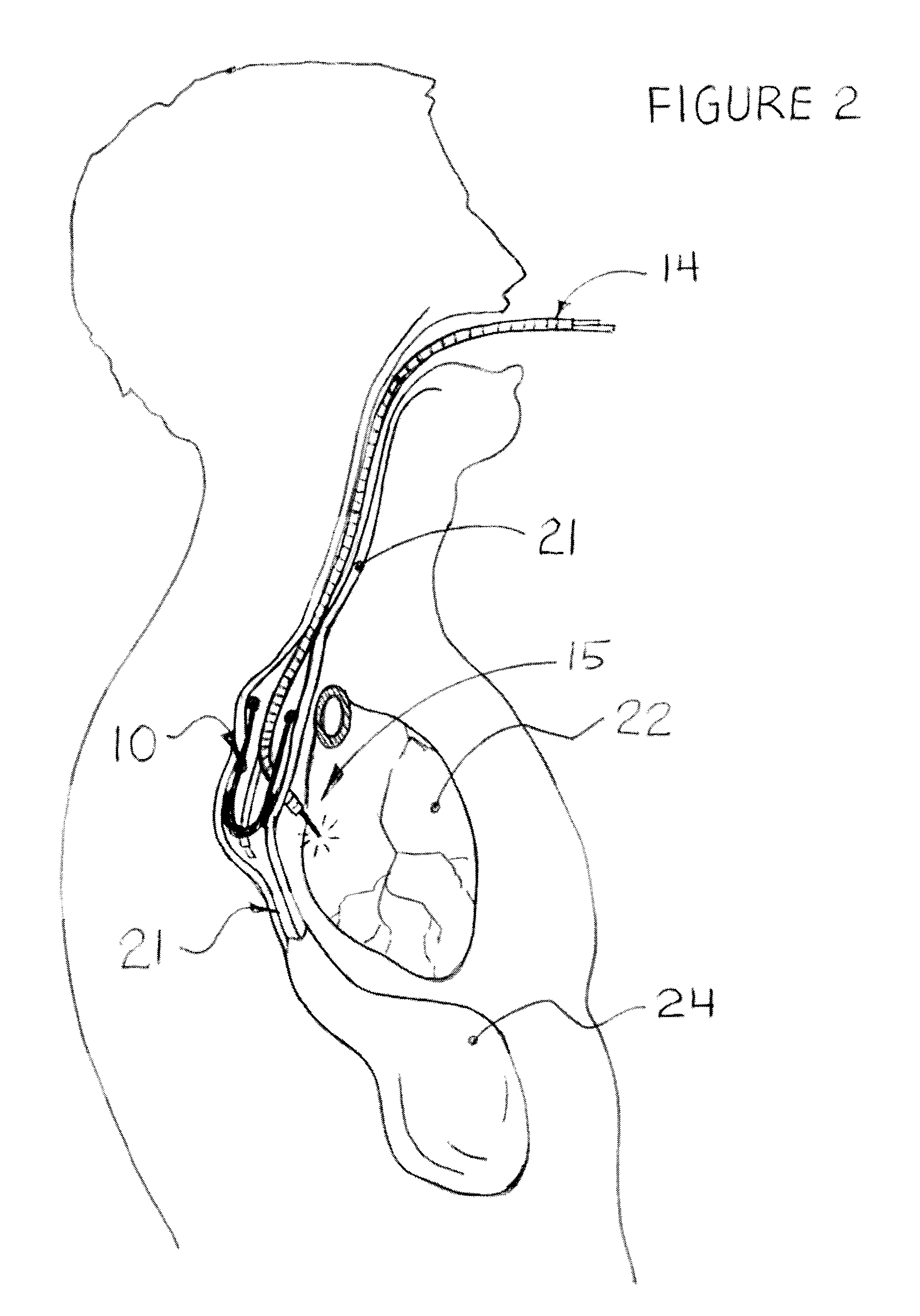
[0005]In one aspect, a liner (also called a “liner device” herein) is used to demarcate the selected areas of the esophagus. The liner is a piece of a material which is placed at the selected site, optionally with the assistance of imaging techniques. The liner is made of a biocompatible material and optionally is degradable in situ. The liner may be affixed to the selected site via any of several methods. These include adhesives, vacuum, mechanical affixing devices, and simple passive positioning. The liner has numerous optional additional properties, including transparency for visibility; prefabricated penetration locations; radioopaque markers or other location aids; and carriage of other devices, including device sensing and control means, taking advantage of the propinquity of the esophagus to the heart and other organs that might be monitored following a procedure.
[0007]The invention comprises a liner device for facilitating the controlled and reversible creation of an opening in the wall of a body lumen, for example the esophagus, through which an endoscopic device can be inserted to perform surgery in adjacent tissues, particularly in the cervical and thoracic regions. The liner device is a structure that lines part of the lumen walls of a hollow body organ or body cavity, in proximity to an organ or a structure outside the lumen structure. The invention deals in particular with the lumen of the esophagus, as a lumen of particular value for the use of the invention. However, any body lumen or cavity can be used for the same techniques, sometimes with certain modifications in each specific case. The lumen liner device allows for elongated surgical devices to be deployed across the lumen wall, to target extraluminal organs or structures for the performance of diagnostic and therapeutic procedures.
[0014]The upper and lower ends of the liner can be closed by diaphragms to prevent any fluid or contamination from entering into the surgical field from above or below. At least the upper diaphragm will typically have a slit or other means to allow for passage of an endoscope. The liner can also have balloons on the proximal and / or distal ends to prevent fluid contamination from above or below.
[0029]A liner device of the invention may serve to isolate a partial or full perforation in the wall of the esophagus, wherein the isolation of the perforated portion of said wall prevents contamination of the perforation site by covering it. Said liner device may be one of a generally planar device and a device that is fully or partially tubular.

Problems solved by technology

In exploring this new area of minimally invasive surgery, it has been found that one of the important but difficult details that need improvement is managing the passage of instruments across the esophageal wall.
Surgery via the esophagus has numerous attractions for thoracic and cervical surgeries, but precision location of the entry site, and reliable and simple closure of the site after surgery each present novel problems and require new approaches.

Method used


Image

  • Methods and apparatus for transesophageal microaccess surgery
  • Methods and apparatus for transesophageal microaccess surgery
  • Methods and apparatus for transesophageal microaccess surgery

Examples


example 1

Transesophageal Microaccess for Spine Treatment

[0092]Cervical and thoracic spine disorders are of special importance due to the complexity of the bony / cartilaginous structures in relation to the spinal cord. For disorders like cervical disk disease, the current surgical solutions are very complex and involve dissection through many layers of tissues, and affect many sensitive vascular, neurological or lymphatic structures before the surgeon is able to reach the target lesion in the disk area between cervical vertebrae. This applies to both the anterior as well as the posterior approach.

[0093]What is needed is a simpler, less invasive and more precise method to reach the cervical disk / vertebral lesion and related structures without the need to dissect through many tissue layers. The posterior “Transesophageal Microaccess” approach is described below. As will be shown, the access to the cervical spine through the esophageal wall is short, wide and relatively direct as the esophagus is...


PUM


Abstract

The current invention describes methods of transesophageal access to the neck and thorax to perform surgical interventions on structures outside the esophagus in both the cervical and the thoracic cavity. It describes a liner device made of a complete or partial tubular structure, or a flat plate, the liner having means to facilitate creation of a side opening, which may include a valve. The liner with its side opening form a port structure inside the esophageal lumen. The port structure allows elongated surgical devices to pass through a perforation across the full thickness of the esophageal wall to outside location, in a controlled way. The elongated surgical devices can be diagnostic scopes, therapeutic scopes, manual elongated surgical devices, robotic arms or the like. After being deployed outside the esophagus, the surgical devices can access structures outside the esophagus, in the neck and thorax in 360 degrees of freedom around the esophageal circumference. These structures can be bony, cartilaginous, spinal, vascular, soft tissue, deep tissues, lymph nodal, cardiac, pulmonary, tracheal, nervous, muscular or diaphragmatic, skin and subcutaneous tissues of the neck, skin and subcutaneous tissues of the anterior chest wall, skin and subcutaneous tissues of the skin of the back, and skin and layers of the breast.

Description

CROSS-REFERENCE TO RELATED APPLICATIONS[0001]This application claims the benefit of the priority of US provisional application No. 61 / 137,690, filed on Aug. 1, 2008, which is incorporated herein by reference in its entirety.FIELD OF THE INVENTION[0002]The present invention relates generally to a minimally invasive approach to perform transesophageal surgical, orthopedic and neurosurgical procedures in the mediastinum, cervical and thoracic cavities. The invention describes devices and methods to create a transesophageal access to said structures and surrounding structures to perform a body of surgical procedures while the heart is beating with or without the need for general anesthesia. A key improvement in access to these spaces is provided by provision of an intra-esophageal liner to facilitate transesophageal passage and to reinforce such passages; and to provide, or to catty means for providing, for the sealing of openings through the esophageal wall to prevent permeation of flu...

Claims


Application Information

Patent Timeline
Patent Type & Authority Patents(United States)
IPC IPC(8): A61B1/00
CPCA61B17/3423A61B17/00491A61B17/3439A61B17/3498A61B2017/00004A61B2017/00278A61B2017/003A61B2017/0641A61B2017/0649A61B2017/22054A61B2017/22067A61B2017/22069A61B2017/306A61B2017/3419A61B17/0218A61B2017/0225
Inventor MESALLUM, SAMEHBLECK, JAMES H.
Owner MICROACCESS
Learn More