Method for treating a biological sample with a composition containing at least one polyol and excluding water

a polyol and composition technology, applied in the field of biological sample treatment, can solve the problems of reducing the extraction ability of nucleic acids or proteins from tissues, unable to use molecular-biological methods, and falsifying molecular-biological analyses or indeed making them entirely impossible, so as to achieve satisfactory stabilization of biological samples

Active Publication Date: 2014-08-05
QIAGEN GMBH
View PDF35 Cites 6 Cited by
  • Summary
  • Abstract
  • Description
  • Claims
  • Application Information

AI Technical Summary

Benefits of technology

[0020]In particular, the present invention was based on the object of providing a method of stabilizing a biological sample which, with the use of the smallest possible amounts, if any, of readily flammable, volatile, carcinogenic, teratogenic, environmentally hazardous or toxic substances leads to a satisfactory stabilization of the biological sample.
[0021]Furthermore, the present invention was based on the object of providing a method of stabilizing a biological sample by means of which both frozen and fresh biological samples can be stabilized at moderate temperatures, as far as possible, for example also at room temperature, without adversely affecting the expression profile or the proteome of the biological sample.
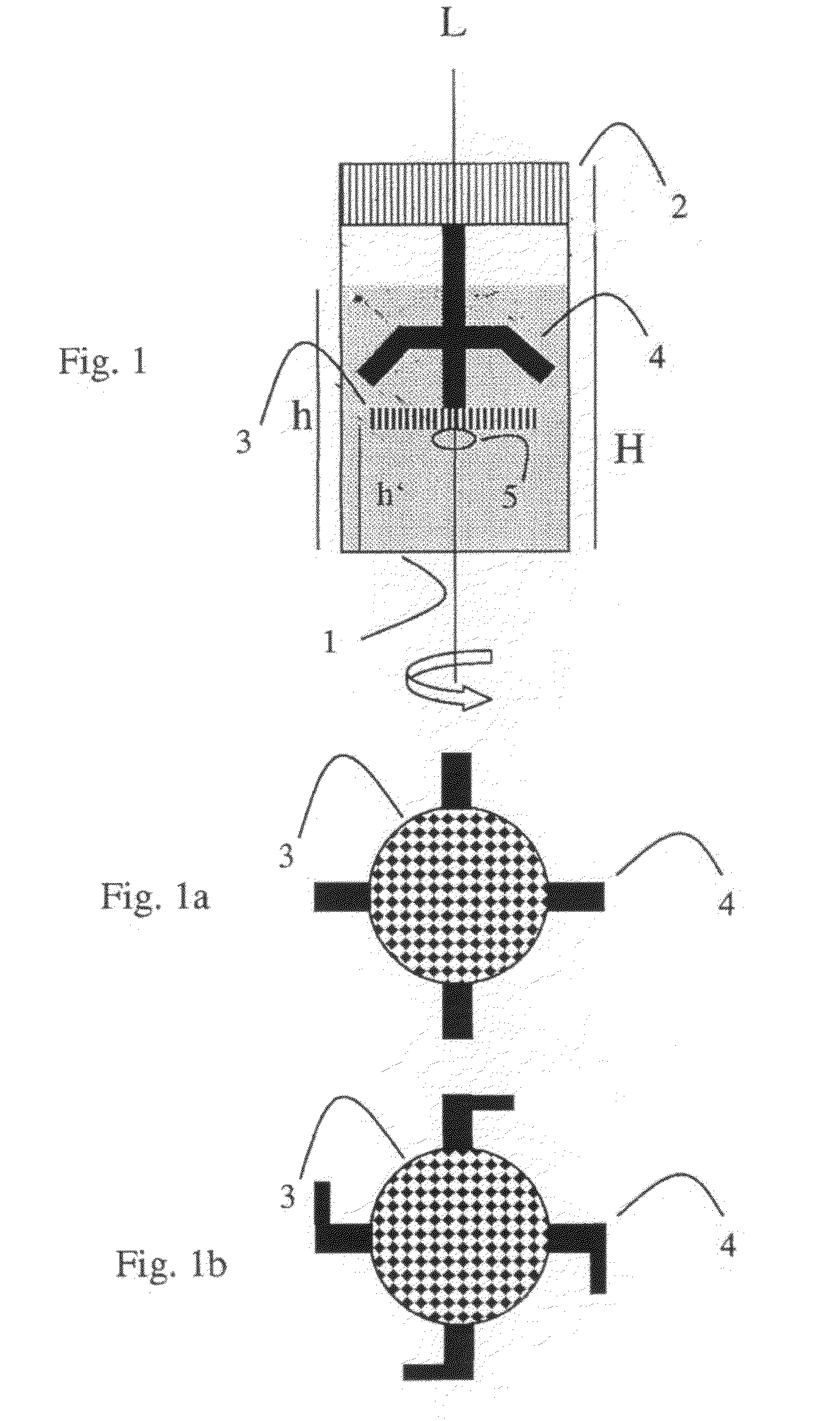
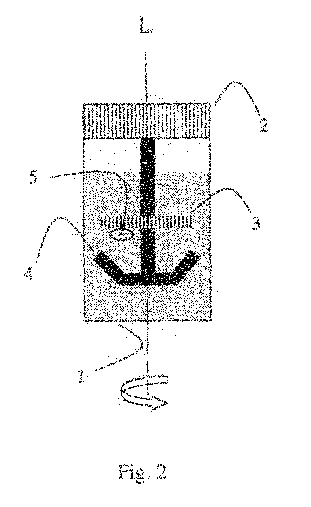
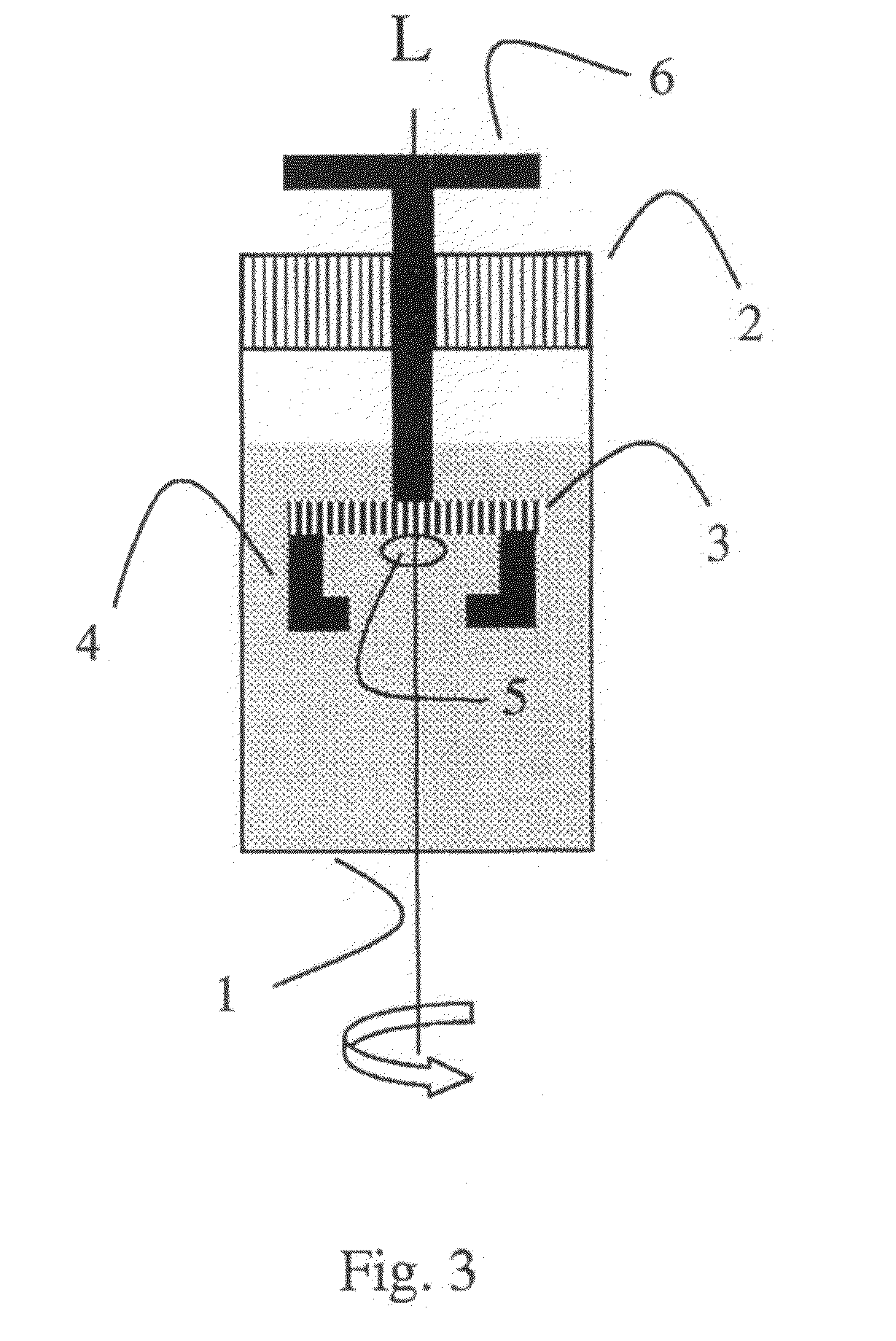
[0024]Furthermore, the present invention was based on the object of providing a device by means of which a biological sample can be stabilized in a simple manner, using the stabilization method according to the invention.

Problems solved by technology

In the course of these developments, one recognized that it is precisely the status (gene expression or protein pattern) of the fresh sample constituents which are important for molecular-biological study which may undergo rapid changes, even directly after the sample has been taken from its natural environment, so that even prolonged storage of the samples in the untreated state, for example as the result of unexpected delays during transport into the laboratory or the like, may falsify a molecular-biological analysis or indeed make the latter entirely impossible.
However, such a stabilization is in most cases unsuitable for the use of molecular-biological methods since the nucleic acids are only very insufficiently stabilized, which only makes possible a qualitative detection, at best, of the nucleic acids or nucleic acid fragments present, but not a quantitative detection.
Moreover, the stabilization with crosslinking stabilizers such as aqueous formaldehyde solution leads to a reduced extractability of the nucleic acids or proteins from the tissues.
Also, aqueous formaldehyde solution is not acceptable for toxicological reasons.
To stabilize nucleic acids in compact pieces of tissue, however, such stabilizing reagents are not sufficient.
Moreover, those reagents and methods with which nucleic acids can be stabilized for the purposes of qualitative detection are, as a rule, not suitable for the simultaneous stabilization of proteins.
Moreover, samples stabilized in this manner cannot be used for histological study since the stabilizer preserves for example the nucleic acids, but not the cell or tissue structures.
However, they are largely unsuitable for use in the stabilization of cell-containing or cell-free body fluids such as, for example, blood, serum or plasma, and also have not as good stabilizing properties in some types of tissue, such as, for example, fatty tissue.
All the above shows that it is particularly difficult simultaneously to stabilize RNA, DNA and proteins in tissue samples and histologically to preserve the tissue samples.
Moreover, work carried out on cells or other biological samples cannot necessarily be applied to compact tissue.
In comparison with other biological samples, the stabilization of nucleic acids in compact tissue samples involves one particular difficulty.
However, all such methods require very complicated logistic requirements since defrosting of the samples during transport, storage or during a wide range of purposes and utilizations must be prevented.
Besides the additional costs for specific sample receptacles and for the permanent cooling of the samples, the use of liquid nitrogen is not only very complicated, but also can only be carried out with specific precautionary measures.
Moreover, a subsequent analysis of the frozen sample material, in particular individual components of the sample, is usually a very difficult endeavor.
This means that samples which have been subjected to defrosting, or incipient defrosting, no longer give reproducible results.
In addition, it is precisely tissue pieces in the frozen state which are very difficult to method, for example divide, manually, or only with complex technical equipment.
However, further analyses, and storage of the sample, in particular at room temperature, are not possible.
The disadvantage of the samples treated with customary transition solutions is that they only remain stable at room temperature over a very short period, which means that the methoding time is only very limited and very readily exceeded in particular when methoding a large number of samples, in particular when cutting and chopping procedures are involved.
Moreover, the transition is only very slow, whereby no direct experiments may follow, and waiting times of in most cases one day result.
Equally, transport of the samples treated thus is not possible at room temperature without the sample being damaged, since not only must the transition take place at temperatures of ≦−20° C., but this must be followed by stable storage of the sample.
Also, the transport of the sample is only possible at ≦−20° C., which requires the use of cooling means, for example dry ice, during transport.
While the use of the traditional transition solutions leads to improvements in sample methoding, such as, for example, chopping or cutting to size, they neither reduce the equipment requirements (since the solution for transition must be precooled at −70 to −80° C., and therefore still requires a suitable cooling device), nor is it possible to stabilize the transition-solution-treated samples at room temperature over a prolonged period.

Method used


Image

  • Method for treating a biological sample with a composition containing at least one polyol and excluding water
  • Method for treating a biological sample with a composition containing at least one polyol and excluding water
  • Method for treating a biological sample with a composition containing at least one polyol and excluding water

Examples


examples

1. Histological Analysis of Samples Treated According to the Invention

[0129]Immediately after the removal of organs, rat liver tissue was treated with in each case 1 ml of the compositions detailed in table 1 and stored for 1 day in the incubator at 25° C. After storage, the tissue pieces are removed from the solutions, transferred into plastic boxes and, following conventional protocols, incubated in an ascending ethanol series and in xylene, and embedded in paraffin. With the aid of a microtome, sections are prepared from the paraffin-embedded tissue, and these sections are stained on the slide with hematoxylin-eosin by customary methods. The stained tissue sections are viewed under the light microscope. The result is compiled in table 1:

[0130]

TABLE 1CompositionResult1,2,3-PropanetriolMorphology retained1,2,6-HexanetriolMorphology retained3-Methyl-1,3,5-pentanetriolMorphology retained25% 1,2,3-propanetriol + 75% 1,2,6-hexanetriolMorphology retained75% diethylene glycol + 25% 1,2,6...


PUM

PropertyMeasurementUnit
temperatureaaaaaaaaaa

Abstract

The invention relates to a method for treating a biological sample, comprising the following method steps: i) preparation of a biological sample and ii) bringing the biological sample into contact with a composition, comprising: (1) 1 to 100 wt. % of at least one polyol and (2) 0 to 99 wt. % of at least one additive, wherein the total amount of components (1) and (2) is 100 wt. %. The invention further relates to biological samples obtained by said method, a method for analysis of a treated biological sample, devices for treating a biological sample, use of said devices, various kits and use of a composition.

Description

CROSS-REFERENCE TO RELATED APPLICATIONS[0001]This application is a United States national stage filing under 35 U.S.C.§371 of international (PCT) application No. PCT / EP2006 / 070267, filed Dec. 29, 2006 and designating the US, which claims priority to European application 05028767.1, filed Dec. 30, 2005.FIELD OF THE INVENTION[0002]The present invention relates to a method of treating a biological sample, to the biological sample obtainable by this method, to a method of analyzing a treated biological sample, to devices for treating a biological sample, to the use of these devices, to a variety of kits, and to the use of a composition.BACKGROUND OF THE INVENTION[0003]For a long time, scientists have focused only on the pathological and / or histological study of biological samples. A preservation and / or stabilization of the samples for such studies was usually carried out, if at all, by placing the samples into formaldehyde solutions and / or by embedding the samples in paraffin. But even ...

Claims


Application Information

Patent Timeline
Patent Type & Authority Patents(United States)
IPC IPC(8): C12Q1/68G01N33/68G01N1/30
CPCY10T436/107497G01N1/31G01N1/30B01L3/508Y10T436/2525Y10T436/108331B01L2300/046
Inventor HOLLANDER, VERA
Owner QIAGEN GMBH
Why Eureka
Try Eureka