Analysis of microbes from microcolonies by maldi mass spectrometry

a mass spectrometry and microbe technology, applied in the field of mass spectrometric analysis of microorganisms, can solve the problems of too many applications, and achieve the effects of less microorganism material, low cost, and high sensitivity

Active Publication Date: 2014-09-16
BRUKER DALTONIK GMBH & CO KG
View PDF13 Cites 10 Cited by
  • Summary
  • Abstract
  • Description
  • Claims
  • Application Information

AI Technical Summary

Benefits of technology

[0019]The disrupted cells of the microbes can be brought into contact with the end of a pin-shaped sample support (sample support pin). The contact surface of the pin-shaped sample support is so small that only microbe proteins of one individual colony are transferred onto the pin-shaped sample support. After the microbe proteins have been transferred, the pin-shaped sample support is preferably inserted into an adapter plate in such a way that the end surface of the pin-shaped sample support is essentially flush with the surface of the adapter plate. Here, “essentially flush” means that a mass spectrometric analysis in a MALDI time-of-flight mass spectrometer with axial ion injection is possible with sufficient resolution. Microbes from different colonies can each be transferred onto one of several pin-shaped sample supports, which are inserted together into an adapter plate and introduced into the mass spectrometer in the adapter plate. It is also possible to transfer microbes from one colony onto several pin-shaped sample supports. The end surfaces of the pin-shaped sample supports preferably have surface areas of less than nine square millimeters, in particular of less than four square millimeters.
[0024]The cell disruption of the microbes, i.e. the destruction of their cell walls, can be effected physically by ultrasound, electromagnetic radiation (e.g. infrared radiation) or high pressure (more precisely the sudden pressure release). It is possible for the positions of colonies on the surface of the nutrient medium to be determined, and colonies selected, and for the ultrasound or infrared radiation to have a localized effect on the selected colonies. For example, narrowly focused infrared radiation from laser diodes can cause the microbes to burst. With an ultrasound micro-tip at 20 to 30 kilohertz, a targeted cell disruption of all microbes of a microcolony can be performed in a very short time without damaging the proteins in the interior of the cell. Mechanical destruction is also possible: with the nitrogen decompression method, nitrogen is first dissolved in the microbes at high pressure; a rapid decompression then causes the microbes to burst.
[0028]The methods according to the invention have the advantage over the prior art that they require less consumable material because transfer tools are not required, and they are therefore lower cost and that they require less microbe material because of the direct transfer of proteins, and offer a high sensitivity because the proteins can be washed rigorously and so freed from all salts, tensides and other material which deteriorates the MALDI sensitivity. Compared to the manual transfer of microbes from different samples for analysis onto a mass spectrometric sample support, transfer via direct contact makes it possible to avoid mistakes in the sample assignation, by automatically transferring an identification label of each sample for analysis to each mass spectrometric sample support used. This identification label is stored on the support of the nutrient medium. This identification label can be combined by positional data and the identification label of the Petri dish. Transferring the microbe proteins with the aid of a protein-adsorptive coating allows MALDI mass spectra to be acquired with minimal signal suppression of the microbe proteins, particularly if the surface is additionally purified after the transfer of the microbe proteins.

Problems solved by technology

This is too long for many applications, particularly for applications in medical diagnostics.
But for bacteria, whose hard cell walls require special cell disruption processes, it has so far only been possible in rare cases to produce mass spectra which are good enough for an identification from only 103 bacteria or less.

Method used


Image

  • Analysis of microbes from microcolonies by maldi mass spectrometry
  • Analysis of microbes from microcolonies by maldi mass spectrometry

Examples


Embodiment Construction

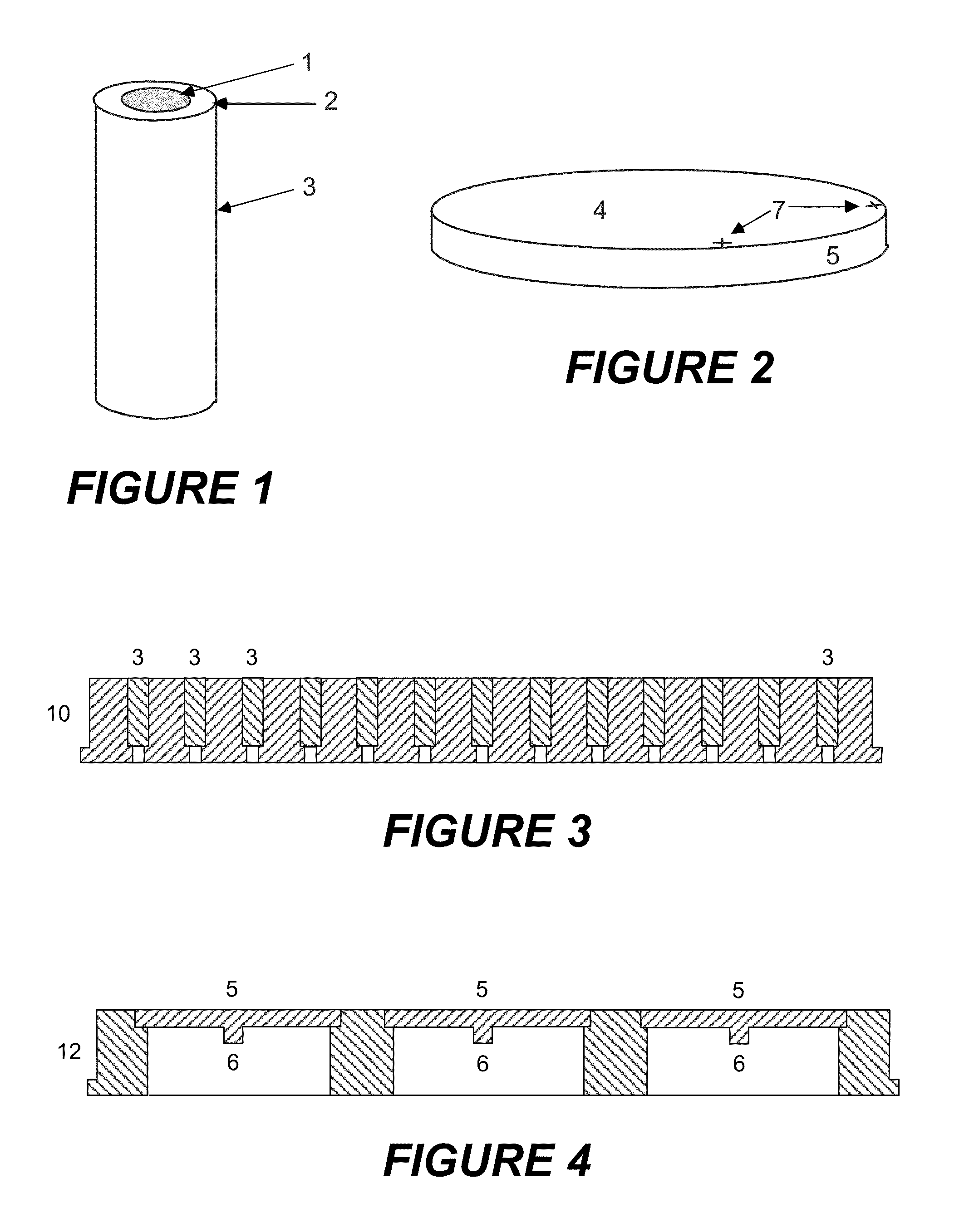
[0035]For the embodiments described below, a sample for analysis with unknown microbes, usually bacteria, is plated (inoculated) in the usual way onto agar in a Petri dish, and cultured in an incubator for six to eight hours at optimum temperature (usually 37° Celsius). Most of the clinically relevant, pathogenic microbes divide after periods of between 15 and 45 minutes (this is called the “generation time”). In six hours, virulent microbes experience 24 generations, slower ones with 45 minutes generation time only about eight generations. After ten generations, the microcolonies should theoretically contain around 103 microbes; after twenty generations, around 106 microbes. The colonies grow on the surface of the nutrient medium; at higher numbers of generations, however, the growth on the agar is so strongly restricted that the theoretical values are not achieved. There are also clinically important exceptions with slow growth which require special treatment.

[0036]The cell disrup...


PUM

PropertyMeasurementUnit
diametersaaaaaaaaaa
diameteraaaaaaaaaa
diametersaaaaaaaaaa

Abstract

The invention relates to the cell disruption of microbes and the preparation of the microbe proteins for mass spectrometric analysis. The cells of microbes from microcolonies are disrupted by physical or chemical means directly on the nutrient medium. The released proteins are then transferred to sample supports by direct contact with their contact surfaces; electrophoresis can be used for assistance. Once the proteins are firmly adsorbed on the contact surfaces, they can be washed with water in order to remove substances which interfere with the ionization process. For analysis by matrix-assisted laser desorption (MALDI), the proteins are prepared on the contact surfaces of the sample supports with matrix substances to form MALDI samples; the sample supports are then introduced into a MALDI mass spectrometer for the acquisition of mass spectra. The microbes are identified by similarity comparisons between the mass spectra of the microbe proteins and similarly obtained reference spectra.

Description

BACKGROUND OF THE INVENTION[0001]1. Field of the Invention[0002]The invention relates to methods for the mass spectrometric analysis of microbes from colonies on surfaces of nutrient media, particularly in a mass spectrometer with ionization by matrix-assisted laser desorption (MALDI).[0003]2. Description of the Related Art[0004]The routine, fast and error-free analysis of microorganisms plays an important role particularly in clinical and extra-clinical infection diagnostics, hygiene monitoring in hospitals or rivers and lakes used for swimming, food analysis, monitoring and control of biotechnological processes, and in microbiological research. The term microorganisms, here called microbes for short, describes all microscopically small organisms, for example unicellular fungi (e.g. yeasts), algae, or protozoae (e.g. plasmodia as malaria pathogens), although the focus of the identification work is usually on bacteria.[0005]The identification of microbes means, in principle, determi...

Claims


Application Information

Patent Timeline
Patent Type & Authority Patents(United States)
IPC IPC(8): G01N30/72C12N1/06C12Q1/04G01N27/62G01N33/543G01N33/68H01J49/04
CPCG01N27/62H01J49/0418C12Q1/04G01N33/5438G01N33/6848C12M1/343G01N1/28G01N33/569
Inventor BO.BETA.MEYER, JENSFRANZEN, JOCHEN
Owner BRUKER DALTONIK GMBH & CO KG
Why Eureka
Try Eureka