

Myogenic differentiation of stem cells and uses thereof
a technology of stem cells and myogenic differentiation, applied in the field of stem cell differentiation, can solve the problems of low conversion rate of techniques, time-consuming, and compromised use of hescs in regenerative medicin
- Summary
- Abstract
- Description
- Claims
- Application Information
AI Technical Summary
Benefits of technology
Problems solved by technology
Method used
Image

Examples
example 1
Materials and Methods
[0073]Reagents
[0074]The reagents were purchased from the following companies: FBS from Biomedia (Drummonville, Québec, Canada); penicillin / streptomycin, trypsin from Gibco (Burlington, Ontario, Canada); MATRIGEL from BD Biosciences (Mississauga, Canada); MTESR1 media and dispase from Stem Cell Technologies (Vancouver, BC, Canada); MB1 medium from Hyclone-Logan (Logan, USA), random primers, Go Tag, Oligo(dT) and RNasin from Promega (WI, USA); mouse monoclonal anti-β chain of spectrin (NCLSPEC1) antibody from Novocastra (Newcastle upon Tyne, UK); mouse anti-MyoD (CS-304) from Santa Cruz; anti-human Myosin Heavy Chain (MHC, Mab 4470) from R&D System; mouse anti-human desmin (clone D33, cat. Nb. M0760) from DAKO (Burlington, Ontario, Canada); mouse anti-SSEA4 (clone MC813, cat Nb. ab16287) from abeam-Abeam (Cambridge, Mass., USA); mouse anti-human CD73 conjugated with APC (clone AD2, Cat Nb. 17-0739-42) from ebioscience (San Diego, Calif., USA); mouse anti CD56 conj...
example 2
Growth Characteristics of Undifferentiated hESCs
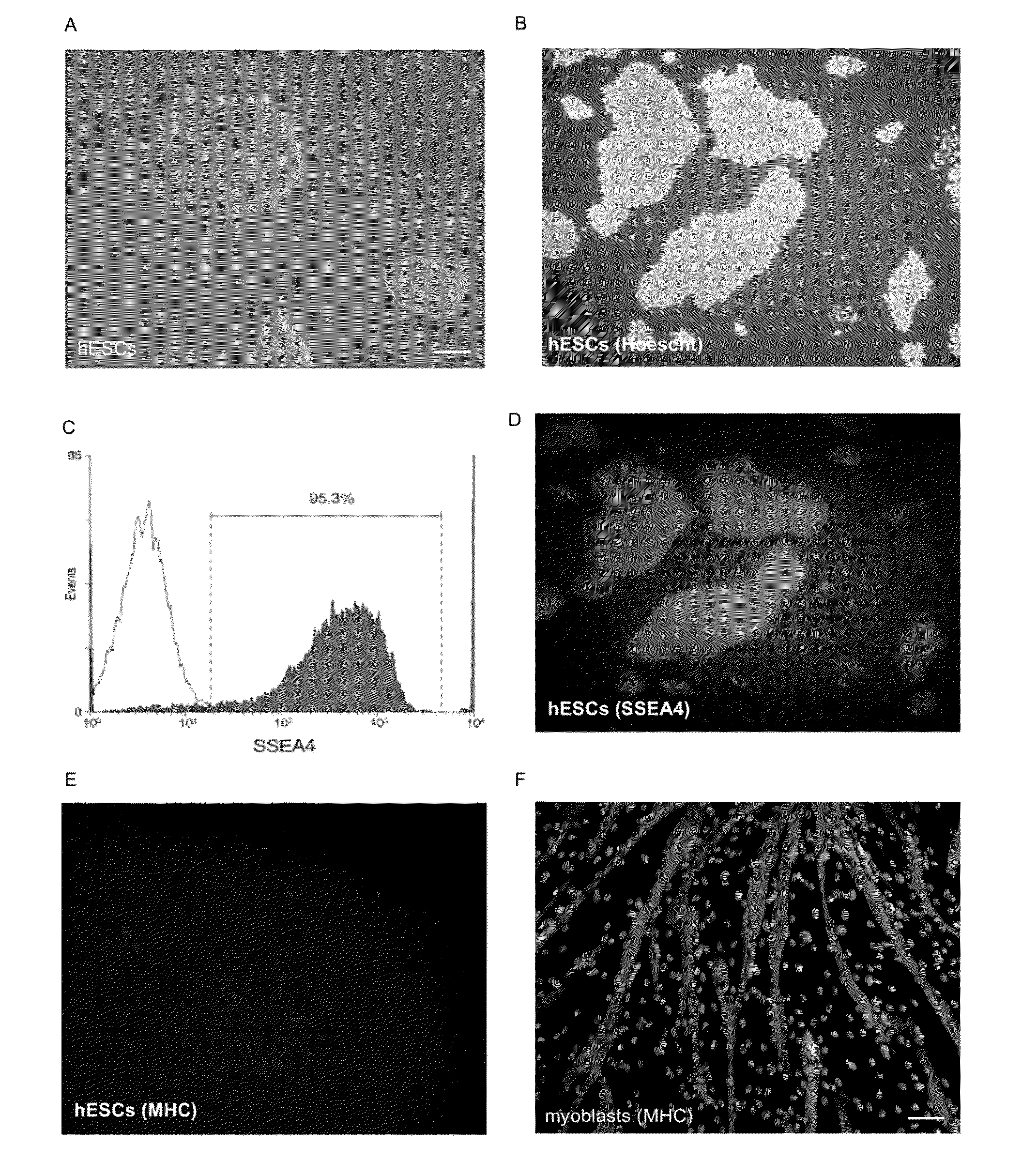
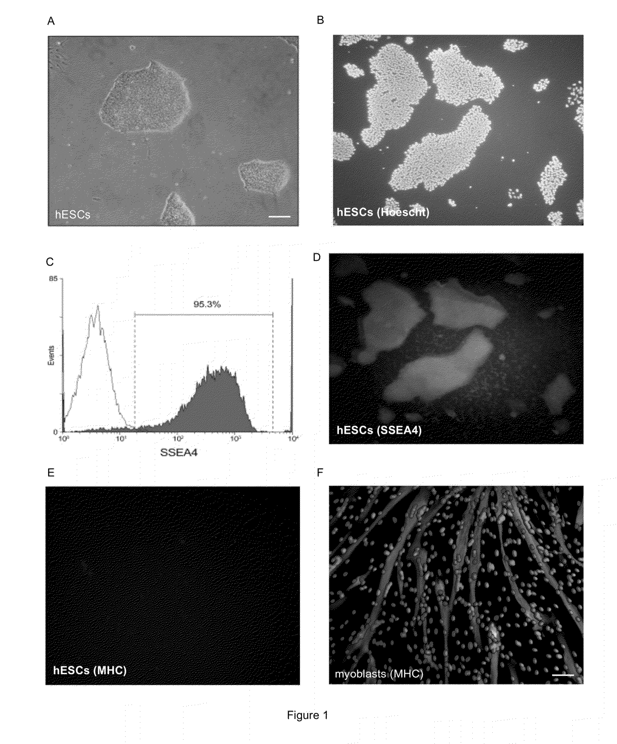
[0115]After a period of mechanical and enzymatic passages, the differentiated cells of the hESC H9 cell line were eliminated and the culture showed characteristics of undifferentiated hESCs. The colonies showed typical characteristics of hESCs grown on MATRIGEL such as a well definite boundary, a high nucleus / cytoplasm ratio and growth in a single layer (FIG. 1A). A FACS analysis for the SSEA4 marker, which is specific to the embryonic stage, confirmed the undifferentiated state of our H9 culture with almost 95.3% of the cells staining positive for this specific antigen (FIG. 1C). Results have been confirmed by immunohistochemistry (FIGS. 1B (Hoechst staining) and D (SSEA4 staining)). At this stage, in contrast to myoblasts (FIG. 1F), the myogenic marker the myogenic marker, Myosin Heavy chain (MHC) was not expressed in hESCs(FIG. 1E). These cells were used for further differentiation experiments.
example 3
Growth in Colonies Block the Myogenic Pathway
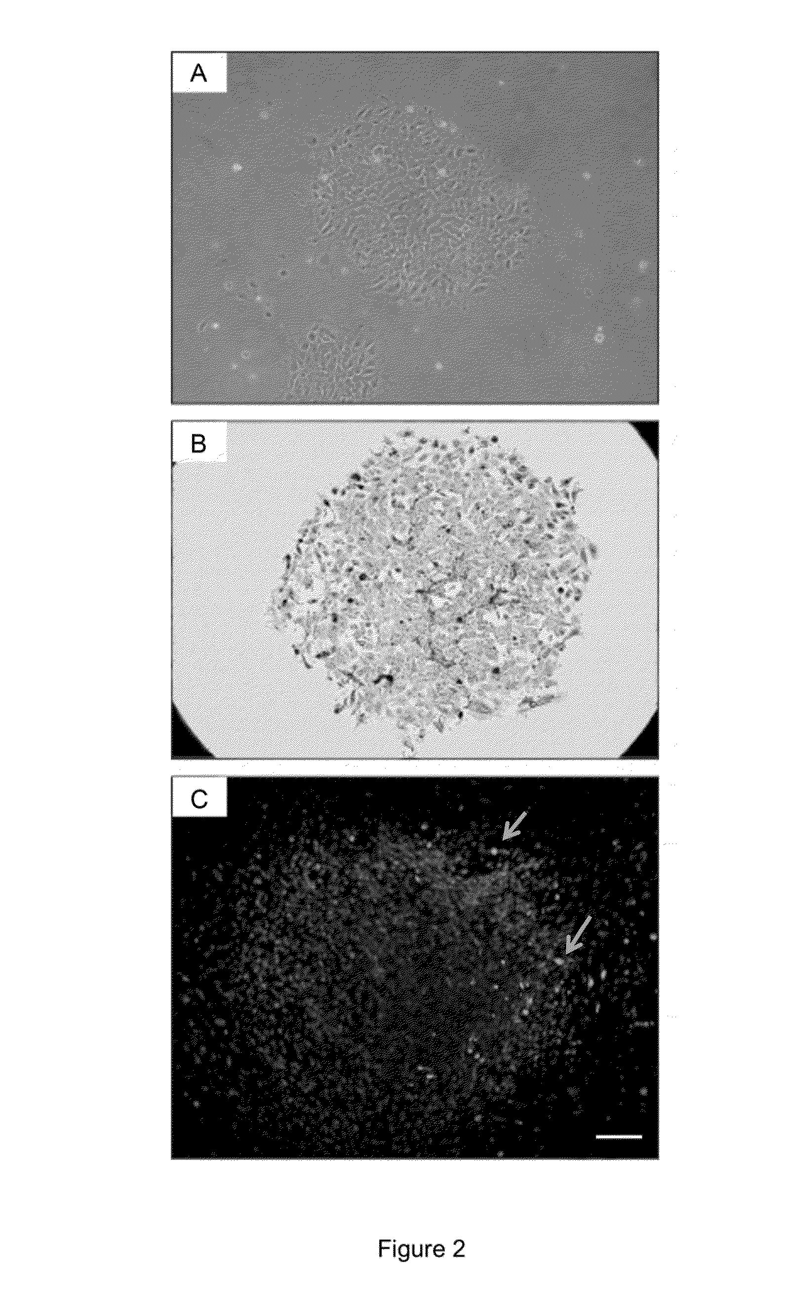
[0116]Initially, hESCs grown in colonies were infected with an adenovirus containing a mouse MyoD gene under a CMV early enhancer / chiken β-actin promotor (ad.CAG-MyoD). The exact MOI could not be established, thus, predetermined / fixed concentrations of Ad.CAG-MyoD and of a control virusfrom 1E5 to 1E8 viral particles per well of a 24 wells plate, were used for infection. An adenovirus (Ad.CAG-GFP) coding for GFP under the same promoter was used as a negative control. As soon as 24 hours after the infection, evidences of differentiation were observed regardless of the virus and of the viral concentration. This could be due to change in the culture medium at the time of viral infection. Colonies started to lose their definite boundary and the cytoplasm of cells expanded (FIG. 2A). However, immunostaining performed five days post-infection revealed that only a few cells of the colonies were infected by the adenovirus (Ad.CAG-MyoD) and expres...
PUM
| Property | Measurement | Unit |
|---|---|---|
| concentration | aaaaa | aaaaa |
| concentration | aaaaa | aaaaa |
| concentration | aaaaa | aaaaa |
Abstract
Description
Claims
Application Information