Methods and materials for the detection of dengue virus infection

a technology of dengue virus and methods, applied in the field of dengue virus diagnosis, can solve the problems of high mortality rate, more severe form of illness, and does not protect against heterologous dengue serotyp

Active Publication Date: 2014-12-30
INBIOS INT
View PDF7 Cites 9 Cited by
  • Summary
  • Abstract
  • Description
  • Claims
  • Application Information

AI Technical Summary

Benefits of technology

[0015]The present disclosure provides monoclonal and polyclonal antibodies, and antigen binding fragments thereof, that are specific for monomeric and oligomeric (primarily dimeric) forms of the dengue virus NS1 polypeptide, together with methods for the use of such antibodies in the diagnosis of dengue virus infection, and kits for use in such methods. The disclosed methods and compositions are effective in the early detection of dengue virus infection and can readily be used in the field, leading to rapid treatment and to control of disease outbreak.
[0020]In certain embodiments, the detection reagent comprises a reporter group selected from the group consisting of: enzymes, substrates, cofactors, inhibitors, dyes, radionuclides, luminescent groups, fluorescent groups, colorimetric indicators and biotin. In the case of an ELISA, the detection antibody can be unlabeled and a goat anti-mouse IgG1 horseradish peroxidase can be employed as a separate detection reagent. Alternatively, the detection antibody can be conjugated to horseradish peroxidase prior to use in the assay. For a lateral flow assay, the detection antibody is preferably labeled with a colorimetric or fluorescent indicator, such as colloidal gold or a fluorescent dye, thereby allowing a user to determine visually whether a test is positive or negative for dengue infection.

Problems solved by technology

However, in the severe form, the infection can progress to hemorrhagic syndrome (referred to as dengue hemorrhagic fever) and fatal hypovolemic shock (referred to as dengue shock syndrome), which has a high mortality rate.
All four dengue serotypes are able to cause clinical symptoms of dengue infection, which provides a life-long immunity to the homologous dengue serotype, but does not protect against heterologous dengue serotype.
Even though the role of NS1 glycoprotein is not clearly defined, some studies have indicated that intracellular NS1 glycoprotein may be indirectly involved in viral replication, with extracellular NS1 glycoprotein being involved in the formation of immunogenic complexes and triggering complement mediated immune response, resulting in a more severe form of illness.
Currently, there is no commercially available vaccine for dengue virus.
As clinical manifestations for dengue virus infections are quite unspecific, it is difficult to affirm diagnosis without laboratory testing.
However, many of these tests have significant disadvantages.
For example, RT-PCR for viral RNA requires expensive laboratory equipment and trained personnel, which makes it hard to use on a large scale or in rural areas.

Method used


Image

  • Methods and materials for the detection of dengue virus infection
  • Methods and materials for the detection of dengue virus infection
  • Methods and materials for the detection of dengue virus infection

Examples


example 1

Preparation of Recombinant NS1 Glycoprotein

[0057]Full-length NS1 protein was prepared using a mammalian expression system. Secreted NS1 was concentrated, purified using glass chromatography, and examined by SDS-PAGE under reducing conditions, with and without heat treatment. No heat treatment would ensure that NS1 dimers can be observed by SDS-PAGE. The results demonstrated the presence of glycosylated monomers, glycosylated dimers and a small fraction of non-glycosylated monomers and non-glycosylated dimers.

example 2

Preparation of Antibodies to NS1 Glycoprotein

[0058]Polyclonal and monoclonal antibodies against the dengue NS1 glycoprotein isolated as described above were prepared as follows.

[0059]30 μg of purified NS1 (prepared as described above) in 50 μl of PBS was emulsified with equal volume of Complete Freund's Adjuvant and injected subcutaneously into a 5 week old female Balb / c mouse. Seven days later, the same amount of NS1 emulsified with Incomplete Freund's Adjuvant was injected intraperitoneally into the same mouse. The injection was repeated once more 21 days after the initial injection. On days 42, 43 and 46, the mouse received intraperitoneal injections of 30 μg of NS1 in 50 μl of PBS without adjuvant. Two days later the animal was sacrificed, and the spleen excised under sterile conditions.

[0060]The spleen was homogenized with scissors under serum free RPMI 1640 medium, and passed through a nylon cell strainer to form a splenocyte suspension. Splenocytes were collected by centrifug...

example 3

Characterization of Antibodies Raised Against NS1

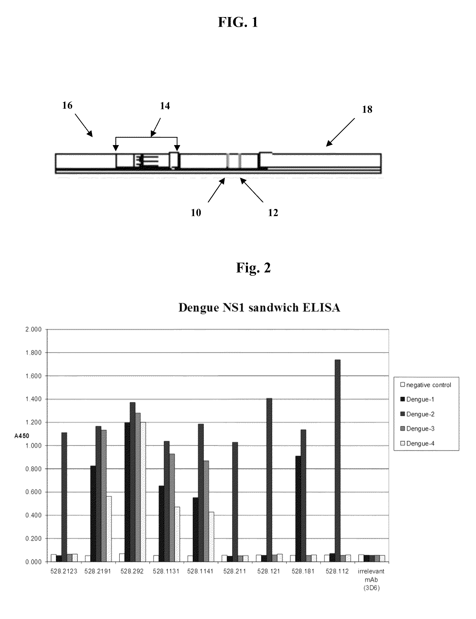
[0066]Affinity purified rabbit polyclonal antibodies to NS1 were coated onto ELISA plates (100 ng per well in 50 μl sodium carbonate / bicarbonate buffer, pH 9.6) overnight at 4° C. The wells were blocked with 200 μl per well of 5% nonfat dry milk in EWB for 1 hour at room temperature, and washed as above. Cell culture supernatants, containing dengue-1, -2, -3 and -4 serotype NS1 were reacted with plate bound rabbit antibodies, followed by incubation with NS1 specific monoclonal antibodies, and developed with goat antibodies specific for mouse immunoglobulin, labeled with horseradish peroxidase. The ELISA results were visualized as above.

[0067]As shown above in FIG. 2, the NS1-specific monoclonal antibodies (mAbs) exhibited at least four classes of specificities: monospecific to dengue-2 (e.g. mAb 528.2123, 528.211, 528.121 and 528.112); all dengue serotype-specific (“pan specific”, e.g. mAb 528.292); dengue-1 / dengue-2 specific (e.g. mA...


PUM

PropertyMeasurementUnit
apparent massaaaaaaaaaa
equilibrium dissociation constantaaaaaaaaaa
volumeaaaaaaaaaa

Abstract

The present invention provides monoclonal antibodies that are specific for the Dengue non-structural glycoprotein NS1 in monomeric and / or oligomeric (primarily dimeric) form, together with methods, including ELISA and lateral flow assays, that employ the disclosed antibodies for the early detection of Dengue virus infection. Diagnostic kits for the detection of Dengue infection are also provided, such kits including the disclosed monoclonal and / or polyclonal antibodies.

Description

STATEMENT REGARDING FEDERALLY SPONSORED RESEARCH OR DEVELOPMENT[0001]This invention was made with government support under grant numbers 1R43AI074172-01 and 5R43AI074172-02 awarded by the National Institutes of Health. The government has certain rights in the invention.FIELD OF THE INVENTION[0002]The present invention relates generally to the diagnosis of dengue virus infection. More specifically, the invention relates to the immunological detection of dengue fever using monoclonal antibodies raised against the dengue glycoprotein NS1.BACKGROUND OF THE INVENTION[0003]Dengue fever is one of most threatening mosquito-borne viral diseases in humans. It is caused by four closely related virus serotypes (DEN1, DEN2, DEN3 and DEN4) of the flavivirus genus within the Flaviviridae family. The dengue virus is transmitted by mosquitoes of the Aedes genus (particularly Aedes aegypti) and causes a febrile illness in tropical and sub-tropical regions. More than two billion inhabitants live in en...

Claims


Application Information

Patent Timeline
Patent Type & Authority Patents(United States)
IPC IPC(8): C07K16/08A61K39/42G01N33/569
CPCG01N2333/185G01N33/56983C07K16/1081C07K2317/33G01N33/54366G01N33/558Y02A50/30G01N33/54388
Inventor RAYCHAUDHURI, SYAMALHOUGHTON, RAYMOND L.MORKOWSKI, STANISLAWSTEVENS, YVONNEBOSE, MOHUANEEDHAM, JAMES WILLIAMCHEN, HONGJING
Owner INBIOS INT
Why Eureka
Try Eureka