System and method for independent manipulation of a fat and a water component in magnetic resonance imaging

water component technology, applied in the field of independent manipulation of a fat and a water component of a magnetic resonance imaging, can solve the problems of inability to achieve optimal clinical segmented inversion recovery protocols, difficult to distinguish between fat and equally bright scar tissue, and poor or no suppression methods when combined with t1-weighted ir. achieve the effect of suppressing fat signal and enhancing visualization of discriminated anatomical elements

Active Publication Date: 2015-05-12
DUKE UNIV +1
View PDF27 Cites 5 Cited by
  • Summary
  • Abstract
  • Description
  • Claims
  • Application Information

AI Technical Summary

Benefits of technology

[0010]A system provides a robust method to independently manipulate a fat and a water component of an MR signal. This system suppresses fat signal in a clinical setting using a timed manipulation of a fat T1-recovery curve that allows substantially complete fat suppression in the presence of an IR pulse, and works for different types of readout. An MR imaging system independently manipulates a fat and a water component of MR signals used for generating image data. An RF (Radio Frequency) signal generator and a magnetic field gradient generator provide an RF pulse and magnetic field gradient sequence for acquisition of an MR signal discriminating between anatomical objects based on longitudinal relaxation time (T1). The sequence comprises, a first pulse sequence for selectively inverting a water component of the MR signal substantially exclusively of fat, a first time delay adjustable to discriminate between different anatomical elements, a second pulse sequence having a resonant frequency selected to invert a fat component of the MR signal substantially exclusively of water and a data acquisition magnetic field gradient for acquisition of the MR signal. An image data processor processes an MR signal acquired to provide a display image with enhanced visualization of discriminated anatomical elements.

Problems solved by technology

Known different fat suppression methods, when combined with T1-weighted IR (for example in contrast-enhanced MR angiography or delayed enhancement) work poorly or not at all.
This can be a problem in image interpretation as it may be difficult to discriminate fat from the other short T1-species present in the image and this is important for making a clinical diagnosis.
In arrhythmogenic right ventricular dysplasia (ARVD), the discrimination between fat and equally bright scar tissue is important and is difficult with known systems.
This method is not optimal in clinical segmented inversion recovery protocols which acquire only a fraction of the data known as a segment (typically 21-29 lines per segment).
This method works poorly at 1.5 T and better, but still not proficiently, at 3 T field strength.
Centric reordering improves this fat suppression method, but is prone to image artifacts.
It is substantially impossible to apply an additional non-frequency selective IR pulse to impart T1-contrast, as the application of both pulses unfavorably alters the image contrast and prevents the suppression of fat signal.
DB preparation is restricted to non-contrast agent applications due to timing limitations.
These methods suppress fat well, but require time-consuming post-processing.

Method used


Image

  • System and method for independent manipulation of a fat and a water component in magnetic resonance imaging
  • System and method for independent manipulation of a fat and a water component in magnetic resonance imaging
  • System and method for independent manipulation of a fat and a water component in magnetic resonance imaging

Examples


Embodiment Construction

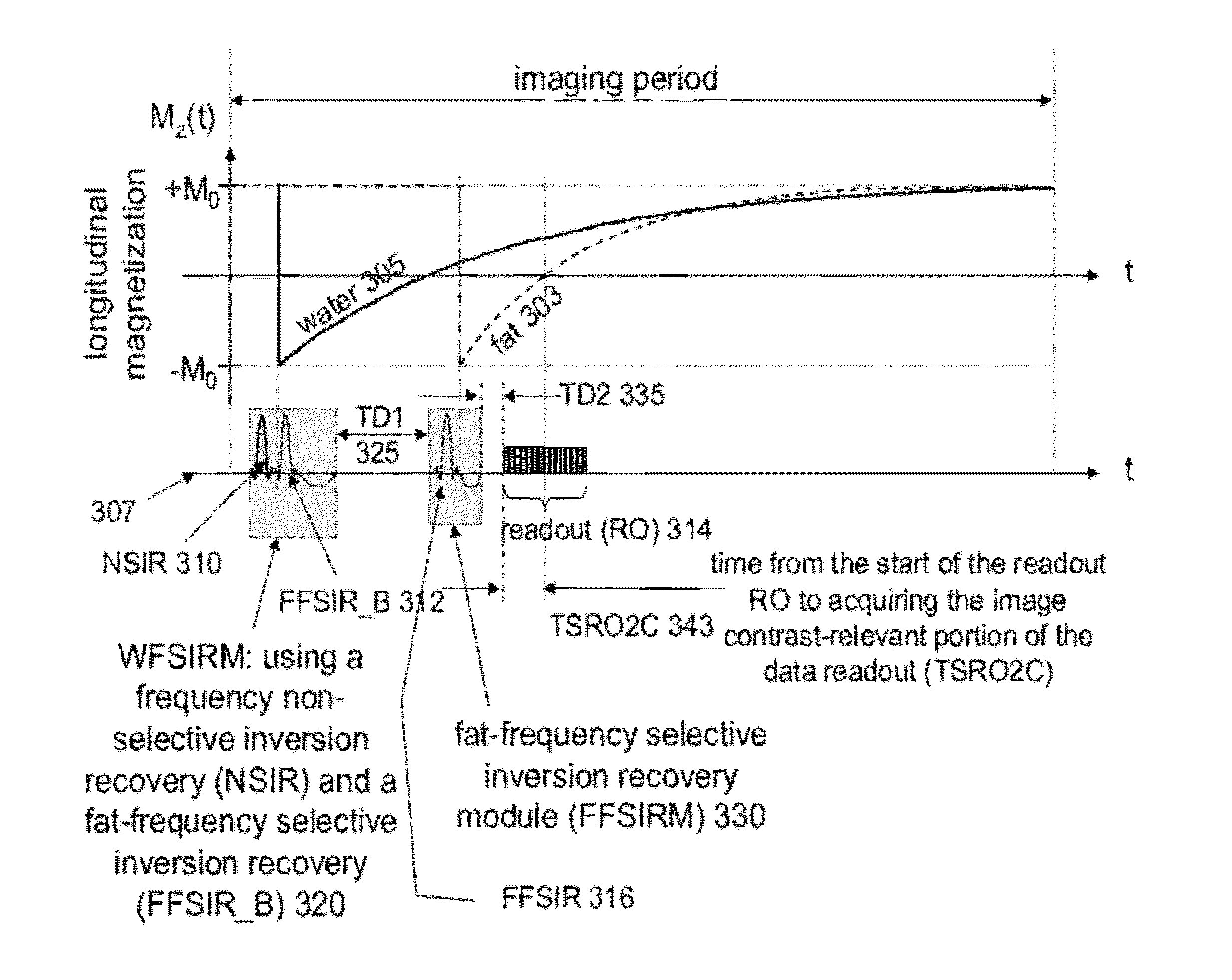
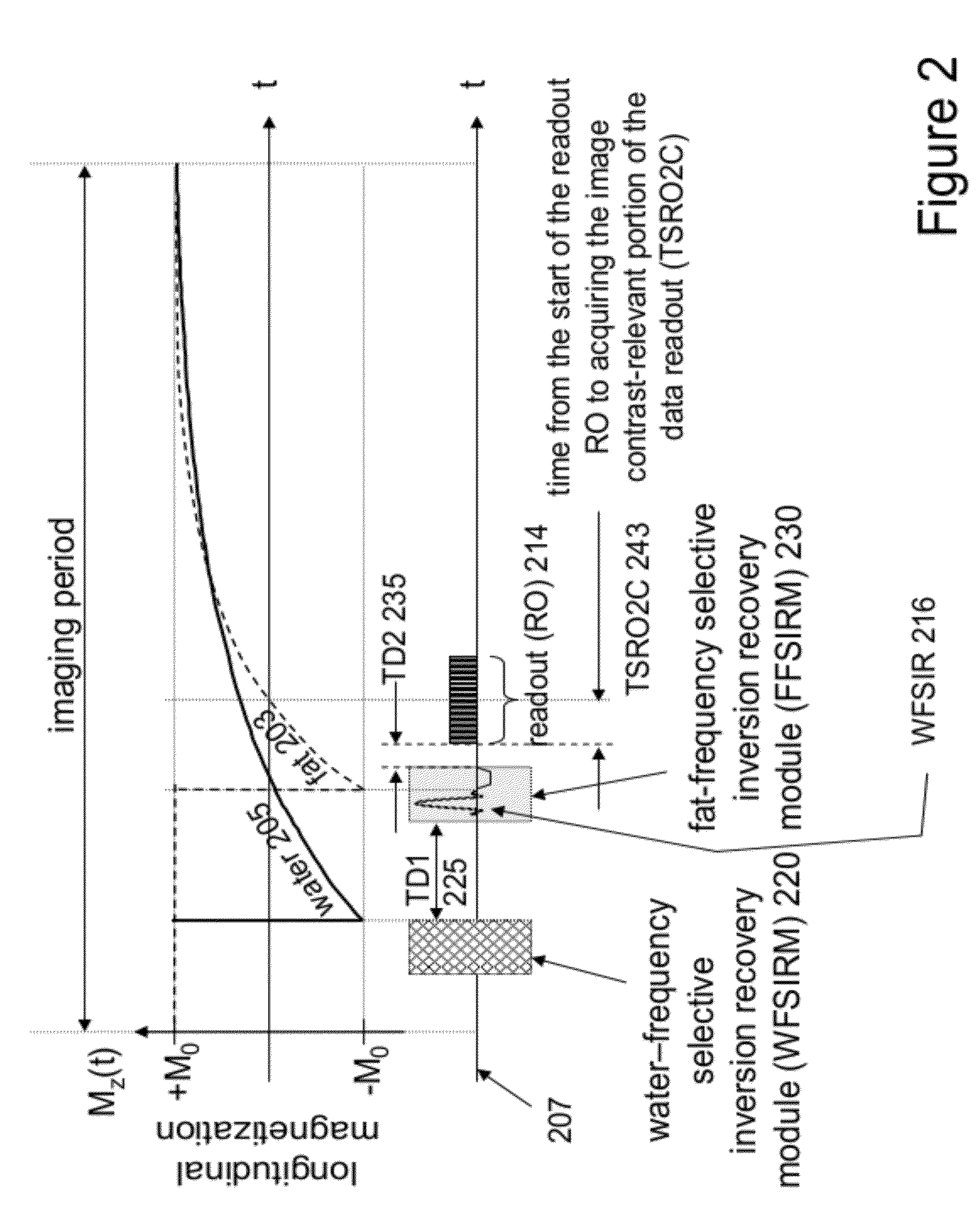
[0023]A system employs a robust method to independently invert the magnetization of an MR water signal and an MR fat signal. The system reliably suppresses an MR fat signal in a clinical setting using a manipulation of a fat T1-recovery curve that enables substantially complete fat suppression in the presence of an RF pulse to invert MR water signal, and works for different types of readout. The system robustly and substantially completely suppresses magnetization and associated signal of fat making fat appear dark (black to dark grey) in magnetic resonance images. The fat suppression works in conjunction with T1-weighted IR imaging. The system in one embodiment, rather than using a suppression pulse (also called “saturation” pulse), uses at least one fat frequency-selective inversion pulse to suppress the fat signal. In one embodiment “fat suppression” (fat nulling) is achieved by selectively restoring the fat magnetization after prior inversion with a frequency non-selective inver...


PUM


Abstract

An MR imaging system independently manipulates a fat and a water component of MR signals used for generating image data. An RF signal generator and a magnetic field gradient generator provide an RF pulse and magnetic field gradient sequence for acquisition of an MR signal discriminating between anatomical objects based on longitudinal relaxation time (T1). The sequence comprises, a first pulse sequence for selectively inverting a water component of the MR signal substantially exclusively of fat, a first time delay adjustable to discriminate between different anatomical elements, a second pulse sequence having a resonant frequency selected to invert a fat component of the MR signal substantially exclusively of water and a data acquisition magnetic field gradient for acquisition of the MR signal. An image shows enhanced visualization of discriminated anatomical elements.

Description

[0001]This is a non-provisional application of provisional application Ser. No. 61 / 436,642 filed 27 Jan. 2011, by W. G. Rehwald et al.FIELD OF THE INVENTION[0002]This invention concerns a system for manipulating a fat and a water component of MR signals used for generating image data and discriminating between anatomical objects based on longitudinal relaxation time (T1).BACKGROUND OF THE INVENTION[0003]T1-weighted inversion recovery (IR) imaging is a common method used in Magnetic Resonance Imaging (MRI) that is frequently performed in conjunction with suppression of fat signal. T1 is the time constant that describes the rate of recovery of longitudinal magnetization. T1-weighted IR is used for imaging different pathophysiologies in different regions of the body, including but not limited to the heart, brain, and vasculature. This method can be used with or without the administration of a T1-shortening contrast agent. Fat suppression methods are used to make fat appear dark in MR i...

Claims


Application Information

Patent Timeline
Patent Type & Authority Patents(United States)
IPC IPC(8): G01V3/00G01R33/48G01R33/56
CPCG01R33/4828G01R33/5607
Inventor REHWALD, WOLFGANG G.JENISTA, ELIZABETH R.KIM, RAYMOND J.
Owner DUKE UNIV
Why Eureka
Try Eureka