Apparatus and method for minimally invasive intracranial hematoma evacuation with real-time assessment of clot reduction

a technology of intracranial hematoma and real-time assessment, applied in the field of minimally invasive surgery, can solve the problems of increased intracranial pressure (icp), elevated icp beyond the normal range can have substantial adverse effects on the health of individuals, paralysis, death, etc., and achieves quick intervention, improved outcomes, and reduced heath care costs

Active Publication Date: 2015-12-15
BLUE BELT TECH
View PDF20 Cites 24 Cited by
  • Summary
  • Abstract
  • Description
  • Claims
  • Application Information

AI Technical Summary

Benefits of technology

[0010]It is an object of some aspects of the present invention to provide improved apparatus and methods for minimally invasive intracerebral hematoma evaluation and evacuation with real time, three-dimensional imaging of the procedure. It is a further object to enable hematoma evaluation and evacuation in a non-operating room setting to enable quicker intervention, better outcomes and reduced heath care cost.

Problems solved by technology

As such, any increase in the volume of the contents of the skull necessarily results in an increase in intracranial pressure (ICP).
While there is some normal variation in ICP on a daily basis, elevated ICP beyond the normal range can have substantial adverse effects on the health of an individual.
These adverse affects can include loss of consciousness, paralysis, coma, difficulty breathing, and death.
Notably, these types of hematomas are dangerous not only because they cause an increase in ICP, but also because they have the capability of shifting the location of other parts of the brain, which can have additional adverse effects on the brain stem.
Intracerebral hematomas (ICH), however, occur within the brain tissue itself and are much more difficult to treat with surgical intervention because they are hard to access without causing additional damage to surrounding brain tissue.
Presently used surgical techniques include craniotomy, stereotactic-guided evacuation, endoscopic evacuation, and catheter evacuation, but each has their problems.
The craniotomy procedure involves removing a portion of the skull so that pressure is relieved and the hematoma can be removed, but it must be performed in a sterile operating room and is likely to induce further trauma to the brain.
Stereotactic-guided evacuation is less invasive than a craniotomy, but requires an enormous amount of equipment, time and cost for a procedure that is ultimately less effective than a craniotomy at reducing the size of the hematoma.
Endoscopic evacuation is not well-known to neurosurgeons, is not well-suited to emergency situations and still requires the use of an operating room and general anesthesia.
Finally, current catheter-based approaches can be performed in the ICU instead of the OR, but necessitate the use of blood thinners and result in slower than desired drainage of the hematoma.
Additionally, repeated catheter placements can cause additional damage to other parts of the brain.
None of these existing surgical techniques is particularly well-suited to the treatment and remediation of an ICH.

Method used


Image

  • Apparatus and method for minimally invasive intracranial hematoma evacuation with real-time assessment of clot reduction
  • Apparatus and method for minimally invasive intracranial hematoma evacuation with real-time assessment of clot reduction
  • Apparatus and method for minimally invasive intracranial hematoma evacuation with real-time assessment of clot reduction

Examples


Embodiment Construction

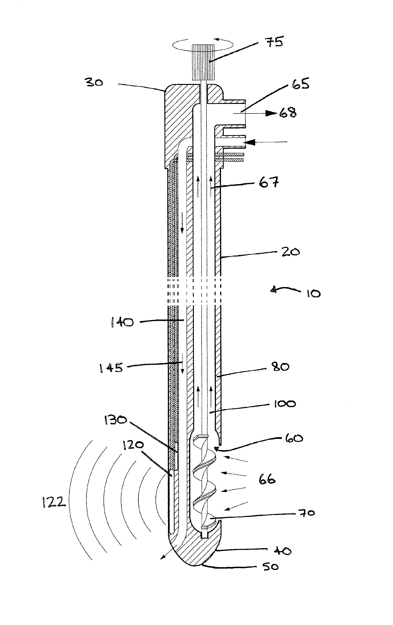
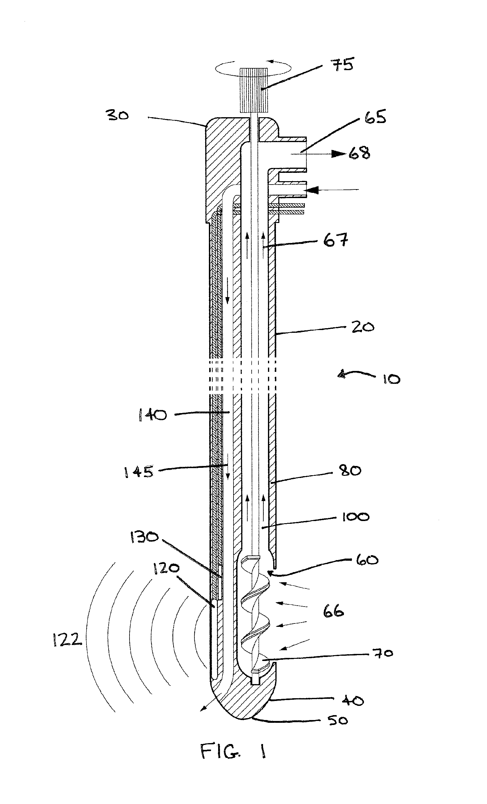
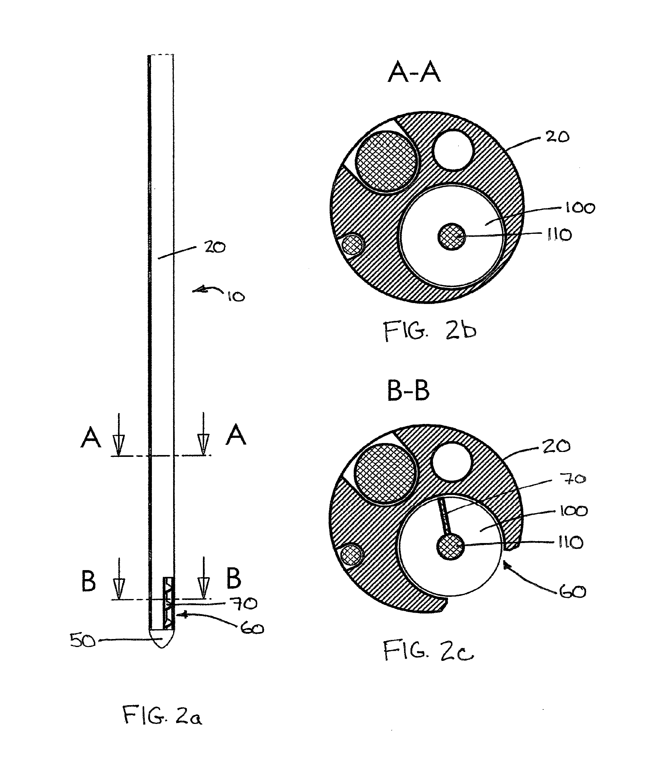
[0020]The present invention provides an improved method and apparatus for the evacuation of intracerebral hematomas. More specifically, an aspect of the present invention is a minimally invasive surgical apparatus within a neuro-navigation system that can provide real-time imaging of the ICH evacuation procedure, including the clot itself. The principal mechanism by which the hematomas are evacuated is an Archimedes screw or auger, which is housed within an apertured lumen and, when placed inside a hematoma and rotated in an appropriate direction, causes the removal of the clotty material. The apparatus also includes ultrasonic imaging capability so that the surgeon can monitor its placement and the evacuation progress in real time. Additionally, an aspect of the invention is that, in a preferred embodiment, the distal portion of the apparatus can house an electromagnetic tracking coil so that the surgeon can use it with a navigation system.

The Probe

[0021]Referring now to FIG. 1, a ...


PUM


Abstract

Method and apparatus for the evacuation of intracerebral hematomas comprises a minimally invasive non-operating room surgical apparatus within a neuro-navigation system that can provide real-time imaging of the ICH evacuation procedure. Apparatus uses an auger housed within an apertured lumen which, when placed in proximity to a hematoma and rotated in an appropriate direction, causes the removal of the clotty material from the hematoma. Apparatus also includes ultrasonic imaging capability and an electromagnetic tracking coil to enable real-time, three-dimensional visualization of the evacuation procedure.

Description

FIELD OF THE INVENTION[0001]This invention relates generally to the field of minimally invasive surgery and, more particularly, to the removal of life-threatening clots from brain tissue.BACKGROUND OF THE INVENTION[0002]Generally, the human skull can be thought of as a fixed volume container. As such, any increase in the volume of the contents of the skull necessarily results in an increase in intracranial pressure (ICP). While there is some normal variation in ICP on a daily basis, elevated ICP beyond the normal range can have substantial adverse effects on the health of an individual. These adverse affects can include loss of consciousness, paralysis, coma, difficulty breathing, and death.[0003]There are many causes of elevated ICP, but the focus of the present application is on intracranial hematomas, which can occur when a vessel in the brain is torn and bleeds. This can occur, for example, as a result of causes as varied as a blow to the head or weakened arterial walls due to a...

Claims


Application Information

Patent Timeline
Patent Type & Authority Patents(United States)
IPC IPC(8): A61B19/00G01R33/28A61B5/06
CPCA61B19/5244G01R33/285A61B5/06A61B17/32002A61B2090/3782A61B5/061A61B17/320783A61B2217/005A61B2217/007A61B90/10A61B2090/364A61B2034/2051
Inventor JARAMAZ, BRANISLAVOH, MICHAEL Y.
Owner BLUE BELT TECH
Why Eureka
Try Eureka