

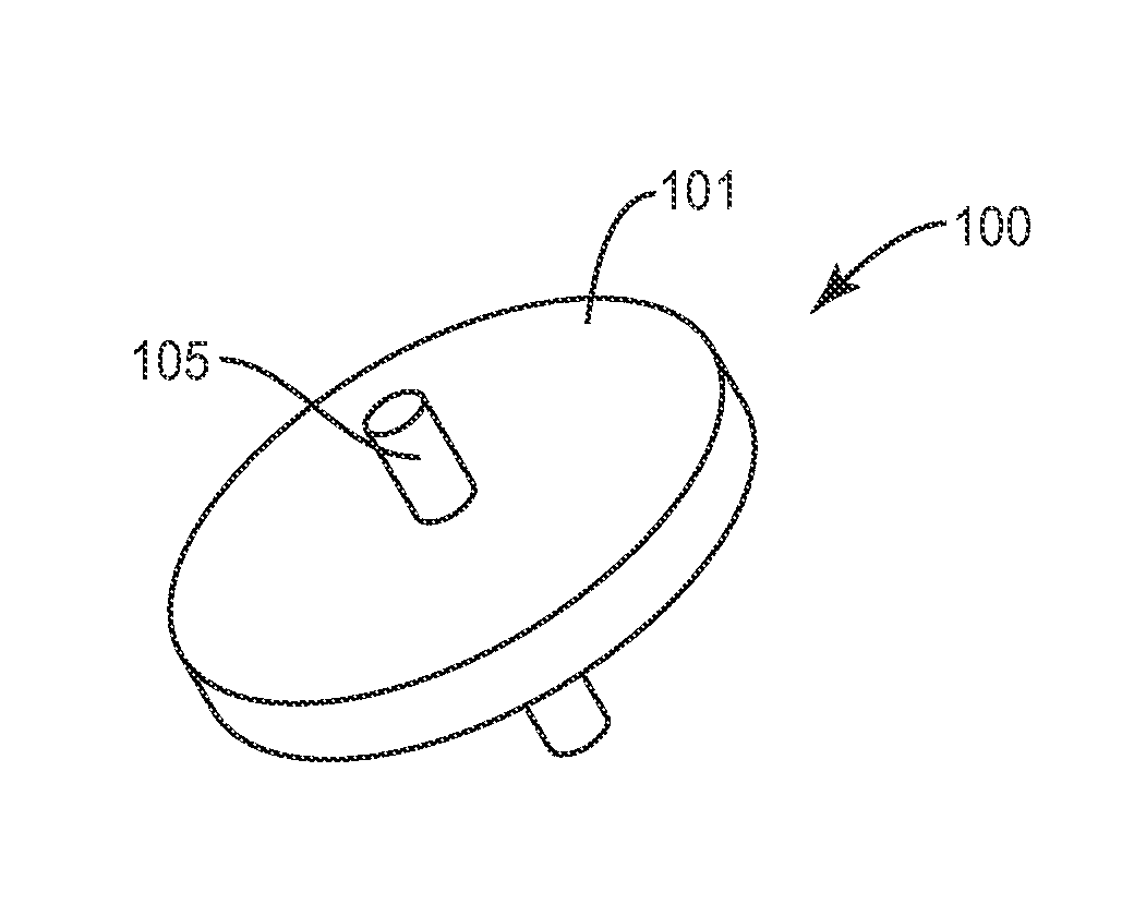
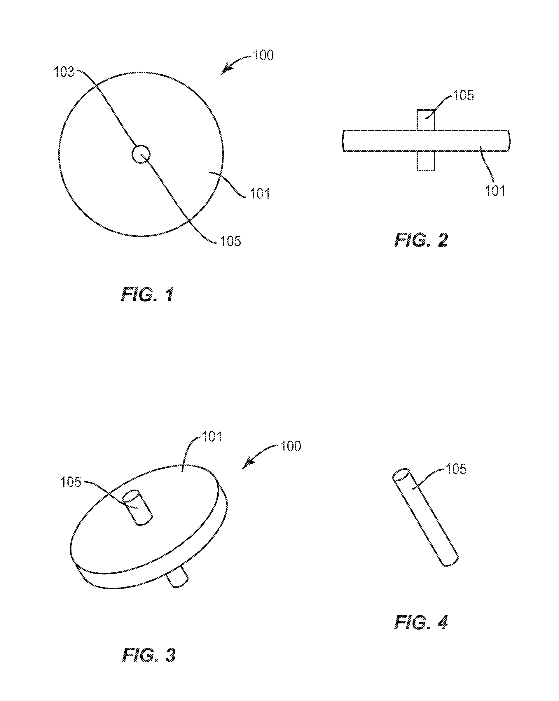
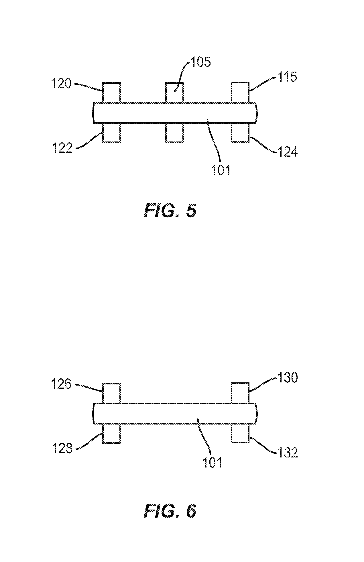
Facet joint implant device
a technology for implanting devices and facet joints, which is applied in the direction of prosthesis, osteosynthesis devices, internal osteosynthesis, etc., can solve the problems of radicular pain, pain and disability for a large segment of the population, and function disruption
- Summary
- Abstract
- Description
- Claims
- Application Information
AI Technical Summary
Benefits of technology
Problems solved by technology
Method used
Image

Examples
Embodiment Construction
[0030]“Bioactive Agent or Bioactive Compound,” as used herein, refers to a compound or entity that alters, inhibits, activates, or otherwise affects biological or chemical events. For example, bioactive agents may include, but are not limited to, osteogenic or chondrogenic proteins or peptides, anti-AIDS substances, anti-cancer substances, antibiotics, immunosuppressants, anti-viral substances, enzyme inhibitors, hormones, neurotoxins, opioids, hypnotics, anti-histamines, lubricants, tranquilizers, anti-convulsants, muscle relaxants and anti-Parkinson substances, anti-spasmodics and muscle contractants including channel blockers, miotics and anti-cholinergics, anti-glaucoma compounds, anti-parasite and / or anti-protozoal compounds, modulators of cell-extracellular matrix interactions including cell growth inhibitors and antiadhesion molecules, vasodilating agents, inhibitors of DNA, RNA or protein synthesis, anti-hypertensives, analgesics, anti-pyretics, steroidal and non-steroidal a...
PUM
| Property | Measurement | Unit |
|---|---|---|
| Length | aaaaa | aaaaa |
| Length | aaaaa | aaaaa |
| Fraction | aaaaa | aaaaa |
Abstract
Description
Claims
Application Information