Method, kit and system for imaging a blood sample

a blood sample and kit technology, applied in the microbiology field, can solve the problems of inaccurate and stably image, inability to accurately and accurately image, and inability to add stain without fixation,

Active Publication Date: 2016-05-03
S D SIGHT DIAGNOSTICS LTD
View PDF59 Cites 50 Cited by
  • Summary
  • Abstract
  • Description
  • Claims
  • Application Information

AI Technical Summary

Problems solved by technology

However, smearing might cause a change in cell morphology.
In addition, with smearing it is difficult to accurately and stably image living cells, for at least the reason that cells dry out quickly and additional staining without fixation is almost impossible.
Manufacturing of chambers having height of this order is not always possible and is relatively expensive.

Method used


Image

  • Method, kit and system for imaging a blood sample
  • Method, kit and system for imaging a blood sample
  • Method, kit and system for imaging a blood sample

Examples


example 1

Detection of Trypanosoma brucei

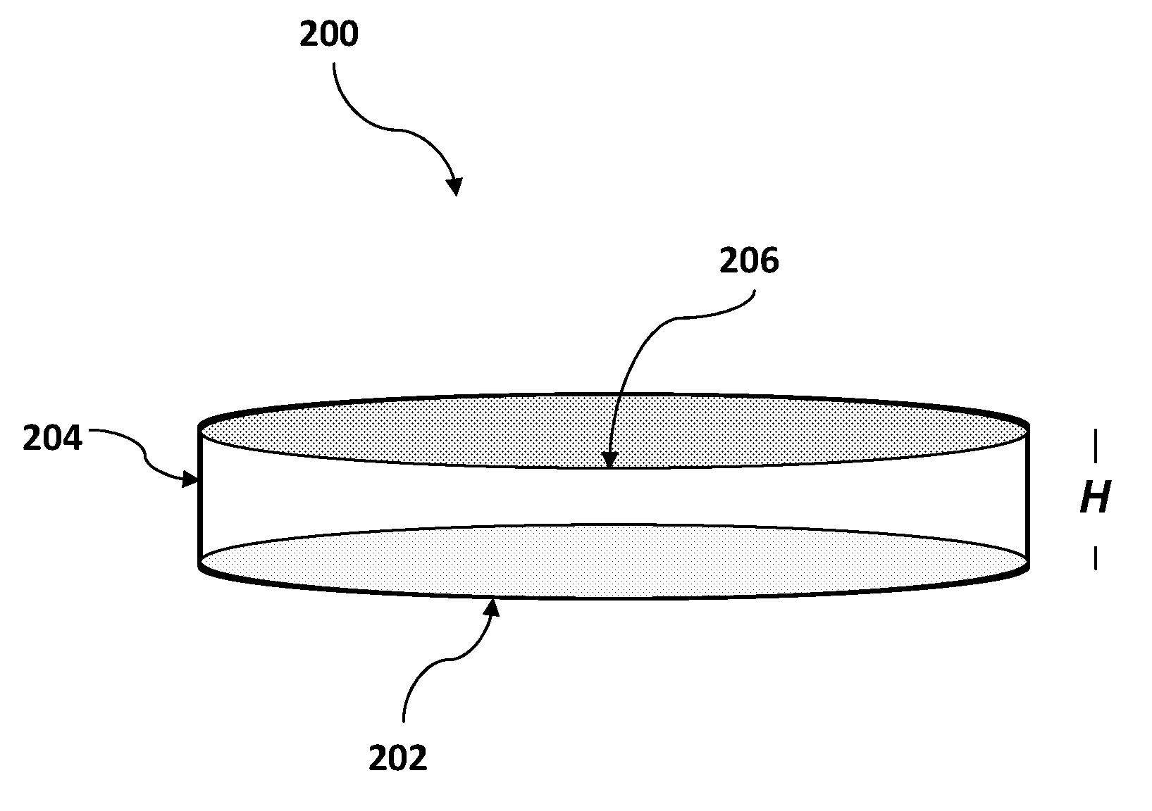
[0183]A mammalian blood sample (human) was assayed as follows for the presence of Trypanosoma brucei. Typically, such blood samples hold between 3,000,000 and 6,000,00 RBC's in each 1 μl (C0). A cartridge having one main chamber was manufactured, whereby the height (H) of the chamber was 100 μM and the chamber could receive a volume of 1 μl.

[0184]The blood sample was diluted 50× (D) to have a surface coverage (F) of between 0.6-0.8 with a 1000 μl solution, containing 1% TRIS-EDTA 1 mM, 15 μl Hoechst 1 μg / μl, 2 μl acridine orange 1 μg / μl, 99% buffered saline. The sample was loaded into the chamber and the chamber was transferred to a microscope stage for imaging in both brightfield and in fluorescence at excitation 370 nm and 475 nm and emissions of approximately 433 nm and 517 nm, and 613 nm using an automated microscopy device. Sedimentation of the sample occurred at about 1 second per 1 μM height of the chamber (H), which is approximately 90 seconds...

example 2

Detection of Plasmidium. falciparum

Staining Solution Composition

[0187]The purpose of this example stain solution is to identify live pathogen (e.g. Plasmodium) inside living blood cells. The solution comprises Hoechst 33342 (excitation 350 nm) and Acridine Orange (excitation 500 nm). The dyes were mixed with saline and Tris-EDTA to create an isotonic solution to keep red blood cells at physiological conditions during the stain and prevent them from lysing. This solution was used as a dilution solution thus potentially providing dyes and diluting the cells in a single step.

Stating Blood Sample for Detection of Plasmodium

[0188]Blood previously mixed with EDTA (or any other anti-coagulant) was diluted in the above stain solution (˜1:100). Within 10 seconds the blood was stained with the chemical dyes and giving off fluorescent signals between 450 nm and 550 nm when appropriately illuminated. The solution mixed with the blood was loaded into a plastic cartridge. After the blood cells ...


PUM

PropertyMeasurementUnit
vertical depthaaaaaaaaaa
heightaaaaaaaaaa

Abstract

Provided is a method for imaging a blood sample and a kit and system for executing the method. The method includes introducing a cell suspension including red blood cells onto a base surface of a carrier having a vertical height (H) being greater than or equal to a vertical depth (h) of the cell suspension when on the base carrier, the cell suspension including a cell concentration (C) being determined by a defined function; allowing the cells in the cell suspension to settle on the base surface of the carrier to form a monolayer of cells thereon; and acquiring at least one microscope image of at least a portion of the monolayer of cells; wherein the at least one microscope image is obtained by a microscope set to Depth Of Field that is not more than 20% of the vertical height of the cell suspension settled on the base surface.

Description

TECHNOLOGICAL FIELD[0001]The present disclosure is in the field of microbiology and in particular to methods relating to cell sample preparations and imaging thereof, for use, inter alia, in diagnosis.PRIOR ART[0002]References considered to be relevant as background to the presently disclosed subject matter are listed below:[0003]Anthony Moody Various rapid diagnostic tests for Malaria parasite in Clinical Microbiology Reviews January 2002 p. 66-78;[0004]Vink J P. et al. An automatic vision-based malaria diagnosis system Journal of Microscopy, 2013, p. 1-13;International patent application publication No. WO 2010 / 116341;[0005]“Counting blood cells with countess Automated Cell Counter” found at http: / / www.lifetechnologies.com / content / dam / LifeTech / migration / files / cell-tissue-analysis / pdfs.par.83996.file.dat / w-082149-countess-applicatio-blood-cells.pdf;[0006]U.S. Pat. No. 4,209,548;[0007]U.S. Pat. No. 4,494,479;[0008]U.S. Pat. No. 6,819,408;[0009]Leif R C. et al Methods for Preparing S...

Claims


Application Information

Patent Timeline
Patent Type & Authority Patents(United States)
IPC IPC(8): G01N21/64G02B21/36G01N33/49G01N1/28G01N1/38G01N15/14G01N15/00
CPCG01N21/6428G01N1/2813G01N1/38G01N15/1463G01N15/1475G01N33/49G02B21/361G01N2015/0065G01N2021/6439Y02A50/30G01N21/6458G02B21/0012G02B21/16G02B21/34G02B21/367C12Q1/04G01N21/6486
Inventor POLLAK, JOSEPH JOELHOURI YAFIN, ARNONSALPETER, SETH J.
Owner S D SIGHT DIAGNOSTICS LTD
Why Eureka
Try Eureka