Retinal pigment epithelial primary cell culture system producing subcellular deposits

a cell culture system and pigment technology, applied in the field of retinal pigment epithelial primary cell culture system, can solve the problems of inability to understand the pathogenic mechanisms involved in macular degeneration and testing new treatments, and inability to accurately model the cell culture system, so as to achieve the epithelial phenotype. , the effect of maintaining the epithelial phenotyp

Inactive Publication Date: 2016-08-23
CURCIO CHRISTINE A +1
View PDF0 Cites 0 Cited by
  • Summary
  • Abstract
  • Description
  • Claims
  • Application Information

AI Technical Summary

Problems solved by technology

Progress towards understanding pathogenic mechanisms involved in macular degeneration and testing new treatments has been hindered by the lack of an accurate model system such as an engineered mouse that exhibits the pathognomonic lesions of the disease, (e.g., drusen and basal linear deposits (BlinD).
Research shows that RPE cells are phenotypically unstable when removed from their host eye and maintained under routine tissue culture conditions.

Method used


Image

  • Retinal pigment epithelial primary cell culture system producing subcellular deposits
  • Retinal pigment epithelial primary cell culture system producing subcellular deposits
  • Retinal pigment epithelial primary cell culture system producing subcellular deposits

Examples


example 1

Establishment of a RPE Primary Cell Culture System

[0061]The methods used to procure animal tissues are approved by the Institutional Review Board at the University of Alabama at Birmingham. RPE cells are dissociated from porcine eyecups, purified and maintained in culture as previously described (citation IOVS 48:1892). Briefly, eyes enucleated from anesthetized animals are transported to the laboratory in sterile ice-cold normal saline. After removal of the anterior segment of the eye and the vitreous, the posterior eyecup is incubated in Leibovitz (L-15) medium; Invitrogen Corp., Carlsbad, Calif.) at room temperature for 30 minutes and the retina detached with forceps and scissors to expose the RPE monolayer. RPE cells are harvested by serial 30-minute incubations at 37° C. in 0.25% trypsin (Sigma-Aldrich, St. Louis, Mo.) in L-15 and collected by repeated gentle trituration with a 1-mL sterile pipette. To facilitate dissociation, trypsin-released RPE cells are incubated with 1% DN...

example 2

Assaying Agents for Modulation of the Production of Subcellular Deposits by the RPE Primary Cell Culture System

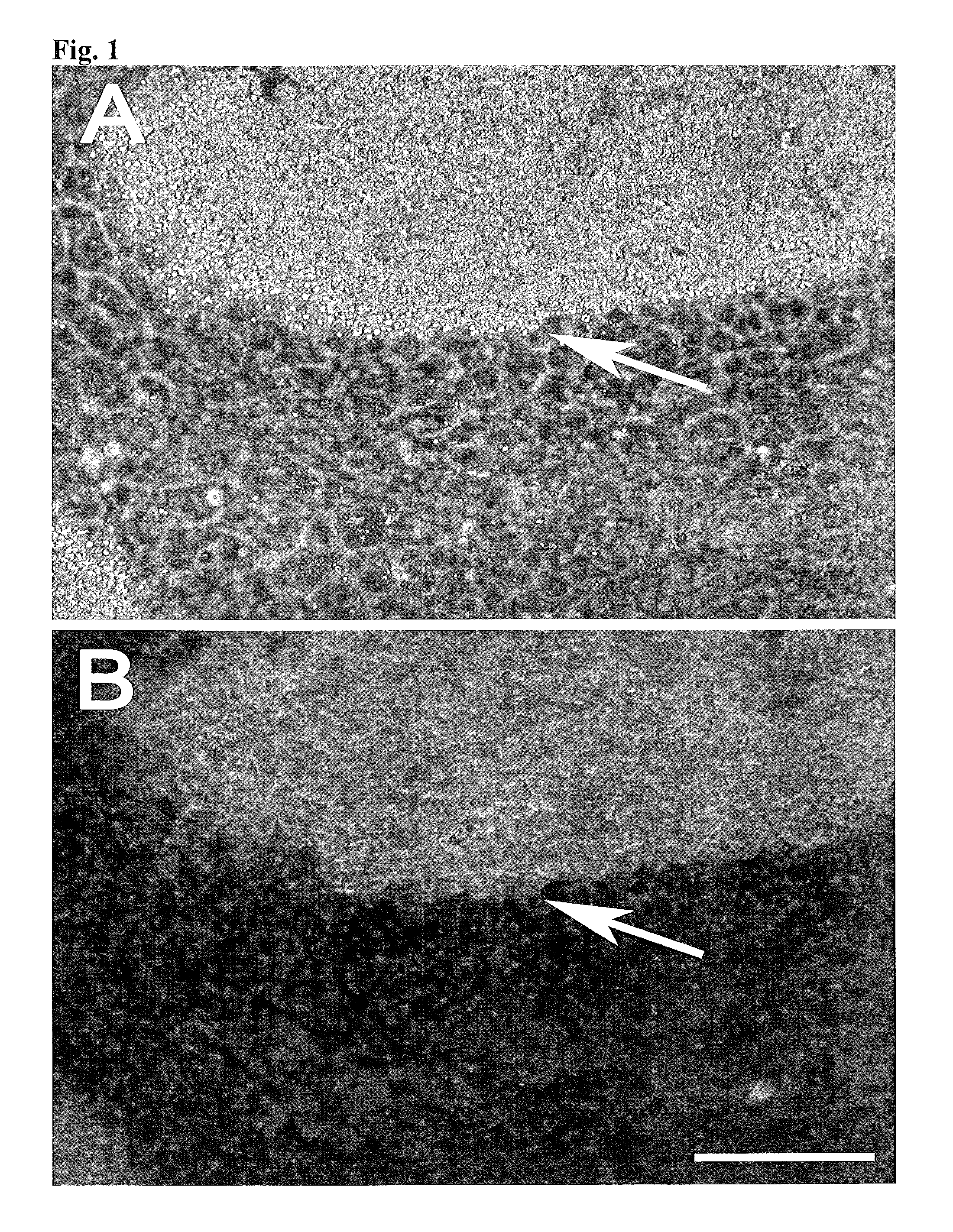
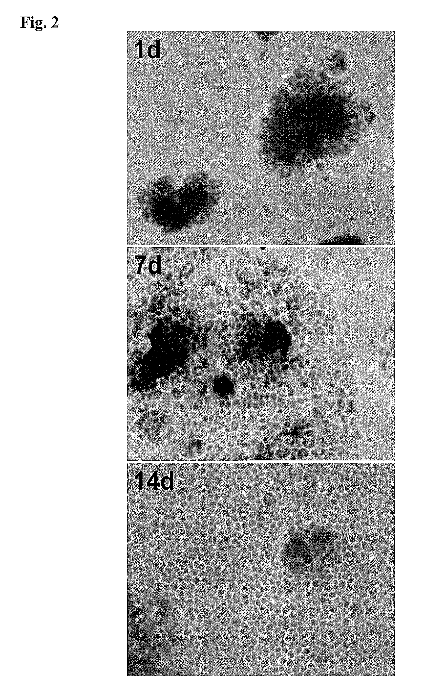
[0063]A RPE primary cell culture system produced as described in Example 1 is contacted with a varying concentration of a candidate agent. The cell culture system is maintained in the presence of the candidate agent for varying periods of time and periodically assayed for changes in the production of subcellular deposits as compared to a control RPE primary cell culture system produced as described in Example 1 but not contacted with the candidate agent. The subcellular deposits produced by the RPE primary cell culture systems are quantified using, for example, dark field photomicrography. Changes that are observed in the RPE primary cell culture system that is contacted with the agent include decreased production of the subcellular deposits by; increased production of the subcellular deposits; prevention of the accumulation of the subcellular deposits; and / or prevention of...


PUM

PropertyMeasurementUnit
sizeaaaaaaaaaa
timeaaaaaaaaaa
timeaaaaaaaaaa

Abstract

The present invention provides a retinal pigment epithelial (RPE) primary cell culture system on a material surface seeded at a high density that produces a layer of subcellular deposits, wherein the RPE primary cells are harvested from non-fetal tissue. The present invention additionally provides methods of making and using the PRE primary cell culture system.

Description

STATEMENT OF GOVERNMENT SUPPORT[0001]This invention was made with government support under Grant No R01 EY06109 awarded by The National Institutes of Health / National Eye Institute. The government has certain rights in the invention.FIELD OF THE INVENTION[0002]The present invention provides a retinal pigment epithelial primary cell culture system and methods of making and using the cell system.BACKGROUND OF THE INVENTION[0003]Age-related macular degenerations (AMD) is a highly prevalent retinal disease in the elderly. The most prominent histopathological and clinical signs of AMD are subcellular lesions (drusen and basal linear deposits), which are neutral lipid-rich subcellular lesions that develop behind the retinal pigment epithelium. Drusen and BLinD are two physical forms—lump and layer—of the same lipid-rich lipoprotein derived debris. Progress towards understanding pathogenic mechanisms involved in macular degeneration and testing new treatments has been hindered by the lack o...

Claims


Application Information

Patent Timeline
Patent Type & Authority Patents(United States)
IPC IPC(8): C12Q1/00C12N5/079G01N33/50C12N5/00
CPCG01N33/5058C12N5/0621G01N33/5035
Inventor CURCIO, CHRISTINE A.GUIDRY, CLYDE
Owner CURCIO CHRISTINE A
Why Eureka
Try Eureka