Cancer diagnosis and imaging

a cancer diagnosis and imaging technology, applied in the field of cancer diagnosis and imaging, can solve the problems of inaccurate and imprecise conventional cancer imaging, improper diagnosis, etc., and achieve the effect of stopping the proliferation of eukaryotic cells

Inactive Publication Date: 2016-08-30
CAVION INC
View PDF81 Cites 9 Cited by
  • Summary
  • Abstract
  • Description
  • Claims
  • Application Information

AI Technical Summary

Benefits of technology

[0009]The present invention provides a method for imaging a disease or condition in a mammal which includes the steps of: administering a therapeutically effective amount of a cell cycle inhibitor to effectively stop proliferation of eukaryotic cells at a cell cycle checkpoint between the G1 and S phase, stopping administration of the cell cycle inhibitor for a period of time, administering a marker to the mammal and imaging the mammal.

Problems solved by technology

Conventional cancer imaging is inaccurate and imprecise.
The reason why conventional cancer imaging is inaccurate and imprecise is because, in part, the metabolic uptake of an imaging marker has a cell cycle-specific mechanism of action.
This variance, which exists between all of the cancer cells throughout the patient, can cause misleading results and improper diagnoses.

Method used


Image

  • Cancer diagnosis and imaging
  • Cancer diagnosis and imaging
  • Cancer diagnosis and imaging

Examples


example 1

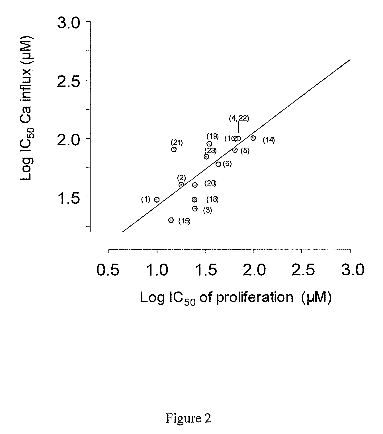
[0041]To measure the correspondence between calcium influx inhibition and proliferation, the ability of several chemical agents to inhibit calcium influx was plotted against the ability of the same agent to inhibit proliferation, as can be seen in FIG. 2 below. These agents were proprietary chemical entities synthesized at the University of Virginia. A least squares correlation line with a slope of 0.98 and R2 value of 0.92 was obtained. Conventionally, a correlation cannot be used to infer causality, but in this case, a Bayesian approach is warranted because calcium influx is necessary a priori for proliferation, such that blocking calcium entry will correspondingly block proliferation. A regression line with a slope very near 1 means that essentially all of the variation in the variables is accounted for by variation in the other, meaning that there is no action of these agents on proliferation other than inhibition of calcium entry.

example 2

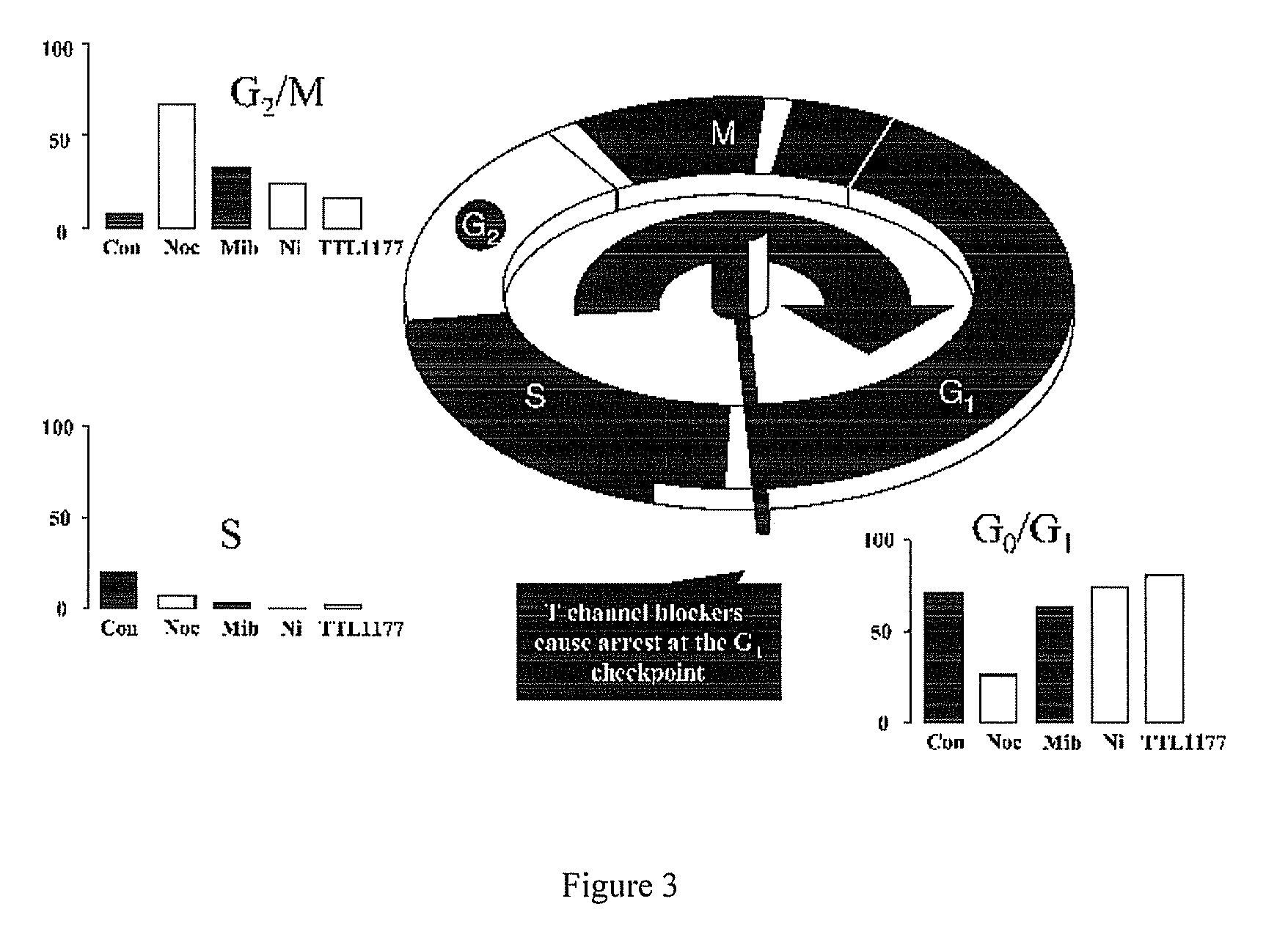
[0042]Cell cycle analysis was performed using flow cytometry and BUdR staining. In FIG. 3, “Con” represents untreated, control cells. All other cell cultures were treated with nocodazole, which interferes with microtubule polymerization and blocks the cell cycle during M phase. Treatment of A10 cells with nocodazole alone blocked cell cycle transit through M phase as determined by flow cytometry. Because A10 cells reside primarily in the cell cycle checkpoint between G0 and G1 (See “Con” in FIG. 3), evaluating the possibility that a pharmacologic agent inhibits transit out of that phase is not as straightforward as determining blockade at other cell cycle checkpoints. Therefore, cell cultures were treated for 24 hours with T channel calcium channel blockers mibefradil (mib), nickel (Ni) or TTL-1177 (a proprietary compound owned by the assignee Tau Therapeutics), before adding nocodazole. Absent an effect on the T channel blockers, cells would be locked at the cell cycle checkpoint b...

example 3

[0043]To measure the efficacy of the cell cycle inhibitor mibefradil at stopping progression through the cell cycle, human lung cancer cells of cell line A549 and human prostate cancer cells of cell line were incubated with 10 μM mibefradil for 48 hours (M48H). As can be seen in FIG. 4, the mibefradil increased the proportion of cells in the G1 phase of the cell cycle by not allowing progression to the S phase, for both cell lines. Further data was collected by incubating the same cell lines with 10 μM of mibefradil for 18 hours followed by a 12 hour washout followed by another 8 hour incubation with mibefradil (M18-12-18h). These results also show a greater proportion of cells in the G1 phase as compared to control cells not incubated with mibefradil.


PUM

PropertyMeasurementUnit
timeaaaaaaaaaa
timeaaaaaaaaaa
timeaaaaaaaaaa

Abstract

The present invention provides a method for imaging a disease or condition in a mammal which includes the steps of; administering a therapeutically effective amount of a cell cycle inhibitor to effectively stop proliferation of eukaryotic cells at a cell cycle checkpoint between the G1 and S phase, stopping administration of the cell cycle inhibitor for a period of time, administering a marker to the mammal and imaging the mammal.

Description

CROSS-REFERENCE TO RELATED APPLICATIONS[0001]This application claims the benefit of U.S. Provisional Application No. 61 / 309,030 filed on Mar. 1, 2010.BACKGROUND OF THE INVENTION[0002]Conventional cancer imaging is inaccurate and imprecise. Typically a patient has a marker, or other chemical compound, injected into their circulatory system, waits about 1 hour and is then imaged using Positron Emission Tomography (PET). The 1 hour time frame is quite short and may not be representative of the general activity of the entire number of cancer cells.[0003]The reason why conventional cancer imaging is inaccurate and imprecise is because, in part, the metabolic uptake of an imaging marker has a cell cycle-specific mechanism of action. More specifically, uptake of the imaging marker is a function of which part of the cycle targeted cell are in. During any given hour, which is the typical wait time between marker administration and imaging, the cancer cells to be imaged may be at any stage of...

Claims


Application Information

Patent Timeline
Patent Type & Authority Patents(United States)
IPC IPC(8): A61K31/4184A61K31/675A61K31/4015A61K45/06A61K33/24A61K51/04
CPCA61K31/4015A61K31/4184A61K31/675A61K33/24A61K45/06A61K51/0491A61K2300/00G16H30/20
Inventor GRAY, LLOYD S.
Owner CAVION INC
Why Eureka
Try Eureka