Fibrosis biomarker assay

a biomarker and fibrosis technology, applied in the field of fibrosis biomarker assay, can solve the problems of increasing tissue size and density, impaired function, and no approved treatment directly targeting the fibrosis mechanism

Active Publication Date: 2017-03-28
NORDIC BIOSCI
View PDF0 Cites 5 Cited by
  • Summary
  • Abstract
  • Description
  • Claims
  • Application Information

AI Technical Summary

Problems solved by technology

Despite its enormous impact on human health, there are currently no approved treatments that directly target the mechanisms of fibrosis 5.
Fibrogenesis is the result of an imbalance in normal ECM regulation that alters the concentration of macromolecules leading to increased tissue size and density, with progressively impaired function.
The imbalance between synthesis and degradation of ECM during fibrogenesis, results from conversion of the low-density subendothelial matrix into matrix rich in interstitial collagens.
However, no evidence for this is provided and there have to our knowledge been no subsequent publications validating this prediction.
Such RIA assays have been commercially available for many years and no reports of their successful use in diagnosing or monitoring any fibrotic disease have appeared.
The patent does not teach the quantitative measurement of peptide fragment carrying neo-epitopes generated during fibrotic disease.
However, the patent does not describe the measurement of connective tissue proteins based on the binding of antibodies to neo-epitopes on said connective tissue proteins.

Method used


Image

  • Fibrosis biomarker assay
  • Fibrosis biomarker assay
  • Fibrosis biomarker assay

Examples


example 1

Collagen Type III Degraded with MMP-9

Method

[0141]Cleavage: Collagen type III isolated from human placenta was dissolved in 10 mM acetic acid (1 mg / ml). The protein solution was then passed through a filter (Microcon Ultracel YM-10) to remove fragment contaminations. MMP-9 was preactivated with 4-aminophenylmercuric acetate (APMA, Sigma) at 37° C. for 3 hours. After activations, collagen type III and MMP-9 were mixed 100:1 and incubated shaking for 3 days at 37° C.

[0142]The solution was analyzed by liquid chromatography / mass spectrometry (LC / MS) and the fragments were identified by performing Mascot Search. The peptide sequences were selected by homology search, ensuring no cross-reactivity to other or related proteins, as well as interspecies cross-reactivity.

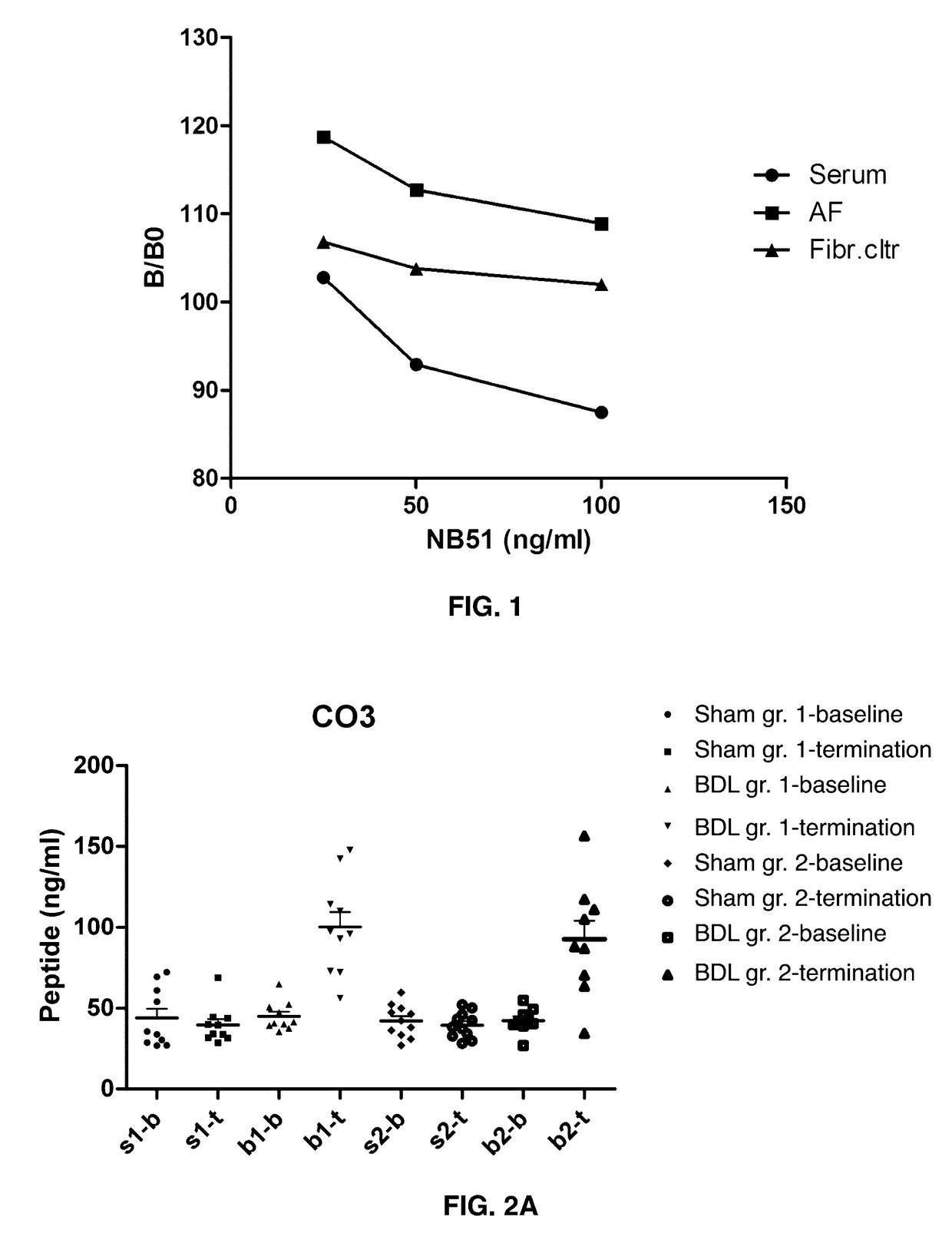
[0143]Antibody design: The peptide sequences were synthesized and conjugated to ovalbumin (OVA). Mice were immunized ever 2-3 weeks, up to five. Antibody titers were checked by screening peptides, both selection and de-selectio...

example 2

CO3 in Biological Relevant Samples

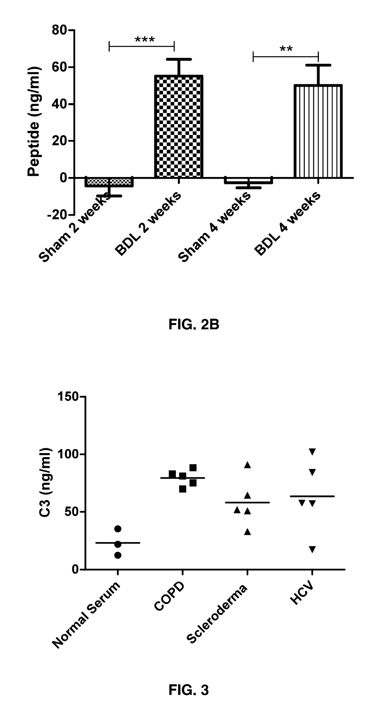
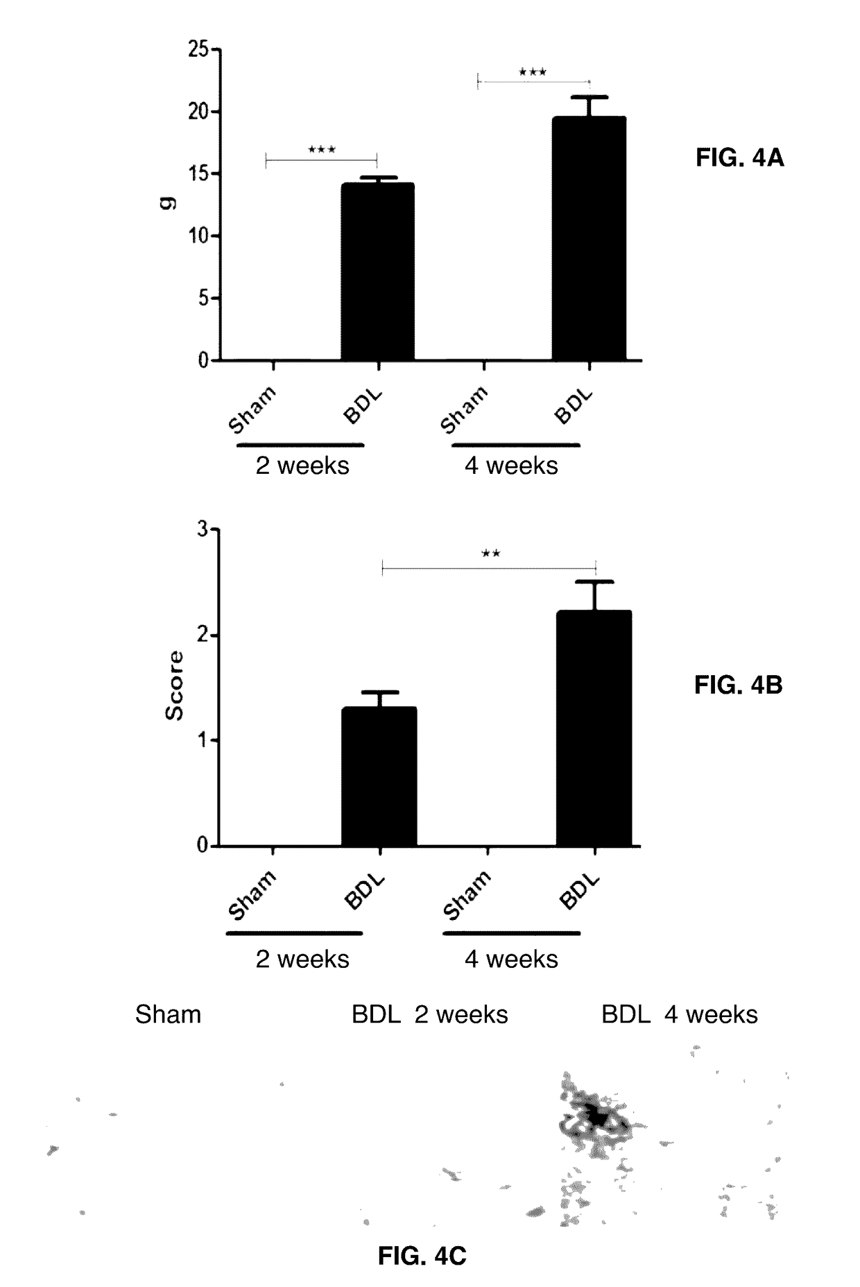
CO3 Levels in Bile Duct Ligated Rats Compared to Sham Operated Rats.

[0146]Method: Forty female Sprague-Dawley rats (6 months old) were housed at the animal research facilities at Nordic Bioscience. The experiments were approved by the Experimental Animal Committee of the Danish Ministry of Justice, and were performed according to the European Standard for Good Clinical Practice (2008 / 561-1450). The rats were housed in standard type III-H cages at 18-22° C. with bedding and nest material (Altromin 1324; Altromin, Lage, Germany) and purified water (Milli-Q system; Millipore, Glostrup, Denmark) ad libitum. Rats were kept under conditions of a 12-hour light / dark cycle.

[0147]Liver fibrosis was induced by common BDL. In short: The rat was anaesthetized, the bile duct found, two ligations were performed around the bile duct followed by dissection between the ligations, the abdomen was closed. In sham operated rats, the abdomen was closed without bile duct ...

example 3

CO3 in Different Fibrotic Diseases (Human Serum)

[0149]CO3 levels were measured in serum from human with three different fibrotic diseases: Chronic obstructed pulmonary disease (COPD), Scleroderma, and Hepatitis virus C (HCV). The serum samples were retrieved from Sera Laboratories International Ltd (SLI Ltd), UK. CO3 levels were increased in the three different fibrotic diseases (FIG. 3).


PUM


Abstract

Provided herein are methods of diagnosis or of quantitation of fibrosis. An immunoassay is conducted to measure neo-epitope containing protein fragments of collagen type III, collagen type I, collagen type IV, collagen type V, or collagen type VI, elastin, biglycan, decorin, lumican, versican, perlecan, neurocan, brevican, fibromodulin, serglycin, syndecan, betaglycan, vimentin, or C-reactive protein naturally present in a biofluid sample obtained from a patient. An above normal elevation of the measured protein fragments in the patient is associated with the presence or extent of fibrosis.

Description

CROSS REFERENCE TO RELATED APPLICATIONS[0001]This application is a divisional of pending application U.S. Ser. No. 12 / 749,652 which is a continuation-in-part of PCT / EP2008 / 064946 filed on Nov. 4, 2008 which claims Convention priority from GB0721713.6 filed in the United Kingdom on Nov. 5, 2007, GB0722748.1 filed in the United Kingdom on Nov. 20, 2007 and GB0802814.4 filed in the United Kingdom on Feb. 15, 2008, and also claims the benefit under 35 U.S.C. §1.119(e) of U.S. Provisional application No. 61 / 211,467 filed on Mar. 30, 2009 and U.S. Provisional application No. 61 / 289,081 filed on Dec. 22, 2009. The entire contents of each of the aforementioned patent applications are incorporated herein by this references.BACKGROUND OF THE INVENTION[0002]Field of the Invention[0003]The present invention relates to assays for biomarkers useful in the diagnosis of fibrosis disease and prognosis of its development, including biomarkers indicative of the risk of developing fibrosis after a chro...

Claims


Application Information

Patent Timeline
Patent Type & Authority Patents(United States)
IPC IPC(8): G01N31/00G01N33/68G01N33/53C12Q1/37
CPCG01N33/6893C12Q1/37G01N2333/4737G01N2333/78G01N2800/085G01N2800/32G01N2800/50G01N33/53
Inventor VEIDAL, SANNE S.KARSDAL, MORTEN A.OERSNES-LEEMING, DIANA J.MICHAELSEN, NATASHA BARASCUKSKJOT-ARKIL, HELENESEGOVIA-SILVESTRE, ANTONIOVASSILIADIS, EFSTATHIOS
Owner NORDIC BIOSCI
Why Eureka
Try Eureka