Microelectrode for recording laboratory animal visual sense electrophysiological reaction
A technology for recording electrodes and experimental animals, applied in applications, eye testing equipment, medical science, etc., can solve problems such as large deviations in measurement results, poor contact, and easy drift, and achieve stable recording waveforms, low manufacturing costs, and repeatable good performance effect
- Summary
- Abstract
- Description
- Claims
- Application Information
AI Technical Summary
Problems solved by technology
Method used
Image
Examples
Embodiment 1
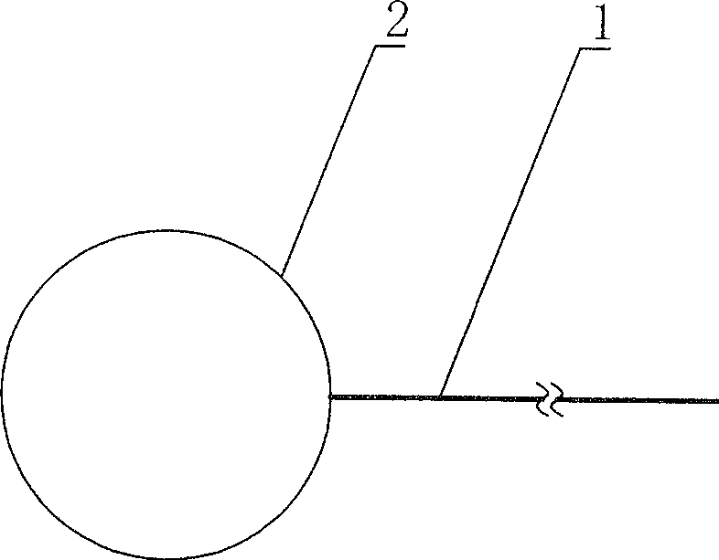
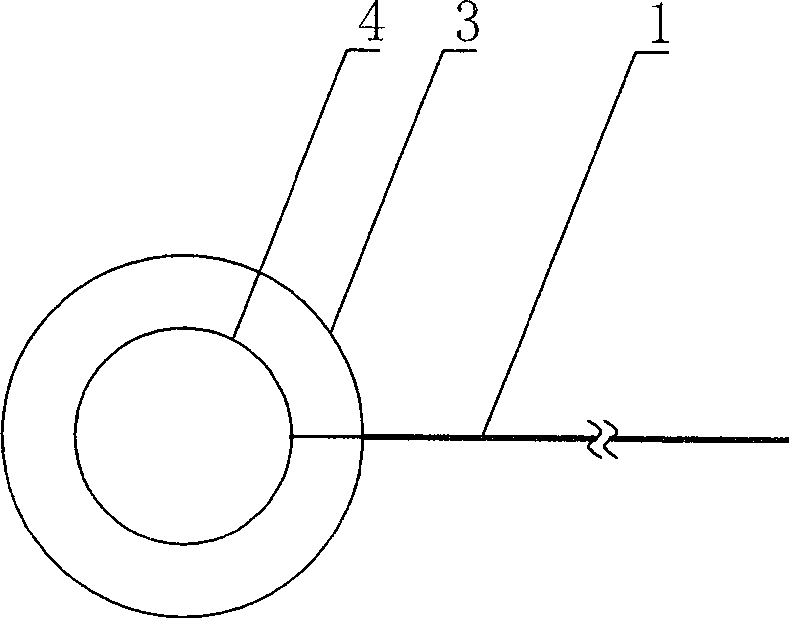
[0043] The recording electrode is a ring-shaped corneal contact electrode, the reference electrode and the ground electrode are needle-shaped electrodes, and the experimental animal is a rabbit.
[0044] Put methyl cellulose (or ofloxacin ointment) on the corneal surface of rabbit eyes. After corneal surface anesthesia, place double-circle contact electrode with inner / outer electrode diameter of 3mm / 5mm respectively, reference electrode and ground electrode needle Electrodes are pierced into the ears on the same side and the top of the head.
[0045] Using the above-mentioned micro-electrodes, in conjunction with the American LKC UTAS-2000 electrophysiological instrument, dual-channel recording of ERG, the recorded waveforms are as follows: Figure 4 Shown.
[0046] It can be seen from the figure that the a→b segment in the displayed waveform curve is basically free of interference clutter (that is, no steep spikes), and the waveform curve is smoother. The repetitive performance o...
Embodiment 2
[0048] The recording electrode is a ring-shaped corneal contact electrode, the reference electrode and the ground electrode are needle-shaped electrodes, and the experimental animal is a BN (Brown Norway) rat.
[0049] The recording electrode adopts a 0.8mm / 1.5mm double-circle contact electrode. After anesthetizing the animal, it is placed directly into the eyeball. The surface is dripped with saline or methylcellulose. The reference electrode and the ground electrode are needle electrodes, respectively, pierce the same side ears. And the top of the head.
[0050] Using the above-mentioned micro-electrodes, in conjunction with the American LKC UTAS-2000 electrophysiological instrument, dual-channel recording of ERG, the recorded waveforms are as follows: Figure 5 Shown.
[0051] It can be seen from the figure that the N1→O4 segment of the displayed waveform curve has no interference clutter, and the waveform curve is smoother, which has a good diagnostic and evaluation value.
[00...
Embodiment 3
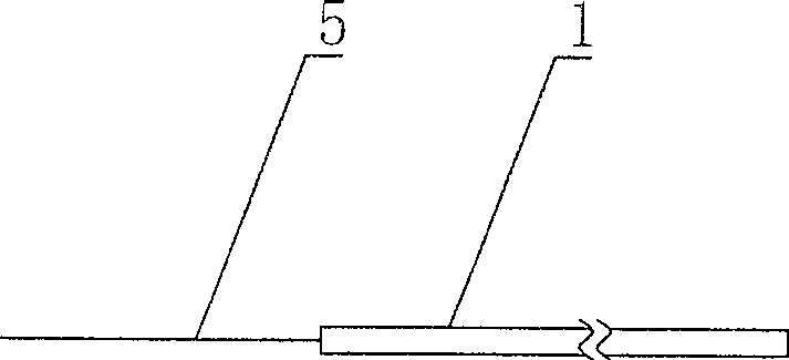
[0054] The recording electrode, the reference electrode and the grounding electrode adopt a metal needle electrode with a 1ml stainless steel needle as the tip for injection, and the experimental animal is a Wistar rat.
[0055] The recording electrode was pierced into the subcutaneous connection between the two ears, and the reference electrode and ground electrode were pierced into the same ear and the top of the head respectively.
[0056] Get the FVEP record waveform as Figure 6 Shown.
[0057] It can be seen from the figure that the displayed waveform curve has no clutter interference and stable waveform, which embodies the advantages of the technical solution of convenient and accurate positioning, easy fixation, good contact, stable recording waveform, and good repetitiveness of the recorded waveform.
[0058] The rest is the same as in Example 1 or 2.
PUM
 Abstract
Description
Claims
Application Information
 - R&D
- Intellectual Property
- Life Sciences
- Materials
- Tech Scout
- Unparalleled Data Quality
- Higher Quality Content
- 60% Fewer Hallucinations
Browse by: Latest US Patents, China's latest patents, Technical Efficacy Thesaurus, Application Domain, Technology Topic, Popular Technical Reports.
© 2025 PatSnap. All rights reserved.Legal|Privacy policy|Modern Slavery Act Transparency Statement|Sitemap|About US| Contact US: help@patsnap.com


