Preparation method of liquid phase protein chip

A protein chip and liquid phase technology, applied in the field of liquid phase protein chip preparation, can solve the problems of low sensitivity, complicated labeling steps, cumbersome operation, etc., to improve sensitivity and detection range, save specimen and reagent consumption, and improve detection efficiency Effect

Inactive Publication Date: 2008-03-19
GUANGZHOU DARUI BIOTECH
View PDF0 Cites 16 Cited by
  • Summary
  • Abstract
  • Description
  • Claims
  • Application Information

AI Technical Summary

Problems solved by technology

These methods have a common defect: they are all single-index detection, the amount of information is small, and when multiple items need to be detected, the amount of specimen is large, the detection cost

Method used


Image

  • Preparation method of liquid phase protein chip

Examples


Embodiment 1

[0039] Example 1: Preparation of liquid-phase protein chip

[0040] 1. Preparation of antibody-conjugated microspheres

[0041] (1) Take 2.5×10 6 Carboxylated microspheres (purchased from Luminex) were placed in a 1.5ml microcentrifuge tube, centrifuged at 13,000 rpm for 4 minutes, and the supernatant was discarded.

[0042] (2) Add 100 μl of deionized water, vortex and sonicate for 20 seconds to resuspend the microspheres. Centrifuge at 13,000 rpm for 4 minutes and discard the supernatant.

[0043] (3) Add 100 μl activation buffer (0.1mol / L NaH 2 PO 4 , pH 6.2), vortexed and sonicated for 20 seconds to resuspend the microspheres. Centrifuge at 13,000 rpm for 4 minutes and discard the supernatant.

[0044] (4) Add 80 μl of activation buffer, vortex and sonicate for 20 seconds to resuspend the microspheres.

[0045] (5) Weigh an appropriate amount of N-hydroxysulfosuccinimide (Sulfo-NHS) and carbodiimide (EDC) respectively, and prepare them with deionized water to a conc...

Embodiment 2

[0074] Embodiment 2: the preparation of the solution required for reaction

[0075] 1. Preparation of Standard Diluent

[0076] Prepare the standard dilution solution according to the following formula: 0.8% NaCl, 0.02% KCl, 0.29% Na 2 HPO 4 , 0.024% KH 2 PO 4 , 1.5-7.5% BSA, 0.01% Tween20, 0.05% sodium azide, pH7.4.

[0077] 2. Preparation of standard products

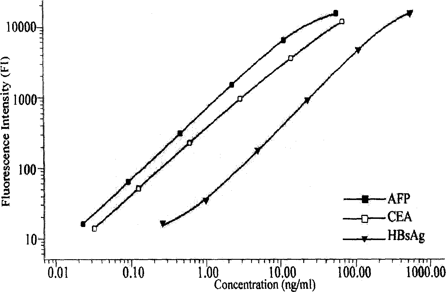
[0078] Take an appropriate amount of stock solutions containing high concentrations of CEA, AFP, and HBsAg antigens, and prepare standard substances 1-6 (Std1-Std6) and standard substance 7 ( Std7) is a standard product dilution without any antigen.

[0079] CEA (ng / ml)

AFP(U / ml)

HBsAg(ng / ml)

Std 1

80

55

1000

Std 2

16

11

200

Std 3

3.2

2.2

40

Std 4

0.64

0.44

8

Std 5

0.128

0.088

1.6

Std 6

0.032

0.022

0.4

Std 7

0

0

0

[0080] 3. Preparation of assay buffer ...

Embodiment 3

[0084] Example 3: The method of using the liquid-phase protein chip

[0085] 1. Vortex and sonicate the antibody-coupled microspheres of CEA, AFP, and HBsAg for 20 seconds to resuspend.

[0086] 2. Take an appropriate amount of microsphere suspension, and use the assay buffer to prepare a diluted microsphere mixture containing 80-100 microspheres with different fluorescent codes per μl.

[0087]3. Add 100 μl / well of assay buffer to the required reaction wells of a 96-well membrane filter plate (MultiScreen MABVN 1.2 μm) to pre-wet, seal the remaining wells with plate sealing film, and vacuum filter to remove the liquid.

[0088] 4. Add 25 μl / well of standard and serum samples (pre-diluted 25 times with standard diluent) to the standard wells (double wells can be set) and sample wells of the filter plate.

[0089] 5. Add 25 μl / well of the diluted microsphere mixture to the above wells.

[0090] 6. Seal the reaction well with a sealing film, and incubate at room temperature fo...


PUM

PropertyMeasurementUnit
Diameteraaaaaaaaaa

Abstract

The present invention relates to the biologic technology field, and discloses a liquid phase albumen chip and the preparation and the usage method thereof which can simultaneously test human serum carcinoma embryonic antigen (CEA), Alpha fetoprotein (AFP), and hepatitis B surface antigen (HBsAg). The present invention couples the specificity antibody of CEA, AFP, and HBsAg on different fluorescence micro-spheres, and uses the test antibody marked by biotin or phycoerythrin to determine the nature quickly and quantitatively analyze the above three indexes with the double antibody sandwich method. The present invention uses the filtering membrane board when testing, and washes the board 3 times after each reaction is finished to increase the signal and improve the sensitivity. The present invention has high sensitivity, strong specificity, stable result, excellent repeatability, and simple operation; 1 micro liter serum sample can test three indexed simultaneously. The present invention is applicable to the health test and the general examination as well as the clinic test of the high risk population, and can facilitate the early diagnosis and the early treatment of the knub.

Description

[0001] Technical field [0002] The invention relates to the field of biotechnology, in particular to a method for preparing a liquid-phase protein chip. Background technique [0003] Biochip is a biological detection technology developed in the early 1990s. 2 Different biomolecular probes (proteins, nucleic acids, etc.) are immobilized at different positions in the form of microarrays on support media such as glass slides, silicon wafers, nylon membranes, gels, or metals. These probe molecules can be mixed with solutions The interaction of target molecules in the reaction (such as hybridization, antigen-antibody binding reaction, etc.), after the reaction is completed, the unreacted molecules are washed away by washing the film. The target molecule can be marked in advance, or it can be stained after reacting with the probe molecule, and then the presence or strength of the reaction signal is detected by signal collection equipment (such as fluorescence microscope, laser co...

Claims


Application Information

Patent Timeline
IPC IPC(8): G01N33/546G01N33/545G01N33/577
Inventor 李明陈玮吴英松
Owner GUANGZHOU DARUI BIOTECH
Why Eureka
Try Eureka